Cerebral Tumor Surgery: Procedure, Recovery and Results

Surgery may aim to remove a tumor, obtain a biopsy, relieve pressure, improve symptoms, or make other treatments more effective. Neurosurgeons use detailed imaging, navigation technology and functional testing to protect important areas of the brain whenever possible.
Key Takeaways
- Surgery may aim to remove a tumor, obtain a biopsy, relieve pressure, improve symptoms, or make other treatments more effective.
- Neurosurgeons use detailed imaging, navigation technology and functional testing to protect important areas of the brain whenever possible.
- A short stay in an intensive care unit is common after brain surgery for close observation and is not necessarily a sign of a complication.
- Recovery can involve temporary fatigue, headaches, swelling and changes in neurological function; rehabilitation may support independence and quality of life.
- Survival after surgery depends mainly on the tumor diagnosis and biology, rather than on the operation alone.
Cerebral tumor surgery is a carefully planned operation to remove all or part of a tumor in the brain, obtain tissue for diagnosis, and reduce symptoms caused by pressure on nearby brain structures. Recovery, results and long-term outlook vary widely with the tumor’s type, location, grade, and the person’s overall health.
Overview: what cerebral tumor surgery involves
Cerebral tumor surgery is an operation performed to remove, reduce, or obtain a sample from a tumor located in the brain. It is often called cerebral tumor resection or cerebral tumor removal. The purpose may be to establish an accurate diagnosis, ease pressure within the skull, improve symptoms, remove as much tumor as safely possible, or prepare for treatments such as radiotherapy, chemotherapy, targeted therapy, or monitoring.
The operation is individualized. Some tumors can be completely removed, while others lie close to areas that control movement, speech, vision, sensation, memory, or breathing. In these situations, the safest and most useful result may be partial removal rather than attempting to remove every visible cell. The pathology report from the removed tissue helps the care team identify the tumor type and guide the next steps.
Not every brain tumor requires an immediate operation. Depending on imaging findings, symptoms, growth pattern, location, and suspected diagnosis, a specialist may recommend surveillance imaging, a needle biopsy, medication, radiation treatment, or another approach. A neurosurgical assessment helps determine which option best balances benefit and safety.
Who may be a candidate for surgery?
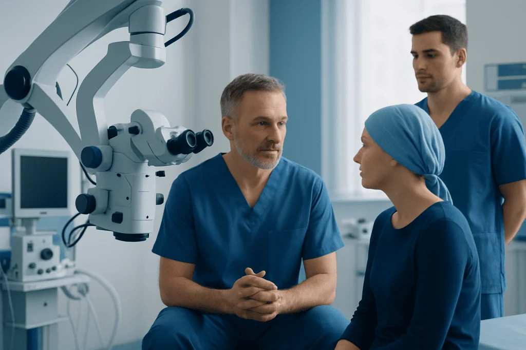
A person may be considered for cerebral tumor surgery when a tumor is causing symptoms, appears to be growing, has uncertain features on imaging, or can be safely approached to reduce its size. Symptoms that can lead to evaluation include seizures, persistent or worsening headaches, new weakness or numbness, balance problems, language difficulties, vision changes, confusion, or changes in personality. Some tumors are found incidentally during imaging performed for another reason.
The decision is usually made by a multidisciplinary team that may include a neurosurgeon, neurologist, neuroradiologist, neuropathologist, medical oncologist, radiation oncologist, anesthesiologist, and rehabilitation professionals. The team reviews MRI or CT scans, the suspected tumor type, its relationship to critical brain structures, and the person’s medical history and goals.
Before surgery, patients may have blood tests, heart or anesthesia assessments, and additional MRI scans for surgical planning. Functional MRI, tractography, or other specialized studies can help map pathways involved in movement or language. In selected cases, an awake procedure is considered so the surgical team can monitor speech, movement, or other functions during removal.
- Good candidates are not defined by age alone; overall health and the expected benefit matter most.
- Some deep, diffuse, or high-risk tumors are better assessed with biopsy or treated primarily with non-surgical therapies.
- Patients should discuss expected benefits, alternatives, and possible changes in neurological function before consenting to surgery.
How is cerebral tumor surgery performed?
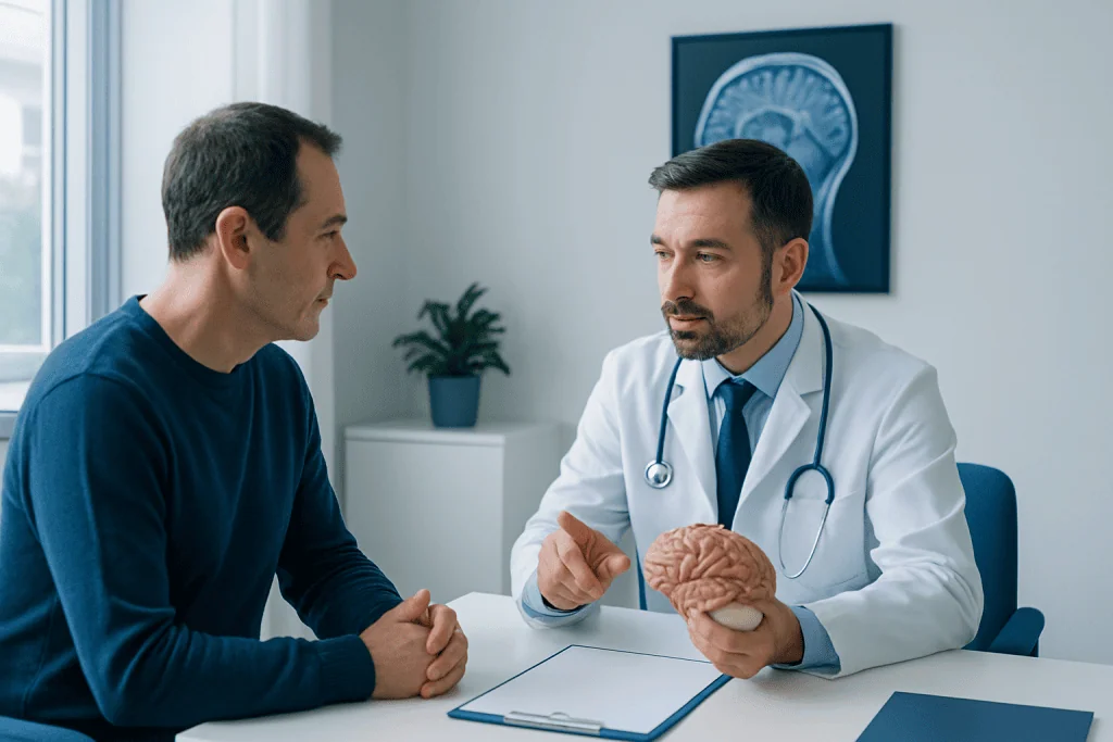
Most cerebral tumor surgery is performed through a craniotomy, in which the surgeon temporarily removes a small section of skull to reach the tumor. The patient is placed under general anesthesia for many procedures. The scalp is prepared, an incision is made, and the surgical team uses detailed preoperative images and image-guidance systems to identify the safest route to the tumor.
During the operation, the neurosurgeon removes tumor tissue while preserving healthy brain tissue and blood vessels as far as possible. Microsurgical instruments, an operating microscope, ultrasound, fluorescence guidance, neurophysiological monitoring, and navigation systems may be used. A specimen is sent to the laboratory, where a neuropathologist examines it and may perform molecular tests that help classify the tumor.
Some tumors close to areas governing speech or movement may be removed during an awake craniotomy. Although the scalp and skull are numbed, the patient can respond to simple tasks during key portions of surgery. This can help the team identify and protect important functions. Other patients may undergo stereotactic biopsy, a smaller procedure that uses image guidance to collect tissue when full removal is not advisable.
After the tumor is removed or sampled, the bone section is usually secured back in place and the incision is closed. The length of surgery varies substantially according to the tumor’s size, location, blood supply, and complexity. An early postoperative scan is commonly performed to assess the surgical area.
What should I expect after brain tumor surgery?
After brain tumor surgery, the patient is transferred to a recovery area or an intensive care unit for frequent checks of alertness, pupils, speech, strength, sensation, and vital signs. Headache, tiredness, nausea, scalp tenderness, and swelling around the face or eyes can occur in the first days. These symptoms are usually monitored and managed with an individualized care plan.
Medication may be used to control pain, prevent or treat seizures, reduce brain swelling, prevent nausea, and lower the risk of blood clots when appropriate. Steroids are sometimes prescribed for swelling and are adjusted carefully by the clinical team. Patients should not change or stop prescribed medicines without medical advice.
Hospital stay often lasts several days, but it may be shorter or longer depending on the operation and recovery. Most people are encouraged to sit up, walk with support, and begin gentle activity as soon as it is safe. Before discharge, the team reviews wound care, medications, warning signs, activity limits, follow-up imaging, and the expected pathology timeline.
Neurological changes after surgery may be temporary or persistent. For example, a person may notice slowed thinking, fatigue, difficulty finding words, weakness, visual changes, or balance problems. Early assessment by physiotherapy, occupational therapy, speech and language therapy, or neuropsychology can help identify needs and create a practical rehabilitation plan.
Recovery timeline, including cerebellum surgery recovery
Recovery is gradual and differs from person to person. During the first one to two weeks, rest, short walks, hydration, regular meals, and avoiding strenuous activity are commonly advised. Many patients experience significant fatigue, which can fluctuate through the day. The incision generally needs monitoring for redness, drainage, separation, or increasing pain, and follow-up appointments allow the team to assess healing and review results.
Over the following weeks, energy and daily function may steadily improve, although recovery can take months. Returning to work, driving, exercise, travel, or caring responsibilities should be discussed with the treating team. Driving restrictions may apply after a seizure or depending on local rules and neurological recovery. People should avoid alcohol or medicines that affect alertness unless their clinician confirms they are safe.
Cerebellum tumor surgery recovery may particularly involve balance, coordination, dizziness, nausea, eye-movement changes, or difficulty with fine motor tasks because the cerebellum helps coordinate movement. Cerebellum surgery recovery can therefore include balance training, walking support, vestibular rehabilitation, and occupational therapy. These symptoms often improve, but the pace depends on the tumor, surgical area, and any pre-existing neurological effects.
Follow-up usually includes a postoperative MRI and further scans at intervals selected by the treating team. The pathology findings may lead to observation alone or to additional care such as radiotherapy, systemic treatment, or enrollment in an appropriate clinical trial. The follow-up plan is tailored to the specific diagnosis.
Benefits, risks and quality of life after surgery
Potential benefits of surgery include relieving raised pressure in the skull, improving seizures or other symptoms, confirming a diagnosis, and reducing the amount of tumor that remains. For many tumors, removing a larger amount safely can improve local control or support the effectiveness of other treatments. However, the goal is always safe, meaningful treatment rather than removal at any cost.
All brain operations carry risks. These can include bleeding, infection, fluid leakage, blood clots, seizures, stroke, swelling, anesthesia-related problems, and wound-healing concerns. Depending on the location, there can also be changes in speech, memory, concentration, personality, vision, sensation, strength, balance, swallowing, or coordination. The surgeon discusses the risks that are most relevant to the individual tumor and planned approach.
What is the quality of life like after brain tumor surgery? Quality of life can be good, especially when symptoms caused by the tumor improve and rehabilitation supports recovery. Some people return to many previous activities, while others need temporary or longer-term help with mobility, communication, thinking skills, mood, or fatigue. Open communication with the care team, early rehabilitation, symptom management, and emotional support can all make a meaningful difference.
Mood changes, anxiety, low mood, sleep disruption, and uncertainty during follow-up are understandable experiences. Patients and families can ask for psychological support, social work guidance, support groups, and rehabilitation services. Acibadem International’s multidisciplinary specialists and JCI-accredited hospitals can assess and treat brain tumors for international patients, with care plans coordinated across neurosurgery, oncology, imaging, pathology, and rehabilitation.
What is the average survival time after brain tumor surgery?
There is no single average survival time after brain tumor surgery because “brain tumor” describes many different conditions. Some tumors are benign and may be cured or controlled for many years after surgery. Others are malignant and require ongoing treatment; their outlook depends strongly on tumor grade, molecular features, location, how much can be safely removed, age, general health, and response to further therapy.
Surgery itself is only one part of the prognosis. The pathology report, including molecular testing when relevant, provides more useful information than the operation alone. For example, different types of glioma, meningioma, pituitary tumors, metastases, and childhood brain tumors can have very different expected courses.
Patients should ask their neurosurgeon or oncologist to explain the diagnosis in plain language and discuss what is known about their individual outlook. It is appropriate to ask about the treatment goal, the recommended follow-up plan, possible additional therapy, and supportive care needs. Prognostic discussions are most accurate after final pathology results are available.
Is it normal to go to the ICU after brain surgery? When to seek medical care
Is it normal to go to the ICU after brain surgery? Yes. A stay in an intensive care unit, high-dependency unit, or specialized neurosurgical observation area is common after a craniotomy. It allows nurses and doctors to perform frequent neurological checks and respond quickly to changes in blood pressure, pain, nausea, breathing, or neurological function. The length of monitoring depends on the procedure and the person’s recovery.
After discharge, patients should contact their surgical team promptly for a fever, increasing redness or drainage from the wound, persistent vomiting, worsening headache, new or increasing weakness, confusion, speech difficulty, vision changes, severe drowsiness, a seizure, chest pain, shortness of breath, or a swollen painful leg. Sudden neurological symptoms, such as facial drooping, one-sided weakness, difficulty speaking, loss of consciousness, or a first seizure, require emergency medical attention.
Follow-up is also important even when recovery appears smooth. Appointments allow the team to review pathology, scan results, medications, wound healing, rehabilitation progress, and future treatment options. Patients are encouraged to bring questions and, if helpful, a family member or trusted support person to consultations.
Frequently asked questions
How long does cerebral tumor surgery take?
The duration varies greatly, often from a few hours to longer for complex tumors. It depends on the tumor’s size, location, blood supply, relationship to important brain functions, and whether specialized mapping is required. The surgical team can provide a more individualized estimate before the procedure.
Will all of the tumor be removed during surgery?
Not always. The aim is to remove as much tumor as is safely possible while protecting essential neurological function. When a tumor is close to critical brain structures or spreads diffusely into surrounding tissue, a partial resection or biopsy may be safer and may be followed by other treatments.
How painful is recovery after brain tumor surgery?
Pain is usually related to the scalp incision, head positioning during surgery, and postoperative swelling rather than the brain tissue itself. Headache and tenderness are common and are treated with an individualized medication plan. Severe or worsening pain should be reported to the surgical team.
When can someone return to normal activities after brain tumor surgery?
Light activity and short walks may begin soon after surgery when approved by the clinical team, but full recovery commonly takes weeks to months. Return to work, driving, exercise, and travel depends on neurological recovery, seizure risk, fatigue, and the type of work involved. A treating clinician should clear these activities individually.
Can brain tumor surgery change personality or memory?
It can, particularly when the tumor or surgical area involves networks responsible for thinking, emotion, language, or behavior. Some changes are temporary and improve as swelling resolves, while others may require rehabilitation or longer-term support. Cognitive assessment and neuropsychological rehabilitation can be helpful when concerns arise.
What happens if pathology shows a malignant brain tumor?
The care team reviews the tumor type, grade, molecular features, and amount removed, then recommends an individualized plan. This may include radiotherapy, chemotherapy, targeted treatments, monitoring, rehabilitation, and symptom management. A multidisciplinary discussion helps coordinate these decisions.
References
- World Health Organization
- National Cancer Institute
- American Brain Tumor Association
- National Institute of Neurological Disorders and Stroke
- European Association of Neuro-Oncology
This article is for general information only and is not a substitute for professional medical advice. Please consult a qualified doctor about your individual situation.
Oncology care in Turkey — second opinion and treatment plan
JCI-accredited · board-certified surgeons · reply within 24h

More from the Health Library


Related Specialists
Dr. Muzaffer Ağır
Orthopedic Surgery & Traumatology
Prof. Dr. Hüsnü Görgen
Gynecology & Obstetrics
Dr. Mehmet Erem
Otorhinolaryngology
Prof. Dr. Mehmet Ali Şehitoğlu
Otorhinolaryngology