

Corneal Graft: An Evidence-Based Patient Guide

A corneal graft may replace the full thickness of the cornea or only the damaged layers. The best procedure depends on which corneal layer is affected and whether cataract or glaucoma is also present.
Key Takeaways
- A corneal graft may replace the full thickness of the cornea or only the damaged layers.
- The best procedure depends on which corneal layer is affected and whether cataract or glaucoma is also present.
- Vision recovery is gradual and can take months; follow-up and prescribed eye drops are essential.
- Rejection is possible but is often treatable when warning symptoms are reported promptly.
- Long-term graft survival varies with the diagnosis, graft type, eye health and ongoing care.
A corneal graft, also called a corneal transplant or keratoplasty, replaces diseased or damaged corneal tissue with healthy donor tissue. It may improve vision, relieve pain from certain corneal disorders, or restore the eye’s protective surface when other treatments are not enough.
Corneal graft overview: what it is and why it is used
A corneal graft is surgery that replaces part or all of the cornea, the clear front window of the eye. The cornea helps focus incoming light and provides a smooth, protective surface. When it becomes scarred, swollen, irregular, infected, or permanently cloudy, vision can become blurred and the eye may be uncomfortable or vulnerable to further damage.
Doctors may recommend corneal graft treatment when glasses, contact lenses, medicines, or less invasive procedures cannot provide adequate vision or protect the eye. The goal may be visual rehabilitation, restoration of the cornea’s clarity and structure, relief of pain, or urgent treatment of severe corneal damage. A graft does not treat every cause of reduced sight, so the condition of the retina, optic nerve and other eye structures also matters.
The donated corneal tissue is carefully screened by an eye bank. Because the cornea has no blood vessels, it can be transplanted without matching blood type in most cases. An ophthalmologist with corneal expertise will discuss whether a graft is appropriate and which approach is most suitable.
How a corneal graft works and the main types
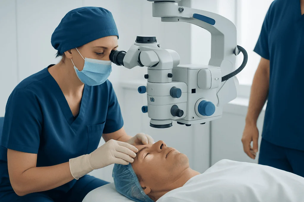
The cornea has several layers. A modern corneal vision graft is usually designed to replace only the layer or layers that are unhealthy whenever possible. Preserving healthy tissue can reduce recovery time, improve stability, and lower some risks compared with replacing the entire cornea.
In penetrating keratoplasty, the surgeon replaces the full thickness of the central cornea with a circular donor button secured using fine sutures. This approach may be needed for deep scars, extensive thinning, severe infection-related damage, or disease affecting multiple corneal layers.
Lamellar procedures replace selected layers. Deep anterior lamellar keratoplasty replaces the front and middle layers while retaining the patient’s healthy inner endothelial layer. Endothelial keratoplasty, including DMEK or DSAEK, replaces the inner cell layer when corneal swelling results from endothelial disease. These techniques are commonly considered for conditions such as keratoconus or endothelial disorders, depending on the individual eye.
In some people, cataract removal and endothelial keratoplasty are performed during the same operation. This is often called a corneal graft triple procedure, typically combining cataract surgery, lens implantation and endothelial graft surgery. Whether combined surgery is advisable depends on the cornea, cataract, eye pressure and the person’s overall visual needs.
Who may be a candidate for corneal graft treatment

Potential candidates include people with corneal scarring after injury or infection, progressive corneal thinning or distortion, inherited corneal disorders, persistent corneal swelling after prior surgery, or graft failure from an earlier transplant. The decision is individualized. Some patients benefit more from corneal cross-linking, specialty contact lenses, cataract surgery, treatment of dry eye, or other measures before transplantation is considered.
Before surgery, the ophthalmologist assesses vision, corneal shape and thickness, corneal cell health, tear film, eyelids, retina and optic nerve. Tests may include slit-lamp examination, corneal topography or tomography, optical coherence tomography, and measurements of eye pressure. Existing glaucoma, inflammation, active infection, severe dry eye, or retinal disease can influence surgical planning and expected results.
Corneal graft and eye pressure require particular attention. Raised eye pressure can damage the optic nerve and may also affect graft health. Some patients need pressure-lowering drops, laser treatment, or glaucoma surgery before or after transplantation. The care team will balance the need to protect both the graft and the optic nerve.
A person is generally not considered ready for an elective graft if there is uncontrolled eye infection or severe active inflammation. Addressing these issues first can improve safety and the likelihood of a stable result.
What happens during corneal graft surgery
Corneal graft surgery is usually an outpatient procedure. Depending on the type of transplant and the patient’s needs, it may be performed with local anesthesia and sedation or with general anesthesia. The eye is cleaned and covered with a sterile drape, and the person should not feel pain during the operation.
For a full-thickness graft, the surgeon removes a precise circular section of affected cornea and replaces it with matching donor tissue. Very fine sutures hold the graft in place. For endothelial procedures, a thin layer of donor tissue is positioned inside the eye and supported temporarily by an air or gas bubble. The patient may need to lie face-up for a specified period afterward so the graft can attach properly.
For certain endothelial transplants, sutures may not be needed. In contrast, full-thickness graft sutures can remain in place for months or longer and may be removed gradually when the cornea is stable. The operation itself varies in duration according to the graft type and whether additional procedures are performed.
After surgery, an eye shield is typically used, and prescribed drops help prevent infection and control inflammation. If cataract is contributing to blurred vision, the ophthalmologist may discuss cataract surgery alone or as part of a combined surgical plan.
How long does it take to recover from a corneal graft?
Recovery from a corneal graft is gradual and varies considerably by procedure. Many people notice early improvement over weeks after endothelial keratoplasty, although vision may continue to refine for several months. Recovery after a full-thickness graft or anterior lamellar graft is often longer, commonly many months and sometimes up to a year or more, because the wound and sutures need time to stabilize.
Immediately after surgery, blurred vision, light sensitivity, tearing, a scratchy sensation and mild redness are common. The eye may be covered with a shield initially. Follow-up appointments are important because the surgeon checks graft attachment, healing, inflammation, stitches when present, and eye pressure.
People should use drops exactly as prescribed, avoid rubbing or pressing on the eye, and protect it from accidental injury. The surgeon will provide individualized guidance about bathing, driving, working, exercise, swimming, lifting and contact sports. New glasses or a contact lens fitting may be considered only after the cornea has become sufficiently stable.
Recovery is not judged only by how clear vision becomes. A healthy, comfortable, stable corneal surface and control of conditions such as glaucoma are also important outcomes. The care plan may include rehabilitation of the ocular surface or visual correction after healing.
How painful is a corneal graft?
A corneal graft is performed with anesthesia, so pain should not be felt during surgery. Afterward, many people experience mild to moderate discomfort rather than severe pain. Common sensations include grittiness, tearing, burning, light sensitivity, or the feeling that something is in the eye, especially in the first few days.
Discomfort is often managed with the aftercare plan recommended by the surgeon. It is important not to self-medicate with eye drops that have not been approved, as some products may delay healing or worsen pressure problems. The ophthalmology team can advise on safe pain relief and how to use prescribed drops.
Increasing pain, marked redness, a sudden decline in vision, or new sensitivity to light should not be assumed to be normal recovery. These symptoms need urgent assessment because they can occur with infection, inflammation, increased eye pressure, graft rejection, or other complications.
Benefits, risks and the typical rate of rejection for corneal grafts
A successful corneal graft can improve corneal clarity, reduce distortion, relieve symptoms caused by a diseased cornea, and help preserve the eye. The degree of visual improvement depends on the original condition and whether other structures, such as the lens, retina or optic nerve, are healthy. Some people still need glasses, specialty contact lenses, or additional treatment after surgery.
Possible complications include infection, bleeding, wound problems, irregular astigmatism, swelling, detachment of an endothelial graft, cataract, and elevated eye pressure or glaucoma. In some situations, a graft can become cloudy or fail over time and may require further treatment or repeat transplantation. Corneal graft removal is not a routine step; when a graft is failing or causing complications, the ophthalmologist first identifies the cause and discusses options, which may include medical treatment, a repair procedure, or repeat grafting.
The typical rate of rejection for corneal grafts cannot be represented by one reliable number. It varies substantially by graft type, underlying disease, previous grafts, degree of corneal vascularization, inflammation, glaucoma, and adherence to steroid drops and follow-up. Endothelial grafts and low-risk first transplants may have lower rejection risk than high-risk or repeat full-thickness grafts, but rejection remains possible with any donor corneal transplant.
Rejection often can be treated if recognized early. Patients are commonly advised to seek urgent eye care for redness, sensitivity to light, reduced vision, or pain. These symptoms are sometimes summarized as the “RSVP” warning signs: redness, sensitivity to light, vision reduction and pain.
Average lifespan, long-term care and when to seek medical care
The average lifespan of a corneal graft varies too widely for one prediction. Some grafts remain clear for decades, while others fail earlier because of the original corneal disease, loss of endothelial cells, rejection, eye pressure problems, infection, injury, or chronic inflammation. Endothelial cell loss occurs gradually over time, which is one reason long-term monitoring remains important even when vision is stable.
Regular ophthalmology reviews help detect subtle changes before they affect vision significantly. Long-term drops may be needed, particularly anti-inflammatory drops, and the prescription should never be stopped or changed without medical guidance. Eye protection during activities with a risk of impact and careful management of dry eye, eyelid disease, diabetes, or glaucoma can support graft health.
When to seek medical care: urgent same-day assessment is appropriate for sudden blurring or loss of vision, increasing redness, significant pain, light sensitivity, discharge, eye injury, or a feeling that the graft has shifted. A prompt review is also important if prescribed drops cause unexpected symptoms or if eye pressure may be elevated. These signs do not always mean rejection, but rapid assessment offers the best opportunity to protect the eye.
Acibadem International’s multidisciplinary eye specialists at JCI-accredited hospitals diagnose and treat corneal conditions for international patients, coordinating care when corneal disease is complicated by cataract, glaucoma or other eye concerns. An individualized ophthalmology consultation can clarify the likely benefits, limitations and aftercare needs of a corneal graft.
Frequently asked questions
What is a corneal graft?
A corneal graft is a transplant procedure that replaces damaged or diseased corneal tissue with healthy donor corneal tissue. It may involve the whole thickness of the cornea or only specific layers, depending on the condition being treated.
How long does it take to recover from a corneal graft?
Recovery depends on the type of graft and the health of the eye. Initial healing may take weeks, while vision can continue improving for months; full-thickness grafts often take longer to stabilize than endothelial grafts. Follow-up visits and prescribed drops are essential throughout recovery.
How painful is a corneal graft?
Anesthesia prevents pain during the procedure. After surgery, a gritty sensation, tearing, light sensitivity and mild to moderate discomfort are common for a short period. Severe or worsening pain should be assessed urgently by an eye specialist.
What is the average lifespan of a corneal graft?
There is no single average lifespan that applies to every person. Some grafts remain clear for many years or decades, while others may fail earlier due to rejection, endothelial cell loss, raised eye pressure, inflammation, infection or the underlying eye condition. Long-term follow-up helps protect graft function.
What is the typical rate of rejection for corneal grafts?
Rejection rates vary by procedure type and individual risk factors, so a single percentage can be misleading. The risk is influenced by previous transplants, inflammation, corneal blood vessels, glaucoma and the original diagnosis. Early reporting of redness, light sensitivity, reduced vision or pain is important because rejection can often be treated promptly.
Can a corneal graft improve vision completely?
A graft can substantially improve vision when corneal disease is the main cause of visual loss. However, the final outcome also depends on astigmatism, cataract, glaucoma, retinal health and optic nerve function. Glasses, contact lenses or further treatment may still be needed after healing.
References
- American Academy of Ophthalmology
- Eye Bank Association of America
- National Eye Institute
- National Health Service
- Mayo Clinic
This article is for general information only and is not a substitute for professional medical advice. Please consult a qualified doctor about your individual situation.
Explore treatments in Turkey — costs, top hospitals & a free quote
JCI-accredited · board-certified surgeons · reply within 24h

More from the Health Library



Related Specialists

Dr. Melisa Ulufi
Inpatient Clinic Physicians Clinical Service
Dr. Ezgi Darıcı
Gynecology & Obstetrics
Prof. Dr. Ayşe Korkmaz Toygar
Neonatal Intensive Care Unit
Dr. Kutluhan Demirarslan
Inpatient Clinic Physicians Clinical Service