Quick answer
Cataract is a clouding of the eye’s natural lens that causes blurred vision, glare, and gradually reduced sight, and it is treated when needed by removing the cloudy lens and replacing it with an artificial one. At Acibadem in Turkey, cataract care includes ophthalmic evaluation, diagnosis, and tailored surgical planning, with lens replacement performed using modern techniques according to the…
What is cataract?
A cataract is a clouding of the natural lens inside the eye. The lens is a small, transparent structure that sits behind the colored part of the eye (the iris) and focuses light onto the retina, the light-sensitive layer at the back of the eye. When the lens is clear, light passes through it easily and vision is sharp. When a cataract develops, proteins within the lens gradually clump together and the lens becomes cloudy. As a result, light is scattered or blocked, and vision becomes blurry, dim, or hazy, often described as looking through a foggy or frosted window.
If you have wondered what is cataract in simple terms, it helps to know that a cataract is not a growth, a film over the eye, or a form of cancer. It is a change within the lens itself, and in most cases it develops slowly over months or years. Cataracts are extremely common, and age is the most important factor: many people begin to develop some lens clouding in middle age or later, and cataract is one of the leading causes of treatable vision loss worldwide. Cataracts can affect one eye or both eyes, though they often do not progress at the same rate in each eye. Less commonly, cataracts occur in younger adults, and some babies are born with them (congenital cataracts) or develop them in childhood.
The medical classification code for an unspecified cataract is ICD-10 H26.9. Doctors also describe cataracts by where the clouding occurs in the lens, for example, in the center (nuclear cataract), around the edges moving inward (cortical cataract), or at the back of the lens (posterior subcapsular cataract). The type and location influence which symptoms appear first and how quickly vision changes.
Symptoms of cataract
Cataract symptoms usually develop gradually, and in the early stages many people notice little or no change in their vision. As the clouding of the lens increases, symptoms become more noticeable and can begin to interfere with everyday activities such as reading, driving, and recognizing faces.
Common cataract symptoms include:
- Blurry, cloudy, or hazy visionoften described as looking through fog or a dirty window
- Increased sensitivity to glarebright sunlight, oncoming headlights, or lamps may feel uncomfortably dazzling
- Difficulty seeing at nightnight driving often becomes harder before daytime vision changes noticeably
- Halos around lightsrings or starbursts around headlights and streetlights
- Colors appearing faded or yellowedthe world may look less vivid or slightly brownish
- Frequent changes in eyeglass or contact lens prescriptionglasses seem to stop working as well as they used to
- Double vision or ghost images in one eyeseeing two overlapping images even when the other eye is covered
- Needing brighter light for reading and other close work
Symptoms can differ depending on the type and stage of the cataract. A nuclear cataract, in the center of the lens, often causes a slow yellowing of vision and, in some people, a temporary improvement in near vision (sometimes called “second sight”) before vision worsens again. A cortical cataract, which starts at the outer edge of the lens, tends to cause problems with glare and light scatter early on. A posterior subcapsular cataract, at the back of the lens, can progress more quickly than other types and often affects reading vision and vision in bright light first; this type is more common in younger people and in those who have taken corticosteroid medications for long periods.
Cataracts do not typically cause pain, redness, or discharge. If you experience eye pain, sudden vision loss, or flashes of light, these symptoms suggest a different problem and should be evaluated promptly, as described in the final section of this article.
Causes and risk factors
The most common cataract cause is the natural aging of the lens. Over time, the proteins and fibers within the lens break down and clump together, gradually reducing its transparency. This process is a normal part of aging, which is why cataracts become increasingly common in later decades of life.
Beyond aging, several factors are known or believed to increase the risk of developing cataracts or to speed up their progression:
- Diabeteshigh blood sugar levels can accelerate changes in the lens
- Ultraviolet (UV) light exposurelong-term unprotected exposure to sunlight is associated with earlier lens clouding
- Smokingtobacco use is linked to a higher risk of cataract
- Heavy alcohol useexcessive drinking over time may contribute to lens changes
- Long-term corticosteroid usesteroid medications taken by mouth, and in some cases by inhaler or eye drops over long periods, are associated with posterior subcapsular cataracts
- Eye injury or previous eye surgerytrauma to the eye can lead to a cataract, sometimes years after the injury
- Other eye conditionslong-standing inflammation inside the eye (uveitis) and certain other eye diseases can promote cataract formation
- Radiation exposureincluding radiation therapy involving the head or eye area
- Family history and geneticscataracts sometimes run in families, and certain genetic conditions increase risk
- Congenital factorssome infections or developmental problems during pregnancy can cause cataracts present at birth
Having one or more risk factors does not mean a person will definitely develop a significant cataract, and some people develop cataracts without any identifiable risk factor other than age. While no method is proven to fully prevent cataracts, doctors generally advise protecting the eyes from UV light with sunglasses, not smoking, managing diabetes carefully, and attending regular eye examinations, as these steps support overall eye health.
Diagnosis
Cataract diagnosis is made by an eye care professional during a comprehensive eye examination. An ophthalmologist (a physician specializing in eye diseases and eye surgery) or an optometrist can identify a cataract using standard, painless tests performed in the clinic. There is usually no need for blood tests or advanced imaging scans to diagnose a typical age-related cataract.
A typical evaluation includes several steps:
- Visual acuity testyou read letters on a chart to measure how clearly you see at various distances, with each eye tested separately.
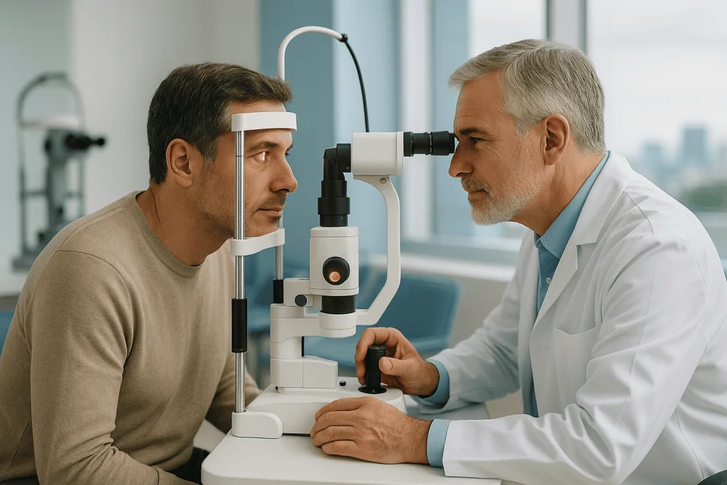
- Slit-lamp examinationthe doctor uses a special microscope with a bright, thin beam of light to examine the front structures of the eye, including the cornea (the clear front window of the eye), the iris, and the lens. This is the key test for seeing the cataract directly and assessing its type, location, and density.
- Dilated eye examinationeye drops are used to widen (dilate) the pupil so the doctor can view the lens and the retina more completely. This also helps rule out other causes of vision loss, such as macular degeneration or glaucoma, that may exist alongside a cataract. Your vision may be blurry and light-sensitive for a few hours after dilation.
- Tonometrya measurement of the pressure inside the eye, usually performed as part of a full examination to check for glaucoma.
In some cases, additional tests are used. Glare or contrast sensitivity testing can show how much a cataract affects vision in real-world conditions, since some cataracts cause more trouble with glare than a standard eye chart reveals. If surgery is being planned, precise measurements of the eye’s length and the curvature of the cornea (called biometry) are taken to calculate the power of the artificial lens that will replace the cloudy one. If the cataract is so dense that the doctor cannot see the retina behind it, an ultrasound scan of the eye may be used to check that the back of the eye appears healthy before surgery.
An important part of cataract diagnosis is deciding how much the cataract is actually affecting your daily life. Doctors combine the examination findings with your own description of your difficulties, with driving, reading, work, or hobbies, to decide whether and when treatment is appropriate.
Treatment options for cataract
Cataract treatment depends on how much the cataract interferes with your vision and daily activities. There is currently no medication, eye drop, or dietary supplement proven to reverse or dissolve a cataract. The only definitive treatment is surgery to remove the cloudy lens and replace it with a clear artificial lens. However, surgery is not always needed right away, and many people manage well for a considerable time without it.
Watchful waiting and non-surgical measures
In the early stages, when symptoms are mild, doctors often recommend monitoring the cataract with regular eye examinations rather than treating it immediately. Practical measures can help in the meantime:
- Updating your eyeglass or contact lens prescription
- Using brighter lighting for reading and close work
- Wearing anti-glare sunglasses and using a wide-brimmed hat outdoors
- Using magnifying aids for fine print
- Limiting night driving if glare and halos make it difficult
These steps do not stop the cataract from progressing, but they can maintain comfortable, functional vision for months or years in many cases. Delaying surgery while a cataract is mild generally does not harm the eye or make later surgery riskier in typical cases, although your doctor may advise earlier treatment in specific situations, such as when a very dense cataract interferes with monitoring another eye disease.
Cataract surgery
When a cataract significantly affects your quality of life, for example, when it interferes with driving, working, or reading despite updated glasses, surgery is usually recommended. Modern cataract surgery is one of the most commonly performed operations worldwide and is typically done as an outpatient procedure, meaning you go home the same day.
The most widely used technique is phacoemulsification. Through a very small incision, the surgeon uses an ultrasound-powered instrument to break the cloudy lens into tiny fragments, which are then gently suctioned out. A clear artificial lens, called an intraocular lens (IOL), is placed in the same position where the natural lens sat. The procedure is usually performed under local anesthesia with the patient awake but comfortable, and it commonly takes well under an hour. In some centers, a laser is used to assist with certain steps of the operation; your surgeon can explain whether this is relevant to your case.
Different types of intraocular lenses are available. Standard single-focus (monofocal) lenses provide clear vision at one distance, and most people still need glasses for reading or other tasks afterward. Other lens designs aim to reduce dependence on glasses or to correct astigmatism (an irregular curvature of the cornea); each option has advantages and trade-offs that your surgeon will discuss with you. If both eyes have cataracts, surgery is usually performed on one eye first, with the second eye treated after the first has begun to heal.
As with any operation, cataract surgery carries some risks, including infection, inflammation, swelling, bleeding, retinal detachment, and, rarely, serious vision loss. Serious complications are uncommon, and most people experience improved vision after surgery, but no procedure can be guaranteed to succeed in every case. Months or years after surgery, some people develop clouding of the thin capsule that holds the artificial lens, sometimes called a “secondary cataract”, which can usually be treated quickly and painlessly with a laser procedure in the clinic.
Cataract care is managed by eye specialists; in the Acibadem network, this condition is evaluated and treated within the ophthalmology department, where ophthalmologists carry out both the diagnostic examinations and surgical treatment described above.
Living with cataract and outlook
For most people, the outlook with cataract is reassuring. A cataract itself does not damage other parts of the eye, and vision lost to a cataract can usually be restored with surgery, provided the rest of the eye, especially the retina and the optic nerve, is healthy. When other eye diseases are present, such as macular degeneration, diabetic retinopathy, or glaucoma, the final level of vision after cataract surgery depends partly on those conditions, and your doctor can help set realistic expectations.
If you are living with an early cataract and have not yet had surgery, small adjustments can make daily life easier: improving home lighting, reducing glare on screens, keeping eyeglass prescriptions up to date, and being cautious with night driving. Because cataracts usually progress slowly, regular eye examinations, often once a year, or as your doctor advises, allow changes to be tracked and treatment to be timed appropriately.
After cataract surgery, most people recover over days to weeks. Vision is often noticeably better within a short time, though it can fluctuate while the eye heals. You will typically use prescribed eye drops for a few weeks, avoid rubbing the eye, and limit heavy lifting, swimming, and dusty environments for a period your surgeon specifies. Follow-up visits check the healing process and update your glasses prescription once vision has stabilized. It is worth remembering that outcomes vary from person to person, and while many people achieve substantially clearer vision, no doctor can promise a specific result.
Frequently asked questions
What is a cataract in simple terms?
A cataract is a clouding of the eye’s natural lens, the clear structure behind the pupil that focuses light. As the lens becomes cloudy, less light reaches the back of the eye clearly, so vision becomes blurry, dim, or glare-sensitive. It is most often a gradual, age-related change and is not a growth or a film on the surface of the eye.
Can a cataract heal on its own or be cured with eye drops?
No. A cataract does not go away on its own, and there is currently no eye drop, medication, or supplement proven to reverse the clouding of the lens. Glasses and lifestyle adjustments can help you cope with early symptoms, but the only established way to remove a cataract is surgery, in which the cloudy lens is replaced with a clear artificial one.
How serious is a cataract if it is left untreated?
A cataract is not usually dangerous in the way that some eye diseases are, but if it is left untreated it typically continues to progress and can eventually cause severe vision loss in the affected eye. In most cases this vision loss remains reversible with surgery, even at an advanced stage, although a very dense cataract can make surgery technically more demanding. Poor vision from an untreated cataract also raises the risk of falls and accidents, particularly in older adults.
What are the first symptoms of a cataract?
Early cataract symptoms often include slightly blurred or cloudy vision, more difficulty seeing at night, increased sensitivity to glare from headlights or sunlight, and colors looking faded. Many people also notice that their glasses prescription changes more often than before. Because these changes develop slowly, they are easy to attribute to normal aging, which is one reason regular eye examinations are valuable.
How long does recovery take after cataract surgery?
Many people notice clearer vision within days of surgery, but full healing generally takes several weeks. During recovery you will usually use prescribed eye drops, protect the eye from rubbing or pressure, and avoid swimming, heavy lifting, and dusty environments for the period your surgeon recommends. Vision may fluctuate while the eye heals, and a new glasses prescription is often given once the eye has stabilized. Recovery times vary from person to person, so follow your surgeon’s individual guidance.
Can a cataract come back after surgery?
The cataract itself cannot come back, because the natural lens has been removed. However, some people later develop clouding of the thin capsule that holds the artificial lens in place, which can make vision hazy again and is sometimes called a “secondary cataract.” This is not a true cataract and can usually be treated with a brief, painless laser procedure performed in the clinic.
Can cataracts be prevented?
There is no proven way to completely prevent cataracts, since aging is the main cause. However, certain habits may support lens health or slow risk factors: wearing UV-blocking sunglasses outdoors, not smoking, limiting alcohol, keeping diabetes well controlled, and eating a balanced diet. Regular eye examinations will not prevent a cataract, but they allow it to be detected early and managed at the right time.
When to see a doctor
You should arrange a routine eye examination if you notice gradually worsening blurry vision, increasing glare or halos around lights, difficulty driving at night, fading colors, or frequent changes in your glasses prescription. These are typical cataract symptoms and deserve professional assessment, both to confirm the diagnosis and to rule out other eye conditions that can cause similar problems.
Seek urgent medical attention, the same day if possible, if you experience any of the following red-flag warning signs, because they may indicate a serious eye problem other than a cataract:
- Sudden loss of vision or a sudden, marked worsening of vision in one or both eyes
- Sudden onset of flashes of light or a shower of new floaters (spots or threads drifting across your vision)
- A dark curtain or shadow spreading across part of your visual field
- Severe eye pain, especially with headache, nausea, or vomiting
- A red, painful eye with worsening vision
- Sudden double vision that appears with both eyes open
- After cataract surgery: increasing pain, increasing redness, worsening vision, or discharge from the operated eye
Prompt evaluation of these symptoms matters because conditions such as retinal detachment, acute glaucoma, or infection after surgery can threaten sight if treatment is delayed. For gradual changes, a scheduled visit to an eye specialist is appropriate; for the warning signs above, do not wait for a routine appointment.
Patient Guides for This Condition
Medically reviewed by the Acıbadem International Medical Board September 2, 2026
See our medical review board →
Update history
- PublishedJune 14, 2026
- Medical review approvedSeptember 2, 2026
- Last content updateSeptember 2, 2026