Eye Examination
An eye examination is a diagnostic assessment of vision, eye pressure, refraction, and overall eye health to detect vision problems or eye diseases early.
Quick answer
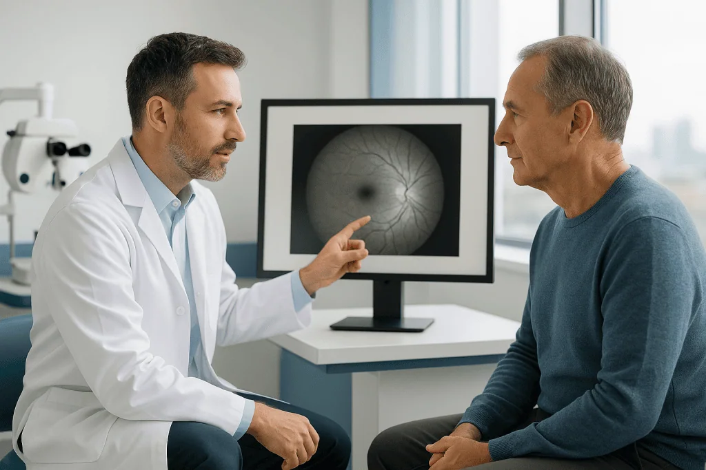
An eye exam is a structured medical assessment of vision and eye health. It typically includes a medical history, visual acuity and refraction testing, eye pressure measurement and a slit-lamp examination, with pupil dilation and retinal imaging added where needed. A routine examination often takes under an hour; a comprehensive assessment with dilation and imaging takes longer and gives a fuller picture of eye health.
Eye Exam: What It Is and Why It Matters
An eye exam is a structured medical assessment of your vision and the health of your eyes. It measures how sharply you see, how your eyes focus and work together, the pressure inside each eye, and the condition of the structures from the eyelids and cornea through to the retina and optic nerve at the back. It serves two purposes at once: correcting vision, updating glasses or contact lenses, and detecting eye disease, often before you notice that anything is wrong.
Vision touches nearly every part of daily life: reading, driving, working, recognising faces, navigating unfamiliar places and feeling confident in your surroundings. When sight changes, even slightly, it is unsettling. Some people worry that blurred vision means a serious eye disease. Others have no symptoms at all but know that diabetes, high blood pressure, age, family history or previous eye surgery places them at higher risk. And for anyone weighing up where to be examined, there is a further question: whether the examination will be thorough, clearly explained and coordinated with the right specialist if a problem is found. This page sets out what a proper eye exam involves so you can judge that for yourself.
The case for regular examination rests on one uncomfortable fact: many eye diseases develop quietly. Glaucoma, diabetic eye disease, macular degeneration, cataracts, retinal tears and certain inflammatory conditions may cause no pain and no obvious visual symptoms in their early stages. By the time vision loss becomes noticeable, some damage may already have occurred. A thorough eye exam can find these conditions earlier, when monitoring, medical treatment, laser procedures, surgery or lifestyle changes have a better chance of preserving vision and slowing progression.
For many people the reasons are more practical. Routine eye exams update a prescription, confirm whether eye strain is caused by refraction, check eye pressure, and establish whether surgery, cataract treatment, refractive surgery or retinal care, is appropriate. At Acibadem, an eye examination is treated as both a diagnostic assessment and a clinical conversation. The aim is not only to measure vision, but to understand your symptoms, medical background, risk factors and expectations, and then to set out the next steps plainly.
What Is a Comprehensive Eye Examination?
A comprehensive eye examination is a full medical evaluation of vision and eye health, not just a check of whether you need glasses. Depending on your age, symptoms and medical history, it can include refraction for a glasses prescription, eye pressure measurement, assessment of corneal health and lens clarity, evaluation of the optic nerve and retina, visual field testing, and a review of eye movement and alignment. Each element answers a different question, and together they build a picture that no single test can provide.
A comprehensive examination can identify cataracts, glaucoma risk, retinal disease, optic nerve problems, corneal disorders, dry eye, inflammation, trauma-related changes and complications of systemic disease. Just as importantly, it can help distinguish where a visual complaint actually comes from: the eye itself, the optic nerve, the brain, a medication side effect or another medical condition. Blur is a symptom, not a diagnosis, and two patients with the same complaint can have entirely different causes.
Is a vision screening the same as an eye test?
No. A basic eye test, reading letters on a chart, identifies reduced visual sharpness, but it says very little about the health of the eye itself. A screening can pass an eye that is quietly developing glaucoma, and it can fail an eye whose only problem is an out-of-date prescription. Screenings are useful as a first filter, particularly for children and drivers, but they do not replace a comprehensive examination in which the internal structures of the eye are actually examined.
The exact tests are personalised. A patient in their twenties who needs a contact lens assessment does not follow the same pathway as a patient with diabetes, a child with suspected lazy eye, or an older adult with worsening night vision. In a hospital environment, ophthalmologists can add imaging, laboratory testing or consultations with other departments when the findings call for them. This flexibility matters most for complex conditions and for second opinions after an unclear diagnosis.
Who May Need an Eye Exam?
Almost everyone benefits from an eye exam at appropriate intervals, but some people need one sooner or more often than others. Common reasons for arranging an examination include blurred vision, double vision, eye pain, light sensitivity, frequent headaches, halos around lights, difficulty seeing at night, floaters, flashes of light, redness that does not settle, distortion of straight lines, or a shadow across part of the visual field. Ophthalmologists regard some of these findings as time-sensitive, sudden vision loss in one eye, new flashes accompanied by a shower of floaters, or a curtain-like shadow across the field of vision, because the conditions behind them, such as retinal detachment, can progress quickly.
Examinations matter just as much for people with no symptoms but a higher underlying risk. That includes people with diabetes, high blood pressure, autoimmune disease or thyroid disease; people with a family history of glaucoma or retinal disease; those with a previous eye injury, long-term steroid use, high myopia or previous eye surgery; and people whose work depends on precise vision. In these groups, the absence of symptoms is not reassurance, several of the conditions being screened for are silent by nature.
Children have their own indications. A child may need assessment if they squint, sit very close to screens, tilt the head, cover one eye, avoid reading, struggle at school, or show visible eye misalignment. In young children, refraction is often measured after cycloplegic drops that temporarily relax the eye’s focusing muscle, because children can otherwise “focus through” a significant prescription and mask it. Childhood conditions such as amblyopia respond far better to treatment when they are found early, which is why paediatric assessment is worth taking seriously even when a child does not complain.
How often should you get your eyes examined?
It depends on your age, risk profile and previous findings, so there is no single interval that fits everyone. Healthy adults with no symptoms and no risk factors typically need routine checks every few years. Children benefit from assessment at key developmental stages. From mid-life onwards, more regular review makes sense as presbyopia, cataract change and glaucoma risk begin to appear. People with diabetes, glaucoma, high myopia or established retinal disease usually need examinations on a schedule set individually by their ophthalmologist, sometimes considerably more often than a healthy person of the same age. The first comprehensive examination usually settles what your own interval should be.
Whatever brings you in, diagnosis begins with a careful history. The ophthalmology team will ask about your symptoms and their duration, previous prescriptions, contact lens use, medications, allergies, surgeries, general medical conditions and family history. The examination then moves through a sequence of measurements and clinical tests, with imaging added where needed to document the retina, optic nerve, cornea or other structures in detail. These results together determine whether your eyes are healthy, whether you need glasses or contact lenses, and whether treatment or follow-up is required.
Conditions an Eye Examination Can Detect or Monitor
Some findings are common and straightforward to correct. Nearsightedness, farsightedness, astigmatism and presbyopia, the refractive errors, cause blurred vision, eye strain and headaches, and are typically managed with glasses, contact lenses or, for carefully selected patients, refractive procedures. Identifying and correcting a refractive error is often all that a younger, otherwise healthy patient needs.
Other findings call for medical or surgical care. Cataracts cause glare, faded colours, difficulty driving at night and progressively cloudy vision. Glaucoma can damage the optic nerve over years, usually without early symptoms, which is why eye pressure measurement and optic nerve evaluation belong in every comprehensive assessment; when it is found, glaucoma treatment ranges from drops through laser procedures to surgery, depending on the stage. Retinal conditions, diabetic retinopathy, macular degeneration, retinal tears, retinal vascular disease and macular oedema, can affect central or peripheral vision and may require imaging and timely treatment to limit their impact.
An eye examination also evaluates dry eye disease, allergies, eyelid disorders, corneal disease, keratoconus, eye infections, inflammation inside the eye, optic nerve swelling, eye movement disorders and complications from systemic illness. For patients preparing for a procedure such as cataract surgery or refractive surgery, the examination establishes candidacy, informs the treatment plan and identifies risk factors that could influence the outcome. For patients with an existing diagnosis, repeated examinations show whether the disease is stable, improving or progressing, information no single visit can provide.
In hospital-based ophthalmology, an eye examination may also form part of a broader medical evaluation. Changes in the retinal blood vessels can offer clues about diabetes or vascular disease. Optic nerve changes may prompt neurological assessment. Eye inflammation is sometimes linked to autoimmune disorders. A detailed examination of the eye can, on occasion, be the first step towards diagnosing a wider health problem.
Finally, it is worth being honest about limits. Not every condition found on examination can be reversed, and some vision loss is permanent despite good treatment. Where that is the case, structured vision rehabilitation helps people make the most of the sight they retain: a different goal from treatment, but a valuable one.
What Happens During an Eye Examination?
A comprehensive examination follows a logical sequence: history first, then vision measurement, then eye pressure, then a detailed look at the front and back of the eye, with imaging added where the findings justify it. Knowing the sequence in advance takes much of the uncertainty out of the visit.
What is usually included in an eye exam?
A typical comprehensive eye exam includes the following core elements, in roughly this order:
- A medical history and symptom review
- Visual acuity testing at distance and near
- Refraction to determine any glasses or contact lens prescription
- Intraocular pressure measurement
- Slit-lamp examination of the front structures of the eye
- Pupil dilation and evaluation of the lens, retina and optic nerve
- Imaging or specialised tests where the findings call for them
Not every visit includes every element. A contact lens review is shorter; a diabetic retinal assessment or a surgical planning visit goes deeper. The clinical team decides the scope based on why you are there and what the early tests show.
Before the Examination
Preparation is usually simple. Bring your current glasses, contact lenses, previous prescriptions, eye drops, a list of your medications, and any prior eye reports or imaging, earlier records allow the physician to compare changes over time, which is often more informative than a single snapshot. If your pupils may be dilated, plan not to drive immediately afterwards and consider bringing sunglasses, because vision can be temporarily blurred and light-sensitive for several hours.
Contact lens wearers may be asked to remove their lenses before certain measurements. For specialised corneal testing or refractive surgery evaluation, a period without contact lenses beforehand is sometimes needed so the corneal surface can return to its natural shape. The clinical team will tell you whether and for how long this applies to you.
Step 1: Medical History and Symptom Review
The examination begins with a conversation. The physician or clinical team will ask what you are experiencing and what matters most to you: clearer reading vision, safer night driving, relief from discomfort, an assessment of disease risk, or confirmation of an existing diagnosis. They will also review systemic conditions such as diabetes, hypertension, autoimmune disease and neurological conditions, along with your medications, because all of these can affect the eyes and change how findings are interpreted.
Step 2: Visual Acuity and Refraction
Visual acuity testing measures how clearly you see at distance and near. Refraction then determines whether lenses can improve your vision and identifies nearsightedness, farsightedness, astigmatism or presbyopia. This usually combines automated measurements with a detailed lens comparison in which you are asked which of two options looks clearer. Refraction matters beyond prescribing glasses: it separates optical blur, which lenses can fix, from disease-related vision loss, which they cannot.
Alongside acuity and refraction, the physician may check how your pupils respond to light, how well your eyes align and move together, and, where relevant for occupation or childhood assessment, colour vision and depth perception. Problems with alignment or eye movement can cause double vision, headaches and reading difficulty even when each eye sees clearly on its own, so these brief checks catch issues that a letter chart alone would miss.
Step 3: Eye Pressure Measurement
Intraocular pressure measurement helps assess glaucoma risk. Pressure is only one part of that assessment: some patients develop glaucoma at normal pressures, while others carry elevated pressure without any optic nerve damage. For this reason, pressure readings are interpreted alongside optic nerve appearance, visual field results, corneal thickness and imaging where needed, rather than in isolation.
Step 4: Slit-Lamp Examination
A slit-lamp microscope lets the ophthalmologist examine the front structures of the eye in fine detail: the eyelids, tear film, conjunctiva, cornea, iris and lens. This is how dry eye, infection, inflammation, corneal scars, cataracts and other visible abnormalities are identified. Special dyes may be used to highlight the tear film or small defects on the eye’s surface.
Step 5: Pupil Dilation and Retina Evaluation
Dilating drops enlarge the pupils so the physician can evaluate the lens, vitreous, retina, macula, blood vessels and optic nerve more thoroughly. Dilation causes temporarily blurred near vision and light sensitivity for several hours, inconvenient, but it gives a substantially better view of the back of the eye. Where wide-field retinal imaging is available, the physician may combine imaging with the dilated examination to document findings and track changes between visits.
Step 6: Imaging and Additional Testing When Needed
Modern ophthalmology draws on diagnostic technologies that provide detailed, objective information. Optical coherence tomography creates cross-sectional images of the retina and optic nerve, helping to detect macular disease, glaucoma-related thinning, swelling or fluid. Corneal topography maps the curvature of the cornea and is useful for keratoconus, astigmatism and surgical planning. Visual field testing measures peripheral vision and is important in glaucoma and neurological conditions. Retinal photography documents the appearance of the back of the eye for monitoring over time. Ultrasound, angiography or other advanced tests may be recommended for specific concerns.
These technologies do not replace the physician’s examination. They support it, providing measurements, images and comparisons that inform clinical decisions. They are most valuable for subtle disease, complex cases and long-term follow-up, where the question is not “is something there?” but “has it changed?”.
How long does an eye exam take?
A routine eye exam may take less than an hour, while a comprehensive examination with dilation and imaging takes longer. Complex visits, paediatric assessments, contact lens evaluations, surgical planning and second-opinion consultations, need additional time. In a hospital setting, scheduling can often be coordinated so that multiple tests or specialist consultations are completed within the same visit, rather than spread across separate appointments.
After the Examination
Most patients return to normal activities immediately. If your pupils were dilated, near vision and light sensitivity are affected for several hours, so reading, computer work and driving may be uncomfortable for the rest of the day. Your physician will explain the findings, whether your prescription has changed, and whether treatment, monitoring or referral to a subspecialist is needed. If a condition is identified, you may receive a written plan covering monitoring intervals, medications, imaging, laser treatment, surgery or further medical evaluation. It is worth asking for a copy of your results and any imaging: a documented baseline makes every future examination more informative, because change over time is often the most important finding of all.
Why Acting Early Matters
Eye disease is usually more manageable when found early. Some conditions, such as refractive errors or dry eye, may never threaten vision permanently but interfere with work, reading and quality of life if left unaddressed. Others, glaucoma, diabetic retinopathy, retinal tears and macular disease, can progress without pain. Delayed diagnosis narrows the range of treatment options and can allow vision loss that is difficult or impossible to reverse.
Early examination matters most for people with risk factors. Diabetes can affect the retinal blood vessels before any change in vision is noticeable. Glaucoma narrows peripheral vision so gradually that patients often do not recognise the loss until it is advanced. A retinal tear can progress to retinal detachment, which is a medical emergency. Cataracts develop slowly, but leaving them unassessed can let vision decline far enough to affect safety, mobility and independence.
Acting early does not always mean treating early. Sometimes the best plan is careful monitoring, and a good ophthalmologist will say so. What a timely examination always provides is information: what is normal, what needs attention, what should simply be watched, and a realistic sense of when the next check should happen.
Benefits of an Eye Examination
Depending on your age, symptoms and risk factors, a comprehensive eye examination offers practical, diagnostic and preventive value.
| Benefit | What It Means for You |
|---|---|
| Clearer vision assessment | Identifies whether glasses, contact lenses or a prescription update can improve daily vision for reading, driving and work. |
| Early disease detection | Helps find conditions such as glaucoma, cataracts, diabetic eye disease and retinal problems before they cause significant symptoms. |
| Risk-based monitoring | Creates a follow-up plan based on your personal risks, including age, family history, diabetes, high myopia or previous surgery. |
| Objective documentation | Imaging and measurements allow physicians to compare changes over time and make more informed treatment decisions. |
| Access to subspecialty care | If a problem is found, you can be directed to the appropriate ophthalmology subspecialist, such as retina, glaucoma, cornea or paediatric eye care. |
Recovery and What to Expect Afterwards
An eye examination does not require medical recovery in the way surgery does, but certain tests can temporarily affect vision or comfort, and it helps to know what is normal.
| Time Period | What Patients Can Expect |
|---|---|
| Day 1 | Most patients resume normal activities. If dilation was performed, near vision may be blurry and the eyes may be light-sensitive for several hours. |
| First Week | Any prescribed glasses, eye drops or follow-up tests can be arranged. Mild irritation from drops or contact lens removal usually resolves quickly. |
| First Month | Patients with new diagnoses may begin monitoring or treatment. Additional imaging, visual field testing or specialist review may be scheduled if needed. |
| Longer Term | Follow-up intervals depend on findings. Healthy eyes may need periodic routine exams, while glaucoma, diabetes or retinal disease require closer monitoring. |
Eye Exam Cost: What Shapes the Charges for Eye Test Appointments
Eye exam cost varies widely because “an eye exam” can mean anything from a brief acuity screening at an optical shop to a comprehensive hospital assessment with dilation, imaging and subspecialist review. The charges for eye test appointments reflect that range: the setting, the depth of testing, the equipment involved and the expertise of the person examining you all move the figure, as does the country and health system you are in. This page deliberately quotes no prices, because a figure detached from the scope of the examination behind it tells you very little.
How much is an eye examination?
There is no single answer, because cost tracks scope. The main drivers are: where the examination happens (an optical retailer, a private clinic or a hospital ophthalmology department); which tests are included (refraction alone, or pressure measurement, dilation, imaging and visual field testing as well); whether a subspecialist reviews the findings; and whether the examination stands alone or forms part of a wider treatment pathway such as cataract or refractive surgery planning. An examination bundled into a surgical pathway is usually priced differently from one billed on its own, so when comparing quotes it is worth asking exactly which tests are included, whether imaging is part of the same fee, and whether a follow-up review of the results is covered or charged separately.
Does health insurance cover eye examinations?
Coverage depends on the plan and the reason for the visit. Many public schemes and insurers do not pay for routine eye examinations for glasses or contact lenses, but they can cover examinations linked to specific medical conditions, diabetic eye checks are a common example, and some plan types add vision benefits. Rules differ between plans and countries and change over time, so the insurer’s own documents are the only reliable source for your situation: routine vision care and medical eye care are often covered under different rules.
How do I get a free eye exam?
In many countries, public health systems or insurance schemes provide free or subsidised eye tests for defined groups, commonly children, older adults, people with diabetes or glaucoma risk, and people on low incomes. Some optical retailers also offer free basic screenings as promotions. Be clear about what you are getting: if you search online for a cheap eye vision test, most of what you find checks visual sharpness and prescription only. That is genuinely useful, but it is not a health examination of the eye, and it will not detect the silent conditions a comprehensive assessment is designed to find.
Can you walk in to get an eye exam?
Often, yes, for a basic eye test. Many optical shops accept walk-ins for acuity checks and refraction. Comprehensive hospital-based examinations are usually scheduled in advance, partly because dilation, imaging and subspecialist time need to be arranged, and partly so that your records and history can be reviewed properly. An initial examination also sometimes reveals the need for further investigation before any treatment can proceed, and that kind of follow-on testing is far easier to absorb into a scheduled pathway than a walk-in visit.
What Influences the Accuracy of the Examination and a Good Result?
A good result from an eye examination means more than receiving a prescription. It means the physician has a reliable understanding of your eye health and you leave with clear next steps. Several factors influence whether that happens.
Complete medical information comes first. Eye findings are frequently linked to systemic disease, medications and family history. Sharing details about diabetes control, blood pressure, autoimmune disease, neurological symptoms, steroid use, previous eye trauma and past surgeries helps the physician interpret results correctly. Withholding or forgetting this information does not just leave gaps: it can actively mislead the interpretation.
Appropriate diagnostic testing matters equally. A simple refraction may be sufficient for a healthy patient with mild blur, but patients with pressure concerns, macular symptoms, diabetes or optic nerve changes may need imaging or visual field testing. The best examination is tailored: not excessive, and not too limited. More tests are not automatically better; the right tests are.
Quality of measurements affects decisions. Dry eye, contact lens wear, corneal irregularities or limited cooperation in young children can all distort refraction or imaging results. Sometimes a measurement must be repeated after the ocular surface has been treated, or after a recommended period without contact lenses, before it can be trusted.
Subspecialty interpretation may be needed for complex conditions. Retina, glaucoma, cornea, paediatric ophthalmology, neuro-ophthalmology and oculoplastic specialists each focus on different aspects of eye disease. When findings are subtle or advanced, review by the appropriate specialist can refine both the diagnosis and the treatment plan.
Follow-up adherence is essential. Some eye conditions require monitoring even when vision feels entirely normal. Visual field tests, pressure checks, retinal imaging or repeat examinations can reveal progression that the patient cannot perceive. A strong plan includes realistic follow-up timing that fits your circumstances, so that monitoring actually happens rather than remaining a good intention.
Your own goals belong in the picture too. A pilot, a surgeon, a designer, a student, a professional driver and an older adult living independently have different visual needs. Understanding lifestyle, occupation and expectations helps the ophthalmologist recommend lenses, treatment timing and preventive strategies that actually fit your life, rather than a generic plan.
Eye Examinations at Acibadem
Patients often seek a hospital-based eye examination when they want a comprehensive evaluation, a second opinion, access to subspecialists, or diagnosis and treatment coordinated within a single setting. At Acibadem, eye examinations are structured as diagnostic pathways: assessment leads to a decision, and the decision is explained. The examination is performed by ophthalmologists working within a hospital environment, which means additional imaging, laboratory testing and consultations with other departments can be arranged within the same pathway when findings require them.
The care model becomes multidisciplinary when a patient’s condition requires it. Eye findings may be reviewed by ophthalmology subspecialists and, where appropriate, coordinated with endocrinology, neurology, rheumatology, oncology, paediatrics or internal medicine. This is particularly relevant for diabetic eye disease, optic nerve disorders, inflammatory eye disease, thyroid eye disease, eye tumours and vision changes related to systemic illness. Collaborative case discussion means complex decisions are not made in isolation.
Diagnostic technology plays its part. Depending on the patient’s needs, physicians may use retinal imaging, optic nerve analysis, corneal mapping, visual field testing, ultrasound-based assessment and other ophthalmic diagnostic tools. These document findings, detect subtle changes and support treatment planning. Having the key tests performed within one organised clinical pathway reduces uncertainty and shortens the distance between examination and decision.
Personalised planning matters more in ophthalmology than in most fields, because eye conditions vary so widely. Two patients with identical complaints of blurred vision may have entirely different causes: a simple refractive error, a cataract, dry eye, a corneal irregularity, retinal swelling or a neurological condition. Acibadem physicians evaluate the underlying cause, the severity of the findings, and the patient’s age and medical background, then recommend a plan that may range from observation through medication and laser therapy to surgery.
A second opinion is a legitimate reason for a hospital-based examination in its own right. When a diagnosis is uncertain, or surgery has been recommended elsewhere, a detailed examination can help establish whether surgery is genuinely necessary, whether the timing is right, and whether other conditions should be addressed first. Sometimes the honest recommendation is to monitor rather than operate. Sometimes additional testing produces a more specific diagnosis that changes the plan entirely. Either outcome is useful: the point of a second opinion is clarity, not confirmation.
Reading Your Results and Planning Follow-Up
By the end of a comprehensive examination, you should understand three things: what is normal in your eyes, what needs attention, and what should simply be watched. If a prescription has changed, you will know why. If a condition has been identified, you will know whether the plan is monitoring, medication, laser treatment or surgery, and on what timescale. If nothing was found, the findings themselves become the baseline against which future examinations are compared, which is a genuinely valuable outcome, not a wasted visit.
The practical question after any examination is continuity. A written report with imaging allows any ophthalmologist who sees you in future to continue monitoring on the recommended schedule, and conditions that need hospital-level treatment can be planned with full information rather than started from scratch. An eye exam is rarely an end point; it is the piece of information that makes every subsequent decision about your vision better informed.
Preparation
- Bring your current glasses, contact lenses, previous eye reports, and a list of medications. If pupil dilation is planned, you may need sunglasses afterward and should consider arranging transport. Contact lens wearers may be asked to remove lenses before testing.
Aftercare
- Most patients return to daily activities immediately after an eye examination. If dilating drops are used, light sensitivity and blurred near vision may last a few hours. Follow the ophthalmologist’s advice on glasses, contact lenses, medications, or further tests if needed.
Frequently Asked Questions
What affects the cost of an eye examination?
The final cost depends on the scope of the examination, the diagnostic tests required, whether an ophthalmologist reviews the results, and whether advanced imaging or follow-up is needed. International patient services and translation support may also affect the package.
How can I get a personalised quote?
You can request a free consultation and share your symptoms, medical history, previous eye reports, and any current glasses or contact lens prescription. The care team can then advise which tests may be appropriate and prepare a personalised quote.
Is a basic vision test enough?
A basic vision test can help identify refractive problems, but it may not detect all eye diseases. If you have symptoms, a medical condition, or a family history of eye disease, a specialist may recommend a comprehensive eye examination.
Will I need advanced eye imaging?
Advanced imaging may be recommended if the specialist needs a detailed view of the retina, macula, or optic nerve. It is commonly considered when there are risk factors or unexplained symptoms, but suitability is decided after clinical assessment.
Can an eye examination be combined with treatment planning?
Yes, if the examination detects cataract, glaucoma, retinal disease, dry eye, or another condition, the specialist may discuss treatment options and any further tests. Any treatment costs would usually be quoted separately after evaluation.
Medically reviewed by the Acıbadem International Medical Board September 13, 2026
See our medical review board →
Update history
- PublishedJune 8, 2026
- Medical review approvedSeptember 13, 2026
- Last content updateSeptember 12, 2026
Trusted care for international patients
From the Acibadem Blog
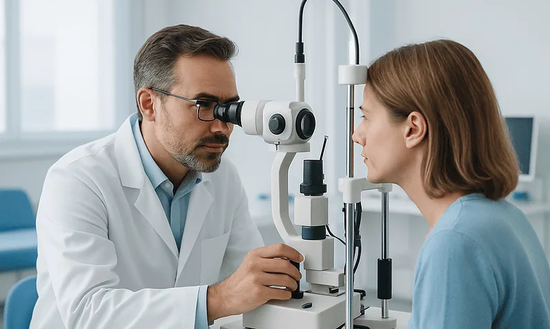
Preparing for an Eye Examination: Glasses, Old Reports, Medication Lists and a Driver
For an eye exam, bring the glasses and contact lenses you actually use, any previous prescriptions or eye reports, a complete list of your…

Why Eye Examinations Look Different When You Have Diabetes or High Blood Pressure
An eye exam for diabetics or for people with high blood pressure focuses on the retina, the light-sensing layer at the back of the…
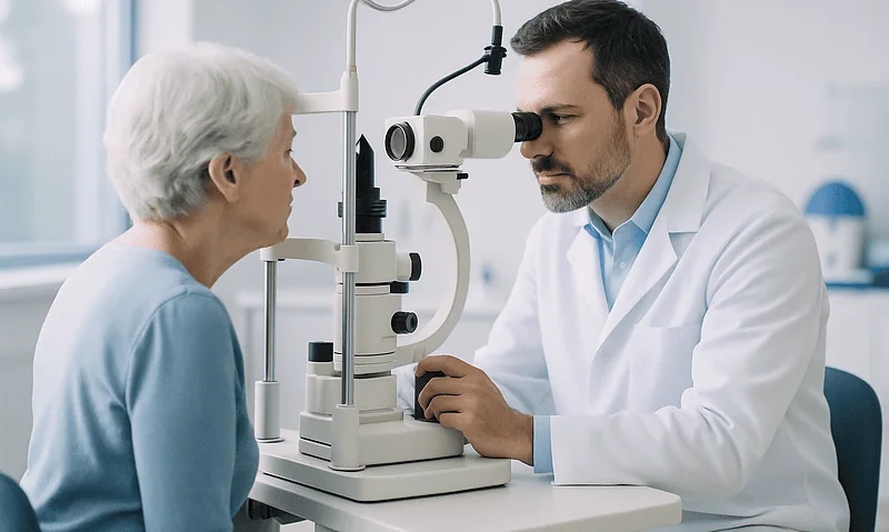
What Happens During a Full Eye Examination: Vision, Pressure, Dilation and Imaging
A full eye examination usually moves through a set order: a conversation about your health and symptoms, a letter-chart test of sharpness, refraction to…