

What Happens During a Full Eye Examination: Vision, Pressure, Dilation and Imaging

Key Takeaways
- A comprehensive eye examination typically takes 45 to 90 minutes, with dilating drops accounting for much of the variation because they need roughly 20 to 30 minutes to work.
- A 20/20 result means you read at 20 feet what a typical eye reads at 20 feet; it measures sharpness only and says nothing about eye pressure, retina or optic nerve health.
- Typical eye pressure is 10 to 21 mmHg, but glaucoma can occur inside that range and pressure can be raised without glaucoma, so the diagnosis rests on the optic nerve, OCT and visual field together.
- Dilation lets the doctor see the retina and optic nerve directly, and its effects commonly last around four to six hours, during which near vision blurs and light feels harsh.
- People with diabetes, a family history of glaucoma, African Americans over 40 and everyone over 60 are advised to have a dilated exam every one to two years even without symptoms.
- Contact lenses temporarily reshape the cornea, so removing them for the period your practice specifies before the visit gives a more accurate refraction and fitting.
A full eye examination usually moves through a set order: a conversation about your health and symptoms, a letter-chart test of sharpness, refraction to find your lens prescription, a pressure measurement for glaucoma risk, a microscope check of the front of the eye, dilating drops so the doctor can see the retina and optic nerve, and often retinal photographs or scans. Most visits take roughly 45 to 90 minutes.
The woman in the chair next to you has a pair of sunglasses in her lap, though it is grey outside. She has done this before. You have not, at least not since a school nurse pointed at a chart and told you to cover one eye. You are wondering about the puff of air everyone mentions, whether the drops will sting, and what exactly the machine with the chin rest is for.
People who ask what happens during an eye exam are usually asking two things at once: what will be done to me, and what is the point of each step. Both deserve a straight answer. An eye examination is not one test but a short sequence of them, each looking at a different part of the visual system, and each piece of equipment earns its place by catching something the others cannot.
This explainer walks through that sequence in the order you will most likely meet it, and is honest about what the evidence does and does not tell us.
What happens during an eye exam: the stations in order
Think of the visit as a corridor of small rooms, each with one job. The exact order varies between practices, and an optometrist (a clinician trained to test vision and detect eye disease) may hand some steps to a technician before the doctor sees you. The sequence below is the common pattern described by MedlinePlus and the Mayo Clinic.
First comes the history: what you have noticed, what medicines you take, what runs in your family. Then the letter chart, which measures visual acuity, the technical term for how finely you can resolve detail at a set distance. Refraction follows, the back-and-forth of lenses that finds the prescription your eyes actually need. A pressure reading, called tonometry, checks the fluid pressure inside the eyeball, because pressure that runs high is a major risk factor for glaucoma, a disease that damages the optic nerve.
The doctor then sits you at a slit lamp, essentially a microscope with a bright, narrow beam, and studies the front of the eye layer by layer. Dilating drops usually go in at this point. Once the pupils have widened, the doctor can look through them to the retina, the light-sensitive lining at the back, and to the optic nerve where it leaves the eye. Many practices add imaging here: a photograph of the retina, or an optical coherence tomography (OCT) scan, which builds a cross-section of the retinal layers using reflected light.
What ties it together is that no single station tells the whole story. A person can read every line on the chart and still have early glaucoma; another can have a blurry prescription and perfectly healthy tissue. The exam is designed so that each test covers a blind spot in the others.
Why the first ten minutes are questions, not tests
The conversation is the part people underestimate, and clinicians rank it near the top. Before any instrument touches you, the doctor wants to know what brought you in, whether anything has changed, and what the rest of your body is doing.
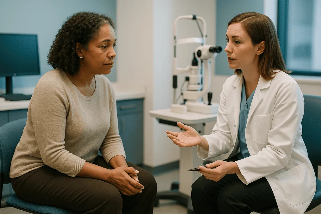
Some of the questions sound unrelated to eyes. Diabetes, high blood pressure and autoimmune conditions all leave marks on the retina, and the Mayo Clinic lists them among the reasons a person may need closer follow-up. A list of your medicines matters because several classes, including some used for rhythm disorders, inflammation and mood, can affect the eye over time; the doctor is not judging your prescriptions, only deciding which structures deserve a longer look. Decisions about any of those medicines stay with the clinician who prescribes them.
Family history carries real weight. MedlinePlus notes that a family history of glaucoma is one of the factors that changes how often adults should be examined and which tests are prioritized. Macular degeneration, a condition in which the central part of the retina deteriorates, also clusters in families.
Then come the everyday specifics. Do you drive at night? Work at a screen for eight hours? Play a sport where a lens could be knocked out? Do headaches arrive at the end of the day? Your answers shape the prescription conversation later, because the “best” correction for someone who reads music on a stand differs from the best correction for a long-haul driver.
Bring your current glasses, even if you dislike them, and any contact lens boxes. The clinician can read the existing prescription directly, which gives a baseline for how much your eyes have shifted. If you have had eye surgery, an injury or a lazy eye as a child, say so early; it changes how some measurements are interpreted.
What the letter chart really measures, and what 20/20 means
The chart on the wall, or its projected or screen-based equivalent, tests visual acuity: the smallest detail you can distinguish at a standard distance. MedlinePlus describes the standard setup as reading rows of letters from about 20 feet, one eye at a time, with the other covered, first without and then with any correction you normally wear.
The famous fraction is simpler than it sounds. A result of 20/20 means you read at 20 feet what a person with typical vision reads at 20 feet. A result of 20/40 means the smallest line you managed is one that a typical eye could read from twice as far away. It is a comparison, not a grade, and it says nothing about how healthy the eye is, only how sharply it is focusing right now.
Several things can knock the number down temporarily. Dry eyes after a long screen day, a smudged lens, tiredness, or simply squinting against glare will all cost you a line or two. That is one reason the test is repeated with lenses in front of your eye during refraction: the clinician wants to know your best corrected acuity, the sharpest you can achieve with the right glasses, because that figure separates a focusing problem from a problem in the eye itself.
Children who cannot read letters are tested with pictures or shapes, or with a matching game in which they point to a card that matches the symbol shown. Very young children may be assessed by watching how their eyes track a moving target. None of this hurts and none of it requires the child to “get it right”; the clinician is watching how each eye behaves, not scoring the child.
Near vision is usually checked with a hand-held card, which becomes more informative after about age 40, when the lens inside the eye stiffens and close focus fades.
Refraction: why you are asked "one or two?" over and over
Refraction is the process of finding the lens power that brings light to a sharp focus on your retina. It is the part of the exam that produces a prescription, and it is also the part that makes people anxious about giving a wrong answer.
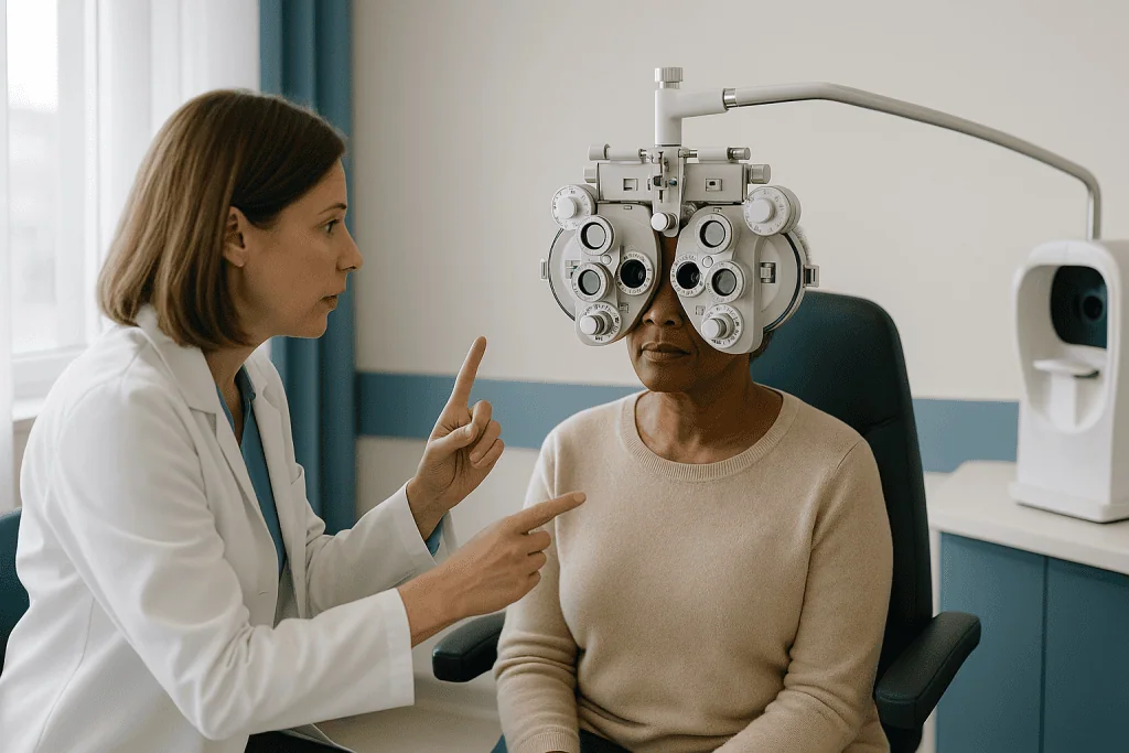
You will sit behind a device called a phoropter, the black mask with dials that swings in front of your face, or the clinician may hold up loose lenses from a trial frame. Lenses flip. You are asked which of two views is clearer, or whether they look about the same. The doctor narrows the choices with each answer, much as a pianist tunes by listening for the beat between two nearly matched notes.
There are no wrong answers in the sense that worries people. When two options genuinely look identical, saying so is useful information; it tells the clinician they have bracketed your best correction. When you hesitate, the pair is usually shown again. Many practices begin with an autorefractor, a machine that shines light into the eye and estimates the prescription in seconds, and use your answers to refine that starting point rather than build from nothing.
The Mayo Clinic notes that refraction can also be estimated with a retinoscope, a hand-held light the doctor sweeps across the pupil while watching the reflex, which is especially useful for small children and for anyone who cannot respond verbally.
For contact lens wearers, refraction is only part of the fitting. The clinician also measures the curvature of the cornea, the clear dome at the front of the eye, because a lens that is too flat or too steep can be uncomfortable or unsafe. If you are being fitted, expect a short additional segment, and expect to be asked to remove your lenses so the corneal surface can be seen without them.
The eye pressure test: the puff of air and the blue light
Tonometry measures intraocular pressure, the pressure of the clear fluid that fills the front of the eye. That fluid is made and drained continuously; when drainage lags, pressure climbs, and sustained high pressure is one of the main risk factors for glaucoma. According to MedlinePlus, a typical reading falls between 10 and 21 millimeters of mercury, though the site is careful to add that pressure can be higher without glaucoma being present and that glaucoma can occur at pressures inside that range.
Two methods dominate. Non-contact tonometry is the puff: you rest your chin, look at a light, and a brief jet of air flattens the cornea for a fraction of a second while the instrument times how the surface responds. It startles more than it hurts. Applanation tonometry is the blue light: a numbing drop goes in, sometimes with a yellow dye called fluorescein, and a tiny flat-tipped probe on the slit lamp gently touches the cornea while the doctor reads the pressure. Because of the numbing drop you feel pressure rather than pain, and most people do not feel the probe at all. MedlinePlus describes both approaches and notes that applanation is generally regarded as the more accurate.
The Mayo Clinic’s glaucoma pages add a third measurement that may accompany tonometry: pachymetry, a quick ultrasound reading of corneal thickness. A thick cornea can make pressure read artificially high and a thin one artificially low, so knowing the thickness lets the doctor interpret the number sensibly.
A single elevated reading is not a diagnosis. Pressure drifts through the day and varies from visit to visit, so a high figure typically leads to a repeat measurement and a closer look at the optic nerve rather than to any immediate conclusion. Whether further testing is needed is a judgment for the examining clinician.
Slit lamp and dilated eye exam: how the doctor sees the back of your eye
The slit lamp is a binocular microscope mounted on a table, with a beam of light that can be narrowed to a thin blade. With your chin and forehead against the rests, the doctor angles that blade through the cornea, the front chamber, the iris and the lens, viewing each in cross-section. Scratches, dryness patterns, early cataract (clouding of the lens) and inflammation all show up here.
To see further in, the pupil has to be wider than it wants to be under bright light. Dilating drops relax the muscle that constricts the pupil, and some also temporarily relax the focusing muscle. The National Eye Institute (NIH) notes that the drops take roughly 20 to 30 minutes to work, which is why you may be sent back to the waiting room mid-visit. They sting briefly, and some people describe a bitter taste as the drop drains through the tear duct into the nose.
Once dilated, the doctor uses a hand-held lens at the slit lamp, or a headset called an indirect ophthalmoscope, to examine the retina, its blood vessels and the optic nerve. This is where the exam earns its reputation as a whole-body check. Diabetic damage appears as tiny bleeds and leaking vessels; long-standing high blood pressure narrows and kinks the arteries; the optic nerve’s cupped center enlarges in glaucoma; the macula, the central patch responsible for reading and faces, shows the deposits of early age-related degeneration.
The NIH describes the dilated examination as the only way to check many of these conditions before they cause symptoms, which is the core argument for having one even when your vision feels fine. Not every visit includes dilation. Some practices substitute wide-field retinal photography for lower-risk patients, and a doctor may skip dilation on a day you need to drive immediately afterward. The choice rests with the examining clinician, so ask before the drops go in if the timing matters to you.
Imaging: retinal photographs, OCT and the visual field test
Imaging has changed what a routine exam can record. The doctor’s view through a dilated pupil is a moment in time; a photograph or scan is a document that next year’s exam can be laid beside.
Fundus photography is the simplest. You look into a camera, a flash fires, and a color image of the retina appears on screen. Wide-field versions capture most of the retina in one shot, sometimes without dilation. The picture shows blood vessels, the optic nerve and the macula, and lets you see what the doctor is describing, which for many people is the most persuasive moment of the visit.
Optical coherence tomography, usually shortened to OCT, uses reflected near-infrared light to build a cross-sectional image of the retina’s layers, in much the way ultrasound maps tissue with sound. You rest your chin, fix on a target, and the scan takes seconds. The Mayo Clinic lists OCT among the tests used to assess the optic nerve fiber layer in glaucoma, and it is also the standard way to measure swelling or thinning at the macula in diabetes and age-related degeneration. There is no radiation and nothing touches the eye.
The visual field test measures peripheral vision, the part you do not consciously notice. You look steadily at a central point while faint lights appear at the edges, pressing a button each time you see one. Glaucoma typically steals sight from the periphery first, which is why the Mayo Clinic includes this test in its glaucoma work-up. It requires concentration and can feel oddly stressful; it is normal to miss some lights, and the software accounts for that.
Not everyone needs every image at every visit. A first exam often includes a baseline set; later visits add scans when history, pressure or the appearance of the nerve calls for them. Which images are ordered, and how they are interpreted, is a decision for your treating team.
Who is usually asked to have a full exam, and who can usually wait
Two groups tend to be told they need a comprehensive dilated examination even when they feel fine: people with a risk factor, and people at an age where silent disease becomes common. The NIH advises that adults with diabetes, adults with a family history of glaucoma, and everyone over 60 should have a dilated exam every one to two years, with African Americans advised to start at 40 because glaucoma is both more common and more aggressive in that group. Adults under 40 with no symptoms and no risk factors are generally asked to come in less often.
The Mayo Clinic frames its schedule by age and gives typical ranges, summarized below. These are intervals for people without symptoms; anyone with a new visual change is asked to come in regardless of the calendar.
| Situation | Typical interval (Mayo Clinic) | What usually drives the visit |
|---|---|---|
| Adults in their 20s and 30s, no symptoms or risk factors | Every 5 to 10 years | Prescription check; baseline health look |
| Ages 40 to 54 | Every 2 to 4 years | Near-vision change; early glaucoma screening |
| Ages 55 to 64 | Every 1 to 3 years | Cataract, macular and pressure monitoring |
| Age 65 and older | Every 1 to 2 years | Age-related disease surveillance |
| Diabetes, glaucoma family history, or African American over 40 | Every 1 to 2 years (NIH) | Dilated retinal and optic nerve check |
Children sit outside this table. Pediatric guidance generally favors a vision check in infancy, again in the preschool years, and before school starts, because a lazy eye is far easier to treat before the visual pathways settle. Contact lens wearers are also asked to return more often than their age alone would suggest, since the cornea needs monitoring for the low-oxygen and infection risks that lenses carry. Your own interval should come from the clinician who examined you, not from a table.
How long does an eye exam take?
The honest answer is: it depends on what is being done, and dilation is the biggest variable. The Mayo Clinic gives a typical range of 45 to 90 minutes for a comprehensive examination, and that spread mostly reflects whether drops are used and whether imaging or a visual field test is added.
A visit that stops at history, acuity and refraction, the kind aimed purely at updating glasses, sits at the short end. Add tonometry and a slit lamp look and you gain a few minutes. Add dilation and you gain the NIH’s 20 to 30 minutes of waiting for the pupils to open, plus the examination itself. Add a visual field test, an OCT and retinal photographs and the visit stretches toward the top of the range, particularly if the doctor sees something that warrants a second scan.
First appointments run longer than follow-ups because everything is being measured for the first time and there is no baseline to compare against. A glaucoma monitoring visit, by contrast, might be brisk: pressure, a nerve scan, a field test, a short conversation.
Children’s exams are unpredictable in a different way. A cooperative four-year-old can be through in half an hour; a tired one may need the tests split across two visits. Clinicians who see children routinely build in that flexibility and would rather stop and come back than force an unreliable result.
Practical planning follows from this. Book a slot when you are not rushing to a meeting immediately afterward. If dilation is likely, treat the appointment as taking up most of a morning or afternoon rather than the hour on the calendar, and arrange your transport accordingly. If you genuinely cannot spare the time on a given day, say so when you check in; the clinician may be able to reorder tests or defer dilation, though that is their call and may mean a return visit.
What not to do before an eye exam
Most of the preparation is about not undoing the measurements before they are taken.
Contact lenses are the main one. Soft lenses gently reshape the cornea while they sit on it, and rigid lenses do so more. A clinician measuring your cornea for a fitting, or trying to get an accurate refraction, wants to see the eye’s natural shape. Practices differ in how long they ask you to go without lenses beforehand, so follow the instruction you were given when you booked and bring your glasses instead. If you were given no instruction, wearing lenses to the appointment is not a disaster; you will simply be asked to take them out.
Eye makeup is worth skipping or keeping light. Mascara flakes into the tear film and shows up under the slit lamp as debris, and heavy liner can make the eyelid margin hard to assess. Eye drops for redness are best avoided on the day, since they constrict blood vessels and can mask the very redness the doctor is trying to evaluate. Lubricating drops are usually fine unless you have been told otherwise.
Do not stop any prescribed medicine, eye-related or not, without speaking to the prescriber. The clinician wants to see your eyes as they are on an ordinary day, medicines included.
There is no need to fast, and no need to rest your eyes in the dark. A long, late screen session the night before may cost you a line on the chart through dryness and fatigue, so it helps to arrive reasonably rested, but this is a nudge rather than a rule.
Bring more than you think you need: current glasses, contact lens packaging, a list of medicines with names rather than descriptions, any previous prescriptions, and sunglasses for the walk out. And if you use insulin or have a condition that can change quickly, a snack and water in your bag make a long visit easier.
The rest of the day after dilation and imaging
The examination itself leaves no mark; the drops do, for a few hours. The NIH says the effects of dilation commonly last around four to six hours, though some people, particularly those with light-colored eyes, notice them for longer, and the drops used for young children are chosen to act for a longer window.
Two things happen while the pupils stay wide. Bright light floods in without the iris to meter it, so daylight feels harsh and screens glare; sunglasses, or the disposable shades the practice may hand you, are the fix. And if a cycloplegic drop was used, one that also relaxes the focusing muscle, near vision blurs. Reading a phone, a menu or a dashboard becomes difficult until the drop wears off. Distance vision is usually less affected, but glare alone can make driving feel unsafe, which is why arranging a lift or public transport is the standard advice.
Work is possible for most people but not pleasant if it involves fine print. Plan the afternoon around tasks that do not need sharp near vision. Numbing drops from applanation tonometry wear off within a shorter window and can leave a gritty sensation; rubbing the eye while it is numb is the one thing to avoid, since you cannot feel whether you are pressing too hard.
Results are often discussed on the day. Refraction produces a prescription you can leave with. Images are usually reviewed with you at the screen. Where a finding needs a second opinion or a further test, the clinician will say so and set out the next step; a request for a repeat pressure reading or a follow-up scan is a normal part of careful practice and not, by itself, a diagnosis.
If the exam was your first, the most useful thing it produced may be invisible to you: a baseline. The photograph of a healthy optic nerve taken today is what makes a subtle change detectable years from now.
What people often get wrong about what happens during an eye exam
“If I can see fine, I don’t need one.” This is the myth the whole dilated examination exists to answer. Glaucoma removes peripheral vision so gradually that the brain fills in the gaps; diabetic retinopathy can bleed for years before central vision dims. The NIH’s position is that many of these conditions have no early symptoms and can only be found by looking at the retina and optic nerve directly.
“The puff of air is the glaucoma test.” Pressure is one risk factor. MedlinePlus is explicit that glaucoma can occur at pressures within the typical 10 to 21 range and that pressure can be raised without glaucoma. The diagnosis rests on the appearance of the optic nerve, the nerve fiber layer on OCT, and the visual field together.
“Giving a wrong answer during refraction will ruin my prescription.” Refraction is designed around uncertainty. Each pair of lenses is a small step, choices are cross-checked, and the autorefractor or retinoscope gives an independent estimate. “They look the same” is a valid and useful answer.
“Dilation is dangerous for the eye.” The drops are uncomfortable and inconvenient, not harmful to the eye’s structures. The rare exception is a person with a very narrow drainage angle, in whom dilation can provoke a pressure spike; that is precisely why the clinician examines the angle at the slit lamp before deciding to dilate, and why the choice sits with them.
“Screens damage children’s eyes, so an exam is about limiting screen time.” The evidence links near work and time indoors to the progression of short-sightedness, but a child’s exam is chiefly about detecting a lazy eye, a squint or a large prescription while the visual system is still adaptable. That window is the reason early checks are recommended, not screen scolding.
“20/20 means perfect eyes.” It means typical sharpness at a distance. It says nothing about pressure, retina, nerve or near vision.
Questions to ask your care team
A good examination leaves time for questions, and the most useful ones are specific. Take this list in, or pick the three that matter most to you.
- Which tests are you planning today, and will any of them involve dilating drops? If so, roughly how long should I expect to be here, and is it safe for me to drive afterward?
- Did anything in my history or my family’s history change what you looked at, or how often you want to see me again?
- What was my pressure reading, and how does it fit with the thickness of my cornea and the look of my optic nerve? Is a repeat reading needed?
- Did you take photographs or scans? Can you show me what a healthy version of this structure looks like beside mine, and can I have copies for my records?
- Is my prescription stable, or has it shifted? If it has shifted, is that ordinary aging or something you want to watch?
- Are there signs of dryness, early cataract or anything at the macula that I should know about now, even if it needs no action today?
- Given my health conditions and medicines, is there anything about my eyes that my other doctors should be told?
- When should I come back, and what symptoms between now and then should bring me in sooner?
- If you have recommended a further test or a referral, what question is it meant to answer, and what happens if it is normal?
Write the answers down or ask for them in writing; dilated eyes and a full afternoon make details slippery. If a term goes past you, ask for it in plain words. Clinicians expect this, and a patient who understands why each test was done is a patient who comes back on time.
Every recommendation that follows from the exam, whether about glasses, monitoring intervals, further imaging or treatment, is a decision for you and the clinician who examined you to make together.
When to call your doctor
The examination described here is a planned, unhurried event. Some eye symptoms are not, and they should not wait for the next routine slot. Contact an eye clinician or emergency services the same day if you notice any of the following, drawing on the warning signs described by MedlinePlus and the Mayo Clinic:
- Sudden loss of vision in one or both eyes, whether complete or a curtain or shadow moving across part of your sight.
- A sudden shower of new floaters, especially with flashes of light, which can signal a retinal tear or detachment.
- Severe eye pain, particularly with a red eye, nausea, halos around lights and blurred vision, a pattern that can indicate a sharp rise in eye pressure.
- Sudden double vision, or a drooping eyelid or pupil that has changed size, which can reflect a problem with the nerves or blood vessels behind the eye.
- Any chemical splash or penetrating injury to the eye.
- Distortion of straight lines, such as door frames appearing bent or wavy, or a dark patch in central vision, which can indicate a change at the macula.
- A red, painful eye in a contact lens wearer, which can be an early sign of a corneal infection.
After a dilated examination, two things are ordinary: light sensitivity and near blur for the hours the NIH describes. Two things are not: pupils that remain widely dilated well beyond that window, and new eye pain or a marked drop in vision in the hours after the visit. Either warrants a call to the practice that examined you.
Between routine visits, a gradual change in your glasses, more frequent headaches after close work, or growing difficulty with night driving are reasons to move your appointment forward rather than emergencies. Whether any of these needs a same-day, next-week or next-scheduled visit is a judgment for the clinician who knows your eyes, so when in doubt, call and describe what you are seeing.
Frequently asked questions
How long does an eye exam take from start to finish?
The Mayo Clinic gives a typical range of 45 to 90 minutes for a comprehensive examination. A visit limited to a prescription check sits at the short end; one that includes dilation, retinal imaging and a visual field test approaches the longer end, largely because dilating drops need around 20 to 30 minutes to take effect before the doctor can examine the back of the eye.
Is there anything I shouldn't do before an eye exam?
Avoid wearing contact lenses for the period your practice specified, skip redness-relief drops on the day, and go light on eye makeup, since all three can distort what the clinician measures or sees. Do not stop any prescribed medicine without talking to the prescriber. There is no need to fast or rest your eyes in the dark. Bring your current glasses, lens packaging, a medicine list and sunglasses.
What should I expect at a first time eye exam?
Expect a longer visit than a follow-up, because everything is measured from scratch. It usually includes a conversation about your health and family history, the letter chart, refraction with flipping lenses, a pressure test, a slit lamp look at the front of the eye and, often, dilating drops and retinal photographs or scans. Nothing is painful; the drops sting briefly and leave you light-sensitive for several hours.
Does the eye pressure test hurt?
No. The non-contact version is a brief puff of air that startles rather than hurts. The applanation version uses a numbing drop before a small probe on the slit lamp touches the cornea, so most people feel light pressure or nothing at all. MedlinePlus lists a typical reading as 10 to 21 mmHg, though a single high or low figure is interpreted alongside corneal thickness and the appearance of the optic nerve.
What is a dilated eye exam and do I need one every time?
A dilated eye exam uses drops to widen the pupils so the doctor can see the retina, its blood vessels and the optic nerve directly, which the NIH describes as the only way to detect many eye diseases before symptoms appear. Not every visit requires it; some practices use wide-field retinal photography for lower-risk patients, and the examining clinician decides based on your age, history and findings.
Can I drive home after my eyes are dilated?
Many people find driving difficult or unsafe while dilated, because glare is intense and, if a focusing-relaxing drop was used, near vision is blurred. The NIH notes the effects commonly last around four to six hours. Arranging a lift, using public transport or scheduling the visit when you do not need to drive is the usual advice, and your clinician can tell you which drops were used.
What if I give a wrong answer during the "one or two" test?
It is very unlikely to matter. Refraction moves in small steps, each choice is cross-checked against the next, and the clinician usually starts from an autorefractor or retinoscope estimate rather than from zero. Saying that two options look the same is a genuinely useful answer, because it tells the doctor your best correction lies between them. If you hesitate, the pair is simply shown again.
What does 20/20 vision actually mean?
It means that at 20 feet you can read the line a person with typical vision reads at 20 feet, according to MedlinePlus. A result of 20/40 means your smallest readable line is one a typical eye could read from 40 feet. The fraction compares sharpness only; it does not measure eye pressure, peripheral vision, near focus or the health of the retina and optic nerve.
How often should adults have a full eye exam?
The Mayo Clinic suggests every 5 to 10 years for symptom-free adults in their 20s and 30s, every 2 to 4 years from 40 to 54, every 1 to 3 years from 55 to 64, and every 1 to 2 years from 65. The NIH advises every 1 to 2 years for anyone with diabetes, a family history of glaucoma, or African Americans over 40. Your own interval should come from your clinician.
Why does the eye doctor ask about diabetes and blood pressure?
Because both conditions leave visible marks on the retina, the only place in the body where small blood vessels can be viewed directly. Diabetes can cause tiny bleeds and leaking vessels; long-standing high blood pressure narrows and kinks retinal arteries. Knowing your history tells the clinician which structures need the closest look and how often you should return, and findings may be shared with your other doctors.
References
- MedlinePlus – Standard ophthalmic exam
- MedlinePlus – Tonometry
- MedlinePlus – Visual acuity test
- National Eye Institute (NIH) – Get a dilated eye exam
This article is for general information only and is not a substitute for professional medical advice. Please consult a qualified doctor about your individual situation.

More from the Blog

Robotic vs Conventional Eye Surgery: Where Computer Assistance Helps and Where It Does Not
Robotic and laser-assisted eye surgery hand certain cutting and measuring steps to a computer-guided device, while the surgeon still plans and completes the operation.…

Life After LASEK: Sunglasses, Screens and Why Surface Healing Deserves Extra Patience
After LASEK surgery, sun protection means wearing UV-blocking, preferably wraparound, sunglasses outdoors from the first day, keeping them on through the weeks when the…

What to Expect From an Intraocular Lens: Distance, Near and the Glasses You May Still Need
An intraocular lens replaces the eye's natural lens, usually after cataract removal, and is set to focus at a chosen distance. Most people see…

Straight Lines Suddenly Look Wavy: Why Macular Degeneration Patients Get Same-Week Reviews
Sudden distortion in macular degeneration, where straight lines bend or ripple over hours to days, can signal that the dry form has converted to…

What Happens During a Retinal Injection Appointment: Numbing, the Injection and Aftercare
During a retinal injection appointment, your vision and eye pressure are checked, the eye is numbed with anesthetic drops or gel, the surface is…

How ReLEx SMILE Reshapes the Cornea Through a Keyhole Incision
SMILE eye surgery works by using a femtosecond laser to cut a thin, lens-shaped disc of tissue, called a lenticule, inside the cornea, then…