Labral Tear Operation: Procedure, Recovery and Results

The labrum is a rim of cartilage that helps stabilize and cushion the hip or shoulder joint. Most labral tear operations are performed with arthroscopy through small incisions.
Key Takeaways
- The labrum is a rim of cartilage that helps stabilize and cushion the hip or shoulder joint.
- Most labral tear operations are performed with arthroscopy through small incisions.
- Surgeons may repair the labrum, remove damaged loose tissue, or reconstruct it when repair is not possible.
- Recovery requires structured rehabilitation and may take several months, particularly after a repair.
- Surgery is considered after a careful assessment and is not necessary for every labral tear.
A labral tear operation is usually a minimally invasive arthroscopic procedure used to repair, reconstruct, or remove unstable parts of a torn labrum in the hip or shoulder. The most suitable approach depends on the tear’s location, the health of the joint, symptoms, activity goals, and whether non-surgical treatment has helped.
Overview: What Is a Labral Tear Operation?
A labral tear operation treats damage to the labrum, a ring of firm cartilage around the socket of the hip or shoulder. In both joints, the labrum deepens the socket, supports stability, and helps the joint move smoothly. A tear may cause pain, clicking, catching, a feeling of instability, or reduced confidence during movement.
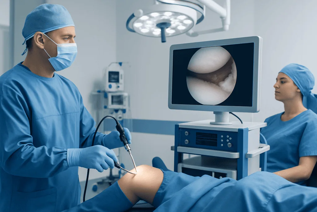
Most procedures are performed arthroscopically. This means the surgeon uses a small camera and slender instruments inserted through several small skin incisions, rather than making one large incision. Depending on what is found inside the joint, the surgeon may repair the torn tissue with anchors and sutures, remove unstable frayed tissue, or reconstruct the labrum using tissue from the patient or a donor.
A labral tear operation is typically considered when symptoms remain limiting despite appropriate non-surgical care, such as activity modification, physiotherapy, and pain-management measures. The procedure aims to improve joint function and reduce symptoms, but results depend on factors including the type of tear, joint shape, cartilage health, rehabilitation participation, and the person’s everyday or sporting demands.
How the Labrum Tears and Who May Be a Candidate
Hip labral tears may develop after twisting injuries, repeated deep hip flexion, falls, or joint-shape differences such as femoroacetabular impingement. In this condition, extra bone can cause repeated contact between the femur and hip socket, which may injure the labrum over time. Hip labral damage can also occur alongside early joint wear or following significant trauma.
In the shoulder, labral tears can result from a dislocation, a direct injury, a fall onto an outstretched arm, heavy lifting, or repetitive overhead activity. Some tears occur at the top of the labrum where the biceps tendon attaches, while others affect the front or lower part of the labrum and may be linked with recurrent shoulder instability.
Good candidates for surgery usually have symptoms that match the clinical examination and imaging findings, and have not improved enough with an individually planned course of conservative treatment. A surgeon will also consider age, activity level, joint stability, bone shape, degree of arthritis, quality of the labral tissue, and readiness to follow rehabilitation instructions. When advanced arthritis is the main source of pain, arthroscopic labral surgery may be less likely to provide meaningful benefit.
Assessment should identify related problems that may need treatment at the same operation. For example, a hip surgeon may address impingement at the time of a labral repair, while a shoulder surgeon may assess instability, tendon injury, or bone loss. This broader joint assessment helps ensure that surgery addresses the underlying mechanical problem rather than only the visible tear.
Assessment and Planning Before Surgery

Diagnosis starts with a detailed discussion of symptoms, past injuries, daily activities, work requirements, and sports participation. The clinician examines movement, strength, joint stability, and areas of tenderness. Because symptoms from the back, muscles, tendons, or nearby joints can resemble a labral tear, a careful examination is important before deciding on an operation.
Plain X-rays are commonly used to assess bone shape, alignment, arthritis, and signs of previous injury. Magnetic resonance imaging, sometimes with contrast injected into the joint, can help show the labrum and surrounding soft tissues. However, imaging results are interpreted alongside symptoms and examination findings; some labral changes are seen on scans in people who have no pain.
Before arthroscopy, the surgical team reviews medical conditions, current medicines, allergies, and previous operations. Patients may be asked to adjust certain medicines before surgery, particularly those that affect bleeding, but changes should only be made with advice from the prescribing clinician and surgical team. A personalized rehabilitation plan is often discussed before surgery so that practical support, transport, work arrangements, and physiotherapy can be organized.
Labral Tear Operation: Step by Step
Labral tear surgery is commonly performed under general anesthesia, sometimes with additional regional anesthesia for pain control. The patient is positioned to give the surgical team safe access to the hip or shoulder. In hip arthroscopy, gentle traction may be used temporarily to create space within the joint; in shoulder arthroscopy, the arm is positioned to allow the surgeon to inspect the joint fully.
The surgeon makes small incisions, called portals, and introduces an arthroscope. The camera sends detailed images to a monitor. Sterile fluid is used to expand the joint and improve visibility. The surgeon systematically inspects the labrum, cartilage surfaces, ligaments, tendons, and other structures, confirming the nature and extent of the injury.
If the labrum is repairable, it is often reattached to the socket with small anchors and strong sutures. Repair preserves the labrum’s contribution to joint stability and load distribution. If there is a small unstable frayed section that cannot be repaired, the surgeon may carefully smooth or remove that tissue, a procedure sometimes called debridement. Reconstruction may be considered when the labrum is too damaged, deficient, or previously treated to be repaired effectively.
Associated mechanical problems may be corrected during the same procedure when appropriate. In the hip, this can include reshaping bone that contributes to impingement. In the shoulder, stabilization procedures may be combined with labral repair when recurrent dislocation or instability is present. Patients can discuss the expected surgical plan and possible intraoperative alternatives with their orthopedic surgeon in advance.
Benefits, Limitations and Possible Risks
The potential benefits of a labral tear operation include less pain, fewer catching or instability symptoms, improved movement, and a return to desired daily activities or sport. Repairing a labrum may also help restore joint mechanics when the tear is a significant source of symptoms. Improvement is usually gradual because repaired tissue needs time to heal and muscles need to regain strength and coordination.
Not every person experiences complete symptom relief. Pain may persist if there is cartilage damage, arthritis, another pain source, incomplete healing, or an ongoing biomechanical issue. A repair may not be possible if tissue quality is poor, and the final procedure can occasionally differ from the original plan based on findings during arthroscopy.
Potential complications include infection, bleeding, blood clots, anesthesia-related problems, stiffness, swelling, numbness or nerve irritation, and ongoing pain. There is also a risk of re-tear, incomplete healing, recurrent instability, or a need for further treatment. Hip arthroscopy can involve temporary traction-related numbness or pressure effects, while shoulder surgery may involve stiffness or a prolonged recovery of strength.
These risks are generally discussed during consent, along with personal factors that may affect recovery. Following wound-care instructions, using prescribed supports, attending follow-up visits, and completing rehabilitation can help reduce avoidable complications and support the best possible recovery.
Recovery Timeline and Rehabilitation
Recovery after a labral tear operation varies by joint, procedure, associated repairs, and individual health. Many patients go home on the day of surgery or after a short hospital stay. Pain and swelling are expected initially and are managed with the care plan provided by the surgical team. Keeping the wound dressings clean and dry, as instructed, is important while the incisions heal.
After hip labral repair, patients commonly use crutches for a period determined by the repair and any bone work performed. The team may set limits on weight-bearing, hip flexion, twisting, or certain exercises during early healing. After shoulder labral repair, a sling is commonly used, and active arm movement is restricted at first to protect the repair. The exact duration differs between procedures and should follow the surgeon’s plan.
Physiotherapy progresses in stages. Early rehabilitation focuses on safe movement, swelling control, and protecting the surgical repair. Later stages build range of motion, muscle control, strength, balance, and sport- or work-specific skills. Returning to desk work may be possible earlier than returning to physically demanding work, lifting, running, contact sports, or overhead sport.
Full recovery often takes several months, and returning to high-impact or competitive activity may take longer. Recovery should be guided by function and clinical assessment rather than by a calendar alone. Any new injury, increasing pain, fever, wound redness, drainage, calf swelling, chest pain, shortness of breath, or sudden loss of function should be reported promptly to the surgical team or assessed urgently when appropriate.
When to Seek Medical Care
Medical assessment is appropriate for persistent hip or shoulder pain that interferes with sleep, work, walking, lifting, exercise, or ordinary activities. Evaluation is also helpful when there is recurrent clicking, catching, giving way, a sensation that the shoulder may dislocate, or reduced movement that does not improve with rest and guided rehabilitation.
Urgent medical care is needed after a significant injury if there is an obvious deformity, inability to move or bear weight through the limb, severe swelling, numbness, weakness, or a suspected joint dislocation. A shoulder that appears dislocated should not be forced back into place outside a healthcare setting.
After surgery, patients should contact their team if pain is worsening rather than gradually improving, if the wound becomes increasingly red or drains fluid, or if they develop fever or other concerning symptoms. Emergency assessment is important for symptoms that could suggest a blood clot or serious complication, such as chest pain, breathing difficulty, or marked swelling and pain in one calf.
Acibadem International’s multidisciplinary orthopedic, anesthesia, radiology, and rehabilitation specialists can assess and treat hip and shoulder labral conditions for international patients in its JCI-accredited hospitals. A qualified orthopedic surgeon can explain whether continued non-surgical care, arthroscopy, or another approach is most appropriate for the individual joint problem.
Frequently asked questions
Is a labral tear operation always necessary?
No. Many labral tears can be managed without surgery, particularly when symptoms are mild or improve with physiotherapy, activity changes, and appropriate pain management. Surgery may be considered when symptoms remain significant and the tear is believed to be a main cause of the problem.
How long does a labral tear operation take?
The duration depends on whether the tear is in the hip or shoulder, the type of repair needed, and whether related problems are treated at the same time. The surgical team can provide a more individualized estimate during preoperative planning.
Will a labral tear heal without surgery?
The labrum has a limited blood supply in some areas, so a tear may not fully heal on its own. Even so, symptoms can sometimes improve substantially with non-surgical treatment by strengthening the muscles around the joint and reducing aggravating movement patterns.
When can someone walk after hip labral tear surgery?
Many people can walk with crutches soon after surgery, but weight-bearing restrictions vary according to the repair and any additional hip procedure. The surgeon and physiotherapist will explain when it is safe to progress to full weight-bearing.
When can someone drive after labral tear surgery?
Driving should wait until the person is no longer taking medicines that impair alertness, can safely control the vehicle, and can perform an emergency stop or steering maneuver without limitation. This timing differs for hip and shoulder surgery and should be confirmed with the treating team.
Can a person return to sports after labral tear surgery?
Many people return to recreational or competitive activity after rehabilitation, but the timing and likelihood of return vary. Return should be based on pain control, movement, strength, joint stability, and sport-specific testing, with guidance from the surgeon and physiotherapist.
References
- American Academy of Orthopaedic Surgeons
- OrthoInfo
- American Orthopaedic Society for Sports Medicine
- National Institute of Arthritis and Musculoskeletal and Skin Diseases
- British Orthopaedic Association
This article is for general information only and is not a substitute for professional medical advice. Please consult a qualified doctor about your individual situation.
Joint and spine care in Turkey — expert assessment & treatment
JCI-accredited · board-certified surgeons · reply within 24h