Pseudotumor Cerebri Treatment: How It Works, Results and What to Expect

Pseudotumor cerebri is also called idiopathic intracranial hypertension (IIH). Protecting vision through prompt eye assessments is the central goal of treatment.
Key Takeaways
- Pseudotumor cerebri is also called idiopathic intracranial hypertension (IIH).
- Protecting vision through prompt eye assessments is the central goal of treatment.
- Weight management and pressure-lowering medicines help many people manage the condition.
- Surgery or stenting may be considered when vision is rapidly worsening or symptoms do not respond to conservative care.
- New, severe or changing visual symptoms require urgent medical assessment.
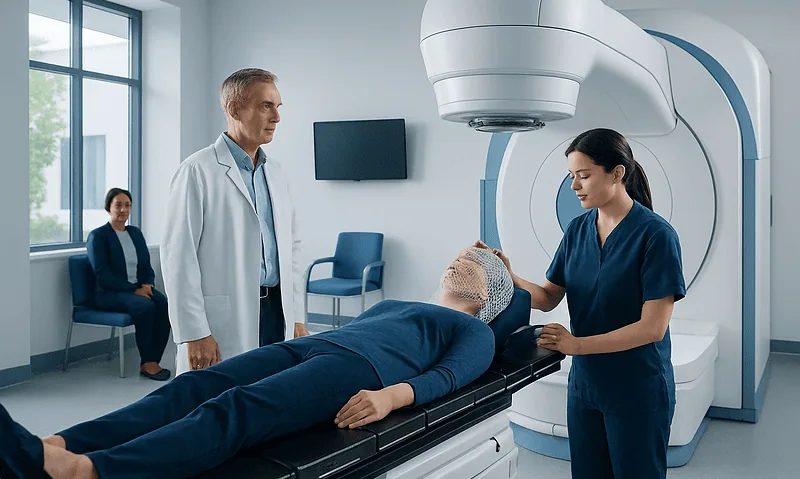
Pseudotumor cerebri treatment aims to reduce raised pressure inside the skull, relieve symptoms and, most importantly, prevent permanent vision loss. Care is tailored to the severity of visual changes and may include lifestyle support, medication, regular eye monitoring and procedures for urgent or treatment-resistant cases.
Overview: How Pseudotumor Cerebri Treatment Works
Pseudotumor cerebri treatment works by lowering the pressure of cerebrospinal fluid, the fluid that surrounds and cushions the brain and spinal cord. Despite its name, pseudotumor cerebri is not a brain tumor. It is more commonly called idiopathic intracranial hypertension (IIH), meaning raised pressure inside the skull without an identified mass, infection or other structural cause.
The treatment plan has two main priorities: protecting eyesight and reducing symptoms such as headache, pulsating noise in the ears and double vision. The optic nerves can become swollen from increased pressure, a finding called papilledema. Because visual loss can sometimes develop without severe pain, regular assessment by an eye specialist is an important part of care.
Management is often coordinated between neurology, neuro-ophthalmology, ophthalmology, radiology and, when needed, neurosurgery. The approach depends on a person’s symptoms, visual field results, optic nerve appearance, overall health and whether vision is stable, gradually changing or deteriorating quickly.
Symptoms, Causes and What Triggers Pseudotumor Cerebri?

Common symptoms include headaches, brief episodes of dimmed or blurred vision, double vision, nausea and a rhythmic whooshing sound in one or both ears that follows the heartbeat. Some people notice neck, shoulder or back pain. Symptoms vary considerably, and headache severity does not always reflect the degree of risk to vision.
In many cases, no single cause is found, which is why the term idiopathic is used. IIH occurs more often in women of childbearing age and is associated with recent weight gain or obesity, although it can affect people of any sex, age or body size. Hormonal and metabolic factors may contribute, but they do not explain every case.
Doctors also review possible secondary causes of raised intracranial pressure. These can include certain medicines, such as vitamin A derivatives, tetracycline antibiotics, growth hormone or changes involving corticosteroids. Conditions affecting blood flow in the veins of the brain, anemia, sleep apnea and some endocrine disorders may also need to be considered. A clinician should review medicines and supplements rather than stopping prescribed treatment without advice.
How Is Pseudotumor Cerebri Diagnosed?

Diagnosis begins with a medical history and examination, including a detailed eye examination. Clinicians assess visual sharpness, color vision, eye movements, visual fields and the optic nerves. Optical coherence tomography, a noninvasive eye scan, may help measure and track optic nerve swelling over time.
Brain imaging, usually magnetic resonance imaging (MRI) and imaging of the brain’s venous channels, helps rule out a tumor, blood clot or another cause of raised pressure. These tests can also show findings that may support IIH, although imaging alone cannot confirm the diagnosis.
A lumbar puncture may then be performed when clinically appropriate. During this procedure, a needle is placed into the lower back to measure cerebrospinal fluid pressure and analyze the fluid for other conditions. It is not simply a treatment test; its results are interpreted alongside symptoms, examination and imaging findings.
Treatment Options, Candidacy and Step-by-Step Procedures
For people with stable vision, treatment commonly begins with an individualized weight-management plan where relevant, headache care and a medicine that reduces cerebrospinal fluid production. Acetazolamide is often used; topiramate may be considered for selected patients, particularly when migraine-like headaches are also present. The choice depends on medical history, possible side effects, pregnancy plans and other medicines.
Weight reduction can lower intracranial pressure and may improve papilledema and symptoms in people for whom excess weight is a contributing factor. Support may involve nutritional counseling, physical activity adapted to symptoms and, for carefully selected individuals, specialist obesity treatment. It should be approached as supportive healthcare rather than blame, as IIH is a complex medical condition.
When vision is threatened, symptoms remain disabling despite appropriate treatment, or pressure needs urgent control, a procedure may be considered. A lumbar puncture can temporarily lower pressure but is usually not a long-term solution. Surgical options include optic nerve sheath fenestration, which creates a small opening around the optic nerve to protect sight, and cerebrospinal fluid shunting, which diverts fluid to another part of the body. In selected cases with narrowing of the brain’s venous sinuses, venous sinus stenting may be evaluated after specialist testing.
Before a procedure, the team reviews eye findings, scans, lumbar puncture results and individual risks. For shunt or optic nerve surgery, preparation includes anesthesia assessment and discussion of expected goals. For venous sinus stenting, catheter-based imaging and pressure measurements may be used to determine whether stenting is appropriate. These procedures are generally reserved for specific clinical situations, not for every person with IIH.
Benefits, Risks and Recovery Timeline
The expected benefit of treatment is stabilization or improvement of optic nerve swelling and vision, along with relief of pressure-related symptoms. Improvement in headache can be less predictable because headaches may continue even after intracranial pressure improves, sometimes due to coexisting migraine or other headache disorders. Continued follow-up helps ensure that each symptom is addressed appropriately.
Medicines can cause side effects, including tingling sensations, taste changes, tiredness, stomach upset or changes in mood and concentration, depending on the medicine used. Clinicians may order blood tests or adjust treatment when needed. Weight-management plans should be safe, realistic and adapted to a person’s medical needs.
Recovery after a lumbar puncture is often brief, though some people develop a temporary post-procedure headache. Recovery from optic nerve surgery, shunt placement or stenting varies by procedure and individual health. Hospital observation may be needed, followed by scheduled eye checks, wound review where relevant and activity guidance from the treating team.
Procedural risks can include infection, bleeding, anesthesia-related complications, low-pressure headaches, shunt blockage or the need for revision. Stenting may involve blood-clotting or vessel-related risks and often requires medication that affects clotting for a period determined by the specialist. The decision is made by balancing these risks against the risk of continuing visual damage from uncontrolled pressure.
Does Pseudotumor Cerebri Ever Go Away?
Pseudotumor cerebri can improve substantially and may enter remission, particularly when contributing factors are addressed and intracranial pressure normalizes. For some people, symptoms and optic nerve swelling resolve after weight reduction, treatment of an associated condition or a period of medical therapy.
However, IIH can be chronic or recur, including after an interval of feeling well. Ongoing follow-up is important until a clinician confirms that optic nerve swelling has settled and vision is stable. People with a previous diagnosis should seek review if typical symptoms return, especially visual changes or pulsating sounds in the ears.
Even when pressure has normalized, headache treatment may still be needed. A personalized plan can distinguish recurring IIH from migraine or another cause of headache, avoiding unnecessary procedures while maintaining appropriate eye protection.
What to Avoid With Pseudotumor Cerebri?
People with pseudotumor cerebri should avoid delaying assessment for new visual symptoms or assuming that a headache is harmless because it resembles a previous one. Vision can be affected with little warning, so planned eye examinations and visual-field testing should not be missed.
It is also sensible to avoid starting vitamin A-containing supplements, retinoid acne medicines, tetracycline antibiotics or other medicines linked with raised intracranial pressure without discussing them with the clinician managing IIH. These medicines can be appropriate for some people, but the potential relationship should be reviewed carefully. Prescribed medicines should never be stopped suddenly without medical advice.
Crash diets, dehydration and unregulated weight-loss products are not recommended. Sustainable changes, supported by a healthcare professional, are safer and more likely to help long term. Alcohol intake, sleep quality and migraine triggers may also be discussed if headaches are a major symptom.
How Serious Is Pseudotumor Cerebri and When to Seek Medical Care?
Pseudotumor cerebri is serious because persistent high pressure can damage the optic nerves and lead to lasting visual impairment if it is not recognized and treated. Most people can be managed effectively with timely monitoring and appropriate treatment, but the level of urgency depends largely on the condition of the vision rather than headache intensity alone.
Urgent medical assessment is needed for sudden or worsening loss of vision, persistent double vision, markedly increasing blind spots, a severe new headache with neurological symptoms, confusion, weakness, fever or vomiting. These symptoms can have causes other than IIH and should not be self-diagnosed.
Anyone with possible IIH symptoms should arrange medical evaluation, especially if blurred vision, brief visual blackouts or pulsating tinnitus occurs. Acibadem International’s multidisciplinary specialists and JCI-accredited hospitals support international patients with diagnostic assessment and treatment planning for conditions involving raised intracranial pressure.
Frequently asked questions
What is the first-line pseudotumor cerebri treatment?
First-line treatment commonly includes close visual monitoring, support with weight management when appropriate and a medicine that lowers cerebrospinal fluid production. The exact plan depends on symptoms, eye findings, other health conditions and the risk to vision. A specialist may also treat coexisting migraine or other headache conditions.
Can pseudotumor cerebri be treated without surgery?
Yes. Many people are treated without surgery through medication, lifestyle support and regular neuro-ophthalmic follow-up. Surgery or stenting is generally considered when vision is worsening, pressure remains uncontrolled or symptoms are severe despite conservative treatment.
How long does pseudotumor cerebri treatment take to work?
The timeline varies. Some people notice symptom improvement within weeks of starting treatment, while optic nerve swelling and visual fields may take longer to stabilize. Regular eye tests provide the most reliable way to monitor whether treatment is protecting vision.
Does losing weight help pseudotumor cerebri?
For people in whom excess weight or recent weight gain is contributing, gradual weight loss can reduce intracranial pressure and improve the condition. It is not the only treatment, and it should be supported by medical monitoring. People should avoid extreme or unsupervised dieting.
Can pseudotumor cerebri cause permanent blindness?
It can cause permanent visual loss if optic nerve damage develops and is not controlled promptly. This is why visual symptoms, optic nerve swelling and visual-field changes require close specialist monitoring. With timely treatment, the risk can often be reduced.
What follow-up is needed after pseudotumor cerebri treatment?
Follow-up typically includes repeat eye examinations, visual-field testing and optic nerve imaging at intervals decided by the care team. Medication effects, weight-management progress and headache symptoms may also be reviewed. Follow-up frequency is usually greater when vision is unstable or papilledema is significant.
References
- National Eye Institute
- National Institute of Neurological Disorders and Stroke
- American Academy of Ophthalmology
- European Headache Federation
- Mayo Clinic
This article is for general information only and is not a substitute for professional medical advice. Please consult a qualified doctor about your individual situation.
Persistent digestive symptoms? Get evaluated in Turkey
JCI-accredited · board-certified surgeons · reply within 24h
Add us as a Preferred Source to see more of our trusted health content across Google Search, AI Overviews and Discover.