Treatment for Broken Eye Socket: How It Works, Results and What to Expect

A broken eye socket is a fracture of one or more bones surrounding the eye and should be assessed promptly after facial trauma. Urgent symptoms include reduced vision, severe eye pain, inability to move the eye normally, marked swelling, nausea with eye movement or increasing drowsiness.
Key Takeaways
- A broken eye socket is a fracture of one or more bones surrounding the eye and should be assessed promptly after facial trauma.
- Urgent symptoms include reduced vision, severe eye pain, inability to move the eye normally, marked swelling, nausea with eye movement or increasing drowsiness.
- Many uncomplicated orbital fractures heal without surgery, but close follow-up is important because swelling can initially mask important signs.
- Surgery may be recommended when there is muscle entrapment, persistent double vision, a sunken eye, a large fracture or associated facial injuries.
- Recovery varies by injury severity and treatment; bruising often improves over weeks, while bone healing and return to normal activity can take longer.
Treatment for a broken eye socket, also called an orbital fracture, depends on whether the injury affects vision, eye movement, the position of the eye, facial sensation or the shape of the orbit. Small, stable fractures may heal with monitoring and protective measures, while fractures causing significant symptoms may need surgery to restore support around the eye.
Overview: treatment for broken eye socket
Treatment for a broken eye socket begins with protecting vision and checking how the eye and surrounding facial bones have been affected. The eye socket, or orbit, is a bony space that supports the eyeball and contains muscles, nerves, blood vessels and soft tissue. A fracture can involve the thin floor, inner wall, rim or several parts of the orbit.
Most orbital fractures occur after a blow to the face, such as a fall, sports injury, road traffic collision or assault. Initial swelling and bruising can be substantial, but the seriousness of the injury is not determined by appearance alone. A clinical examination and usually a CT scan help doctors decide whether observation, specialist follow-up or repair is the safest approach.
The main aims of treatment for eye socket fracture are to preserve sight, ensure the eye can move normally, prevent the eye from settling backward in the socket, and address associated injuries to the cheekbone, nose, eyelids or sinuses. Care commonly involves emergency clinicians, ophthalmologists, maxillofacial or plastic surgeons, and sometimes ear, nose and throat specialists.
Is a broken eye socket serious?
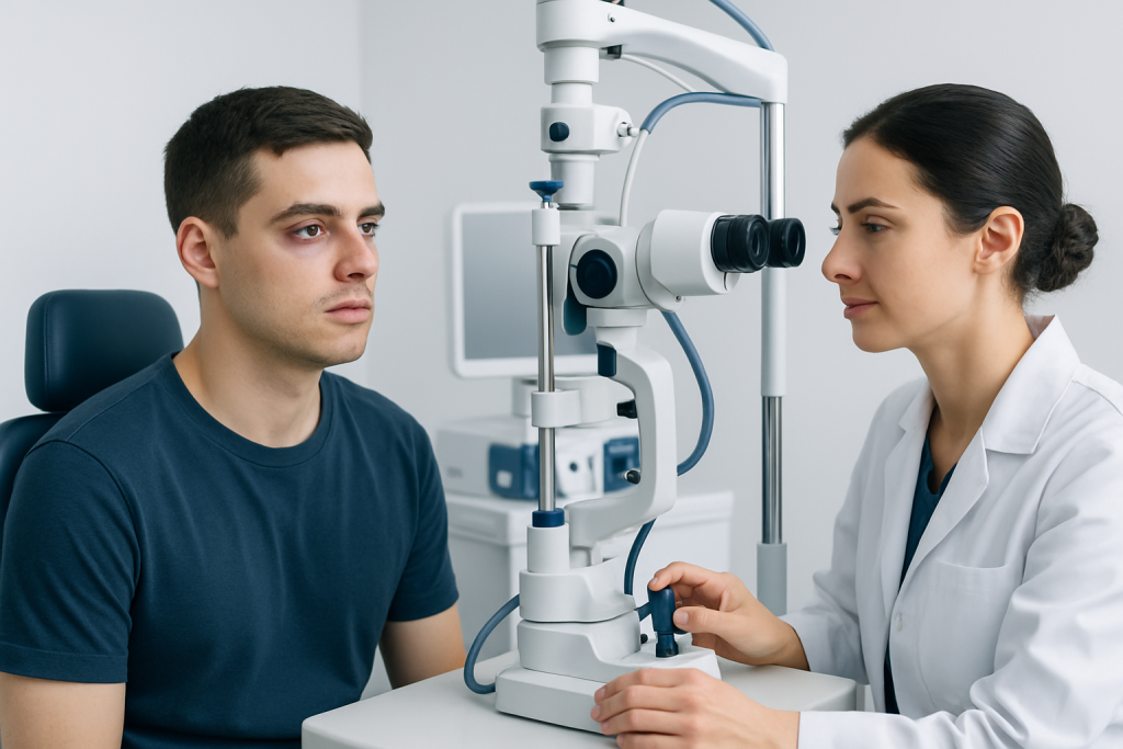
A broken eye socket can be serious because the orbit sits next to the eye, optic nerve, brain and facial sinuses. Some fractures are small and stable, while others can trap an eye muscle or soft tissue, change the position of the eye, damage nearby structures or occur alongside head injuries. Prompt medical assessment is therefore important after a significant injury around the eye.
Possible signs include pain, swelling, bruising, numbness of the cheek or upper lip, double vision, pain or nausea when looking up or down, reduced ability to move the eye, or a sensation that the eye has sunk backward. Bleeding in or around the eye and changes in pupil shape or vision require urgent attention.
Children need particularly prompt assessment. Their bones can bend and then spring back, trapping tissue in what may look like a relatively minor injury. This may cause restricted eye movement, double vision, nausea or vomiting despite limited bruising.
A fracture does not always cause permanent problems. Early evaluation helps identify the smaller group of injuries that need urgent treatment and supports safe monitoring for those likely to heal without an operation.
What do doctors do for a broken eye socket?

Doctors first assess visual acuity, pupils, eye pressure when appropriate, eye movements, facial sensation and the position of the eye. They also look for cuts, globe injury, bleeding behind the eye, facial bone instability and signs of concussion or other trauma. A thin-slice CT scan of the facial bones is commonly used to show the location and extent of the fracture.
Early care may include cold compresses, pain relief, wound care and advice to keep the head elevated. Doctors commonly advise avoiding nose blowing, forceful sneezing and activities that increase pressure in the sinuses, because air can pass through a fracture and worsen swelling around the eye. A clinician will advise on medicines and whether antibiotics or other treatment are appropriate in the individual situation.
If the fracture is uncomplicated, care may consist of observation and repeat examinations after swelling settles. If there are concerning findings, the patient may be referred for orbital fracture surgery or another facial reconstruction procedure. The timing depends on the injury; some problems require urgent intervention, while others can be reassessed over several days once swelling allows a clearer evaluation.
Associated injuries may require additional care. For example, doctors may evaluate cheekbone fractures, nasal injury, eyelid wounds, dental trauma or head injury at the same time. A coordinated plan helps protect both eye function and facial structure.
Can a broken eye socket heal on its own?
Yes, a small, non-displaced broken eye socket can often heal on its own without surgery. This is more likely when vision is normal, the eye moves freely, double vision is absent or improving, the eye has not changed position and imaging shows no large defect or trapped tissue. Follow-up remains important because some changes become clearer as swelling goes down.
Bone healing is only one part of recovery. Doctors also monitor the muscles, nerves and soft tissues that allow the eye to move and maintain its position. Persistent double vision, a visibly sunken eye, significant numbness or cosmetic asymmetry may indicate that further treatment should be considered.
People should not try to determine the severity of an orbital fracture based on bruising alone. They should follow the clinician’s activity guidance, avoid touching or pressing on the eye, and attend all planned reviews. Contact sports and activities with a risk of facial impact should be avoided until a treating clinician confirms it is safe to resume them.
How surgery for an eye socket fracture works
Surgery is considered when the fracture causes muscle or soft-tissue entrapment, persistent troublesome double vision, significant movement restriction, a large orbital defect, progressive sinking of the eye or combined facial fractures. The decision is individualized. Surgeons weigh examination findings, CT imaging, symptoms, the likelihood of natural improvement and the person’s overall health.
Orbital fracture repair is generally performed under general anesthesia. The surgeon often uses an incision hidden inside the lower eyelid or through another carefully selected route, depending on the fracture location. The displaced tissues are gently released from the fracture, and the missing bony support may be rebuilt with an implant or graft chosen for the anatomy and surgical plan.
The goal is not simply to place material in the orbit. The surgeon restores the volume and contour needed to support the eye while avoiding injury to the eye muscles, nerves and surrounding structures. In more complex trauma, repair may be coordinated with treatment of the cheekbone, nasal bones, jaw or eyelids.
Before surgery, patients may have a detailed eye examination, CT review, anesthesia assessment and discussion of expected benefits and limitations. It is important to tell the clinical team about blood-thinning medicines, allergies, long-term conditions and any previous eye surgery.
How long does it take to recover from an eye socket fracture?
How long for a broken eye socket to heal depends on the fracture pattern, whether surgery is needed, associated injuries and the individual’s general health. Swelling and bruising commonly begin to settle over one to two weeks, although residual discoloration, numbness or mild discomfort may last longer. Bone healing often takes several weeks.
After surgery, early follow-up checks wound healing, vision, eye movement and the position of the eye. Temporary swelling, tightness, tearing, numbness around the cheek or upper lip, and blurred vision from swelling or ointment can occur. Double vision may improve gradually as swelling resolves, but its course varies and should be monitored by the surgical and eye-care teams.
Most people can return to light daily activities as advised by their clinician, but strenuous exercise, swimming, heavy lifting and contact sports may need to be postponed. The precise timeline is individualized, particularly for people with complex fractures or work involving physical risk.
Recovery should be guided by scheduled reviews rather than by appearance alone. New or worsening visual symptoms, increasing pain, fever, discharge from a wound, or a growing difference in eye position should be reported promptly.
Benefits, risks and aftercare
When surgery is appropriate, possible benefits include releasing trapped tissue, improving eye movement, reducing persistent double vision, restoring orbital support and improving facial symmetry. However, no procedure can guarantee that all symptoms will resolve, especially when the original injury has affected muscles, nerves or the eye itself.
Potential risks include infection, bleeding, scarring, persistent or new double vision, altered facial sensation, asymmetry, implant-related problems, eyelid position changes, need for further treatment and, rarely, injury that affects vision. The surgical team discusses the risks relevant to the individual fracture and explains how these are minimized.
Aftercare commonly includes keeping the head elevated, using cold compresses as advised, avoiding nose blowing and attending follow-up visits. Patients should use prescribed medicines exactly as directed and ask their clinician before restarting supplements or medicines that may affect bleeding.
Acibadem International’s multidisciplinary specialists and JCI-accredited hospitals evaluate and treat orbital fractures for international patients, coordinating eye care and facial trauma expertise when needed.
When to seek medical care
Anyone with a suspected broken eye socket after facial trauma should seek medical assessment as soon as possible, even if symptoms seem mild. This is especially important when there is double vision, numbness, pain with eye movement, worsening swelling or a change in the shape or position of the eye.
Emergency care is needed for reduced or lost vision, severe eye pain, a bulging eye, inability to move the eye, unequal pupils, persistent vomiting after an eye injury, confusion, loss of consciousness, clear fluid from the nose, uncontrolled bleeding or a deep cut near the eye. These symptoms can indicate injury beyond a simple fracture.
People who have already been assessed should contact their care team if symptoms worsen rather than improve, if new visual changes develop, or if they have concerns during recovery. Timely reassessment can help prevent complications and provide reassurance when healing is progressing normally.
Frequently asked questions
What is a broken eye socket called medically?
A broken eye socket is usually called an orbital fracture. It may affect the orbital floor, medial wall, orbital rim or multiple bones around the eye. The term does not necessarily mean that the eyeball itself is injured, but the eye must always be examined carefully.
Do all eye socket fractures need surgery?
No. Many small, stable fractures can be managed without surgery when eye movement, vision and eye position are normal and imaging does not show a concerning defect. Surgery is more likely when tissue is trapped, double vision persists, the eye is displaced, or the fracture is large or complex.
Why should someone not blow their nose after an orbital fracture?
The orbit is close to air-filled sinuses, and a fracture can create a pathway for air to move into tissues around the eye. Nose blowing may increase swelling and discomfort and can rarely cause more serious pressure-related problems. A clinician can advise when it is safe to resume normal nose blowing.
Can double vision after a broken eye socket go away?
Double vision may improve as swelling decreases, particularly in the first days or weeks after injury. Persistent, severe or worsening double vision needs assessment because it can be related to muscle entrapment, nerve injury or a change in orbital shape. The treatment plan depends on the cause and examination findings.
How long should contact sports be avoided after an orbital fracture?
The timeline varies with the fracture, treatment and the risk of another impact. A treating surgeon or eye specialist should give individualized clearance before returning to contact sports or high-risk activities. Returning too early may increase the chance of reinjury while the bones and tissues are still healing.
Will numbness in the cheek go away after an eye socket fracture?
Numbness of the cheek, upper lip or upper teeth can occur when a nearby facial nerve is bruised or affected by the fracture. It often improves gradually, but recovery can take weeks or longer and may be incomplete in some cases. Ongoing or worsening numbness should be discussed at follow-up.
References
- American Academy of Ophthalmology
- American Association for the Surgery of Trauma
- Merck Manual Professional Edition
- National Health Service
This article is for general information only and is not a substitute for professional medical advice. Please consult a qualified doctor about your individual situation.
Joint and spine care in Turkey — expert assessment & treatment
JCI-accredited · board-certified surgeons · reply within 24h
Add us as a Preferred Source to see more of our trusted health content across Google Search, AI Overviews and Discover.

More from the Health Library


Related Specialists

Dr. Emir Akıncıoğlu
Urology
Dr. Cenk Ersemiz
Acibadem Life Clinical Service
Assoc. Prof. Dr. Fatih Şimşek
Anesthesiology
Assoc. Prof. Dr. Mehmet Barburoğlu
Interventional Neuroradiology