

Cholesteatoma and Mastoid Surgery: Why Does This Growth Behind the Eardrum Need Removal?
Key Takeaways
- A cholesteatoma is trapped skin behind the eardrum, not a tumor, but it erodes bone continuously and the NHS states that surgery is usually required because it does not resolve on its own.
- The growth sits within millimeters of the hearing bones, the facial nerve, the balance organs and the brain, which is why MedlinePlus lists hearing loss, facial paralysis, vertigo and meningitis among untreated complications.
- Canal wall up surgery preserves normal ear anatomy but hides any regrowth, while canal wall down lowers recurrence at the cost of a cavity that needs periodic cleaning in clinic.
- Removing the disease takes priority over hearing reconstruction, so surgeons sometimes stage the operation and rebuild the hearing bones in a later procedure once the ear is confirmed clear.
- The NHS advises roughly one to two weeks off work, several weeks of keeping the ear dry, and checking with the surgeon before flying, with full healing described in months rather than weeks.
- Because residual and recurrent disease are recognized realities, a follow-up MRI or second-look operation around a year later is a planned part of care, not a sign of failure.
A cholesteatoma is a pocket of skin cells trapped behind the eardrum that slowly enlarges and erodes the tiny hearing bones and surrounding skull. It does not clear up on its own and rarely responds to medication, so surgeons usually remove it through mastoid surgery to stop infection, protect hearing and prevent rare but serious complications such as facial nerve damage or meningitis. Follow-up checks are routine because it can return.
The appointment was supposed to be about a stubborn, smelly discharge from one ear, the kind that soaks a pillowcase and shrugs off two courses of drops. Instead, the ENT specialist swung the microscope away, drew a small pouch on the back of a form and said a word the patient had never heard: cholesteatoma. Then came the second surprise. The fix was not a stronger prescription. It was an operation on the bone behind the ear.
That conversation happens quietly in clinics every week, and it usually raises the same question in the same tone of disbelief. Why does a bit of skin need surgery? The honest answer is that this particular skin is in the wrong place, behaving like a slow-motion drill inside a space the width of a fingertip.
This article walks through cholesteatoma mastoid surgery the way a good surgeon would if the clinic were not running forty minutes late: what the growth is, why watching it is rarely safe, what actually happens in the operating room, and what the weeks afterward tend to look like.
What is a cholesteatoma, and why does skin end up in the wrong place?
A cholesteatoma is a collection of skin cells and debris that builds up in the middle ear, the air-filled chamber just behind the eardrum. Despite the name, it is not a tumor and it is not cancer. Think of it as a small cyst lined with the same kind of skin that covers the outside of your ear canal, except it is sealed in a place that has no exit.
Skin anywhere on the body sheds constantly. On your arm, dead cells simply fall away. Inside a closed pocket behind the eardrum, they have nowhere to go, so the pouch fills and swells like a sock stuffed with lint. The trapped material is a comfortable home for bacteria, which explains the persistent, often foul-smelling discharge that first brings many people to a clinic.
Most cholesteatomas are acquired rather than present from birth. According to the NHS, the usual starting point is a eustachian tube that does not work well. The eustachian tube is the narrow channel linking the middle ear to the back of the nose, and its job is to equalize pressure. When it stays blocked, negative pressure gently sucks part of the eardrum inward, creating a retraction pocket that collects skin. Repeated ear infections in childhood, a previous perforation, or past ear surgery can also let skin migrate through the eardrum. A smaller group, congenital cholesteatoma, is present at birth and is often discovered in children who have never had an ear infection.
Why does location matter so much? The middle ear houses the three smallest bones in the body, the malleus, incus and stapes, which relay sound from the eardrum to the inner ear. Millimeters away sit the facial nerve, the balance organs and the thin plate of bone separating the ear from the brain. A growth here has very little room to be harmless.
Why does a cholesteatoma need removal instead of watching and waiting?
The uncomfortable truth about cholesteatoma is that it does not stop. Unlike many benign lumps that reach a certain size and settle, this one keeps recruiting fresh skin cells, and the inflammation around it releases enzymes that dissolve bone. The process is slow, often measured in years, but it is relentless.
MedlinePlus lists the consequences plainly. The hearing bones erode, producing conductive hearing loss, which means sound cannot physically reach the inner ear. The bony wall protecting the inner ear can be breached, causing dizziness and permanent nerve deafness. The facial nerve, which runs through the middle ear in a bony tunnel barely thicker than a pencil lead, can be exposed and damaged, leading to weakness on one side of the face. In the most serious cases, infection tracks upward into the skull, causing meningitis or a brain abscess. These complications are uncommon in countries with good access to ear care, but they remain the reason surgeons treat this condition with respect.
Medication has a limited role. Antibiotic drops can quiet the infection for a while and are often used to dry the ear before an operation, but they cannot remove skin that is already trapped. The NHS states directly that surgery is usually needed, because the growth will continue to enlarge and cause damage if left alone.
There is a real cost to waiting. The longer a cholesteatoma grows, the more bone it consumes, and the less there is left to reconstruct. A pocket caught early may be removed with the hearing bones intact. The same pocket found five years later may have hollowed out the mastoid and destroyed two of the three bones. In this condition, earlier is genuinely simpler.
How do doctors diagnose a cholesteatoma before mastoid surgery?
Diagnosis usually begins with a story: months of discharge from one ear, a gradual muffling of hearing, sometimes a sense of fullness or mild earache. The examination that follows is deceptively simple. An ENT specialist looks into the ear with a microscope or a small camera and, after cleaning away debris, may see a pearly white mass or a deep pocket in the upper part of the eardrum, the area called the attic. That view is often enough to make the diagnosis.
What the eye cannot see is how far the growth has spread. That is where imaging comes in. A CT scan, a detailed X-ray that builds cross-sectional pictures, maps the bony landscape: how much of the mastoid is involved, whether the hearing bones are intact, and whether the thin bone over the facial nerve or the inner ear has been eroded. Surgeons use this map to plan the approach and to warn patients about specific risks. Cleveland Clinic notes that MRI is sometimes added, particularly after previous surgery, because certain MRI sequences can distinguish cholesteatoma from scar tissue when a CT scan cannot.
Hearing tests are the other pillar. A pure-tone audiogram measures how loud a sound must be at each pitch before you hear it, and separates conductive loss (a mechanical problem in the middle ear) from sensorineural loss (a problem in the inner ear or nerve). Recording this before surgery matters for two reasons. It establishes a baseline against which any change can be measured, and it tells the surgeon whether hearing reconstruction is realistic.
Some people are surprised that a swab of the discharge is not the key test. Cultures may guide which antibiotic drops to use, but they do not diagnose the underlying problem. The skin pocket is the disease; the infection is a symptom of it.
What actually happens during cholesteatoma mastoid surgery?
Cholesteatoma mastoid surgery is performed under general anesthesia, so you are asleep throughout. The NHS describes the operation as usually taking two to three hours, though complexity varies with the size and reach of the growth. The surgeon works through a microscope or endoscope, using instruments small enough to handle bones the size of a grain of rice.
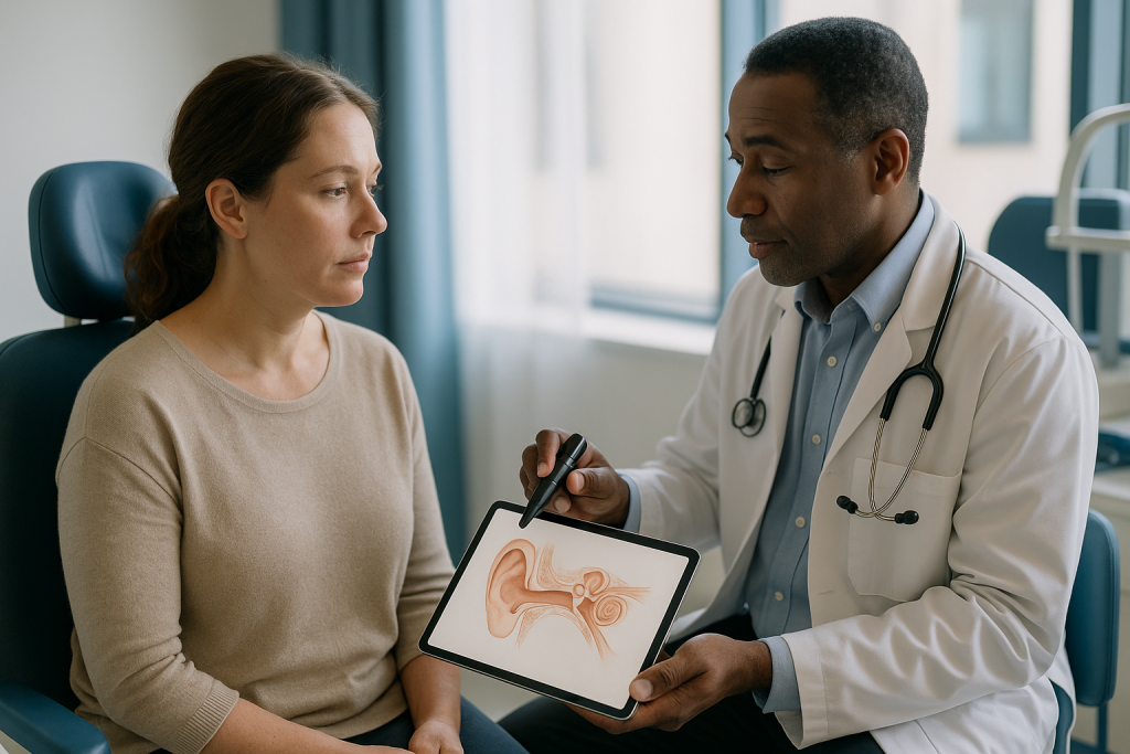
Access comes first. Depending on the extent of disease, the surgeon may work through the ear canal, make a small cut in the crease behind the ear, or both. The mastoid is the honeycomb of air cells inside the bony bump you can feel behind your earlobe. A mastoidectomy, the removal of some of that honeycomb with a fine drill, opens a window onto the middle ear from behind and lets the surgeon follow the cholesteatoma wherever it has crept.
Removal is the heart of the procedure and also the most painstaking part. Cholesteatoma is not a neat lump that lifts out in one piece. Its lining, called the matrix, spreads as a thin film into crevices around the hearing bones, over the facial nerve and into recesses that are difficult to see directly. Every fragment left behind can regrow. Surgeons often use angled endoscopes to peer around corners, and many use a nerve monitor that sounds an alert if instruments come close to the facial nerve.
Reconstruction follows. The eardrum is repaired, usually with a graft of the patient’s own tissue, and if the hearing bones have been damaged, the surgeon may rebuild the chain in the same sitting or plan to do so later. The ear canal is packed with dressing material to hold the graft in place, and a padded bandage goes over the ear. Most people go home the same day or after one night.
Mastoidectomy for cholesteatoma: canal wall up or canal wall down?
One of the first decisions a surgeon makes is what to do with the bony wall that separates the ear canal from the mastoid. Keeping it intact is called a canal wall up (or intact canal wall) mastoidectomy. Removing it to merge the two spaces into a single open cavity is a canal wall down mastoidectomy. Neither approach is universally better; each trades one set of advantages for another, and the choice depends on how far the disease has spread and how the ear is likely to behave afterward.
| Feature | Canal wall up | Canal wall down |
|---|---|---|
| Ear anatomy afterward | Near-normal ear canal and shape | Larger open cavity behind the canal |
| Visibility of any regrowth | Hidden; needs a second-look operation or MRI | Cavity can be inspected in clinic |
| Risk of residual or recurrent disease | Generally higher, because pockets can hide | Generally lower |
| Long-term ear care | Usually minimal once healed | Regular cleaning of the cavity; water precautions often lifelong |
| Hearing reconstruction | Often more straightforward | Possible, but the shape of the ear changes the options |
| Typical use | Smaller or well-defined disease, good eustachian tube function | Extensive disease, revision surgery, only hearing ear, poor follow-up access |
Cleveland Clinic describes both variants along with a third, the simple mastoidectomy, which clears infected mastoid cells without entering the middle ear and is mainly used for mastoiditis rather than cholesteatoma. In practice, surgeons increasingly blend techniques, for example removing the wall and then rebuilding it with bone or cartilage so the ear keeps its shape while remaining easier to check.
Ask which approach is planned and why. The reasoning tells you a great deal about how extensive your disease is and what follow-up will look like.
Can hearing be rebuilt during the same operation?
Hearing is usually the thing people care about most, and it is also the outcome surgeons are most careful not to promise. The reason is anatomical. Cholesteatoma removal and hearing restoration have partly competing goals: the first demands that every scrap of skin be cleared, even if that means removing a hearing bone the skin has wrapped itself around; the second depends on those very bones being present and mobile.
When the ossicles, the three tiny hearing bones, are intact and free of disease, hearing after surgery often improves simply because the eardrum has been repaired and the middle ear is no longer full of debris and infection. When one or more bones has eroded, the surgeon can bridge the gap with a prosthesis, a small implant of titanium or a similar material, or with reshaped cartilage or bone from the patient’s own ear. This rebuilding is called ossiculoplasty.
Whether to do it in the first operation or in a later one is a judgment call. Cleveland Clinic and the NHS both note that surgery may be staged, with the priority in stage one being safe, complete removal and a dry ear, and reconstruction reserved for a second procedure once the surgeon can confirm there is no residual disease. Placing a prosthesis into an ear that later needs to be reopened risks disturbing it.
A realistic conversation before surgery covers three possibilities: hearing may improve, may stay about the same, or may worsen. The last is uncommon but real, particularly when the cholesteatoma has already reached the inner ear. For people whose hearing cannot be adequately rebuilt, conventional hearing aids or bone-conduction devices remain effective options afterward. The primary aim of the operation is a safe ear; better hearing is the hoped-for bonus.
Who is usually offered cholesteatoma mastoid surgery, and who is asked to wait?
Almost everyone with a confirmed cholesteatoma is eventually offered cholesteatoma mastoid surgery, because the natural history of the disease is progression. Still, the timing and urgency vary, and there is a small group for whom surgeons deliberately hold back.
Surgery moves quickly up the list when there are warning features: a facial nerve that is starting to weaken, new vertigo suggesting the inner ear is being breached, severe pain or swelling behind the ear indicating spreading infection, or any sign that infection may be reaching the brain. MedlinePlus lists these among the complications that make prompt treatment essential. Children are generally treated without long delay, partly because childhood disease tends to be more aggressive and partly because the years of potential hearing loss ahead of them are many.
Who waits? Someone with an active, wet infection may be asked to use drops and have the ear cleaned for a few weeks first, because operating on quiet, dry tissue is technically easier and lowers the risk of the graft failing. People with significant heart or lung conditions may need optimization before a general anesthetic. A very elderly person with a small, stable, dry cholesteatoma in a well-monitored ear is sometimes managed with regular microscopic cleaning in clinic rather than an operation, accepting a controlled risk in exchange for avoiding surgery. The NHS notes this is an option in selected cases where surgery is judged inappropriate.
There is also the person whose only hearing ear is the affected one. Here surgeons weigh the risk of the disease against the risk of the operation with particular care, and often choose the approach that maximizes visibility of any regrowth even at some cost to anatomy.
None of these decisions can be made from an article. They depend on your scan, your hearing test and your wider health, and they belong to you and your treating team together.
Is cholesteatoma removal painful?
This is one of the most searched questions about the operation, and the answer is more reassuring than most people expect. During surgery you feel nothing, because you are under general anesthesia. Afterward, the dominant sensations are usually pressure, fullness and a dull ache behind the ear rather than sharp pain. The NHS describes discomfort after cholesteatoma surgery as generally mild and manageable with ordinary pain relief suggested by the care team.
Why is the pain milder than the scale of the operation suggests? Bone drilling sounds dramatic, but the mastoid contains no pain-sensitive lining comparable to a joint or an abdominal organ. Most postoperative soreness comes from the skin incision behind the ear and from the packing in the ear canal, which feels like having an unusually firm earplug in place. Tenderness when chewing is common for a few days because the jaw joint sits directly in front of the ear canal.
Some sensations are odd rather than painful. The ear feels blocked and hearing on that side is muffled until the packing is removed. You may hear your own voice booming, or notice clicking and popping as the ear heals. Taste on one side of the tongue can be altered for weeks because a small taste nerve runs through the middle ear and is sometimes stretched or divided; Cleveland Clinic lists this as a recognized side effect. Mild dizziness on standing is not unusual in the first day or two.
Pain that is increasing rather than easing, especially after the first week, is not part of normal healing and should be reported. What people do not need to fear is the operation itself; the discomfort of chronic infection, with its constant discharge and blocked hearing, is often worse than the recovery.
Cholesteatoma surgery recovery: what the first days and weeks usually look like
Cholesteatoma surgery recovery follows a fairly predictable arc, though every surgeon’s protocol differs slightly and your team’s instructions always take precedence over any general description.
The first 24 hours are about rest. Most people go home the same day or the next morning with a padded dressing over the ear, which is typically removed within a day or two. You may feel groggy from the anesthetic, slightly unsteady, and aware of a firm packing in the ear canal. Nausea is possible and usually short-lived.
Days two to seven are the low-energy stretch. The NHS advises taking around one to two weeks off work or school, avoiding strenuous activity and heavy lifting, and keeping the ear dry. Blowing your nose forcefully is discouraged because it pushes air through the eustachian tube against the fresh graft; sneezing with the mouth open is the usual workaround. A small amount of blood-stained or yellowish fluid on the cotton wool placed in the outer ear is expected.
Weeks two to six center on the first clinic visit, usually at around two to three weeks, when the packing is removed and the surgeon inspects the graft. Hearing often remains muffled until this point and sometimes for several weeks after, as the middle ear slowly clears of fluid and swelling. The NHS notes that the ear needs to be kept dry for several weeks, and swimming is off the table until the surgeon confirms the eardrum has healed.
Flying deserves a specific mention. Pressure changes in an aircraft cabin stress a healing eardrum, and the NHS advises checking with your surgeon before any air travel after the operation. Full internal healing, with the graft fully incorporated and hearing settled, is generally described in terms of two to three months rather than weeks.
Why is a second look or follow-up scan so often part of the plan?
Perhaps the least intuitive part of cholesteatoma care is that the operation is often not the end. Surgeons routinely plan a second-look procedure or a follow-up MRI scan around a year later, and understanding why makes the plan feel less alarming.
There are two distinct ways cholesteatoma can come back. Residual disease means a microscopic fragment of the skin lining was left behind, hidden in a recess the surgeon could not fully see, and has quietly regrown. Recurrent disease means the original problem, a poorly functioning eustachian tube pulling the eardrum inward, has created a brand-new pocket. Both are recognized realities of this condition. The NHS states that cholesteatoma can return after surgery and that a further operation is sometimes needed to check the ear or remove regrowth.
The canal wall up technique, which preserves normal anatomy, is the one most likely to require a second look, because the closed middle ear and mastoid cannot be inspected directly in clinic. Traditionally that meant a second, shorter operation. Increasingly, a specialized MRI sequence called diffusion-weighted imaging can detect regrowth without surgery, because cholesteatoma restricts the movement of water molecules in a way that scar tissue does not. Cleveland Clinic mentions MRI as a tool for exactly this purpose. If the scan is clear, surgery may be avoided; if it shows a suspicious spot, the surgeon knows where to go.
People who have had a canal wall down mastoidectomy trade this uncertainty for a different obligation: the open cavity needs to be cleaned in clinic, often once or twice a year, indefinitely. Wax and skin debris that would normally migrate out of a healthy ear canal collect in the cavity instead.
Either way, the message is the same. Follow-up is not a sign that something went wrong. It is how this disease is kept in check.
What are the risks of mastoid surgery, and what are the alternatives?
Every surgeon discusses risks before this operation, and the list sounds frightening if the context is missing. The context is that the same structures at risk from surgery are the structures the cholesteatoma itself is steadily destroying. The question is never whether there is risk, but which risk is smaller.
Cleveland Clinic and the NHS list the recognized complications. Hearing may worsen rather than improve, most often when the disease has already reached the inner ear. Tinnitus, a ringing or buzzing, can appear or intensify. Dizziness is common for days and occasionally persists longer. Facial nerve weakness is the complication surgeons fear most; temporary weakness from swelling around the nerve is uncommon, and permanent paralysis is rare, particularly with intraoperative nerve monitoring. Altered taste on one side of the tongue is fairly frequent and usually recovers over weeks to months. The graft can fail, leaving a hole in the eardrum that may need repair. Infection, bleeding and the general risks of anesthesia apply as they would to any operation. A rare but serious risk is a leak of the fluid surrounding the brain if the thin bony roof of the mastoid is breached.
What are the alternatives? Genuinely curative-intent treatment without surgery does not exist, since no drug removes trapped skin. The realistic alternative is conservative management: regular suction cleaning of the pocket under a microscope in clinic, antibiotic drops for flare-ups and careful monitoring. The NHS acknowledges this pathway for people who are not fit for surgery or who have very limited disease. It controls symptoms but does not remove the risk of progression, and it requires reliable attendance.
Weighing these options is exactly what the pre-operative consultation is for, and the decision rests with you and your surgical team, informed by your scans, your hearing and your priorities.
Is cholesteatoma hereditary? Does it run in families?
People diagnosed with cholesteatoma often ask whether their children should be checked, and parents of an affected child wonder whether they passed something on. The short answer is that cholesteatoma is not considered a classic inherited condition, but the tendencies that lead to it can cluster in families.
The mechanism explains the nuance. Acquired cholesteatoma, the common form, grows out of eustachian tube dysfunction and repeated middle ear infections. Both of those have a familial component. The shape of the skull base, the angle and length of the eustachian tube, and the tendency toward allergic inflammation of the nasal lining are all partly genetic, which is why glue ear and recurrent ear infections often affect several siblings. Certain conditions that alter mid-face anatomy, such as cleft palate and Down syndrome, carry a higher rate of chronic ear disease and therefore of cholesteatoma; MedlinePlus notes the link between chronic ear infections and cholesteatoma formation.
Congenital cholesteatoma, the rarer form present from birth, appears to arise from skin cells that were misplaced during fetal development of the ear. It is not usually linked to a family history, and most cases occur in children with no affected relatives.
What this means in practice is modest. There is no genetic test, and routine screening of relatives is not part of any mainstream guideline. Reasonable steps are simpler: if a child in a family with a history of ear disease has persistent hearing difficulty, recurrent infections or discharge that does not settle, an ear examination is worthwhile. Early detection matters far more than lineage.
What cholesteatoma is not is contagious, and it is not caused by anything a parent did or failed to do. It is a consequence of anatomy and inflammation, not of care.
What people often get wrong about cholesteatoma and mastoid surgery
Misunderstandings about this condition tend to fall into a few recognizable patterns, and correcting them makes the treatment plan easier to accept.
The first is that cholesteatoma is a form of cancer. It is not. It is misplaced skin, benign in the cellular sense, and it does not spread to distant organs. Its danger lies entirely in location and persistence.
The second is that the right antibiotic will clear it. Antibiotic drops treat the infection that thrives in the debris; they do not remove the debris or the skin lining that produces it. Any quiet period on drops is temporary. The NHS is explicit that surgery is usually required.
The third is that a dry, painless ear means the problem has gone. Cholesteatoma can sit silently for years, eroding bone without discharge. A comfortable ear is not necessarily a safe one, which is why surgeons rely on examination and imaging rather than symptoms alone.
The fourth is that the operation is mainly about hearing. Restoring hearing is a hoped-for outcome, but the primary purpose of cholesteatoma mastoid surgery is safety: stopping erosion toward the facial nerve, inner ear and brain. Some people will hear less well immediately after surgery and still have had a successful operation.
The fifth is that one operation settles everything. Because of residual and recurrent disease, follow-up is built into the plan and a second procedure is sometimes needed. That is a feature of the disease, not evidence of a failed surgeon.
Finally, some people believe that cotton buds, swimming or poor hygiene caused their cholesteatoma. Water and wax habits have nothing to do with it. Blame the eustachian tube, not the bathroom shelf.
Questions to ask your care team
A good consultation leaves you knowing not only what will be done but why. These questions are the ones ENT surgeons tend to welcome, because the answers shape realistic expectations.
- How extensive is my cholesteatoma on the scan, and which structures is it close to or already touching?
- Which approach do you plan, canal wall up or canal wall down, and what led you to that choice for my ear?
- Are my hearing bones intact? If not, will you rebuild them in this operation or in a later one?
- What is the most likely effect on my hearing, and what is the worst realistic outcome?
- Will you use facial nerve monitoring during the surgery?
- How long will I have packing in my ear, and when is my first follow-up visit?
- What activities should I avoid, and for how long: swimming, flying, exercise, nose-blowing?
- Will I need a second-look operation or an MRI, and roughly when?
- If the disease comes back, what would the next step look like?
- What symptoms after surgery should make me call the department the same day?
Bring a written list. Bring someone with you if you can, because two sets of ears retain more than one, particularly when the topic is losing hearing. Ask whether the department has a written information sheet on the specific procedure planned; many do, and having the details in print reduces the anxiety of half-remembered instructions.
One more question is worth asking of yourself rather than the surgeon: what matters most to you? For some people it is the best possible hearing, for others it is the lowest chance of a second operation, for others it is avoiding lifelong water precautions. Different techniques serve those priorities differently, and a surgeon who knows what you value can tailor the plan accordingly.
When to call your doctor
Before surgery, a cholesteatoma that has been diagnosed and is awaiting treatment can still cause trouble. Contact your ENT team promptly, or seek urgent care, if you develop weakness or drooping on one side of the face, sudden severe dizziness or spinning, a rapid drop in hearing, severe headache with fever or a stiff neck, or redness, swelling and tenderness over the bone behind the ear. MedlinePlus identifies these as signs that the disease may be affecting the facial nerve, inner ear, mastoid or the lining of the brain, and they change the timing of treatment.
After surgery, most recovery is uneventful, and the surgical team will give you specific contact instructions. As a general guide, call the same day if you notice:
- New facial weakness, a lopsided smile or difficulty closing the eye on the operated side
- Fever, or increasing rather than easing pain behind the ear
- Heavy bleeding, or bright red blood soaking through the dressing rather than light staining
- Clear, watery fluid dripping steadily from the ear or nose, especially if it tastes salty, which can indicate a leak of the fluid around the brain
- Severe or worsening vertigo, or vomiting that prevents you keeping fluids down
- Sudden complete loss of hearing in the operated ear where some hearing had been present
- Foul-smelling discharge returning weeks after the ear had become dry
Cleveland Clinic and the NHS list these among the warning signs that need review rather than waiting for the scheduled appointment. A brief phone call that turns out to be unnecessary costs nothing; a delayed call about facial weakness or a fluid leak can matter a great deal. When in doubt, ring.
Frequently asked questions
Is cholesteatoma removal painful?
The operation itself is painless because it is done under general anesthesia, and afterward most people describe pressure, fullness and a dull ache rather than sharp pain. Soreness comes mainly from the small incision behind the ear and the packing in the canal, and the NHS describes it as generally mild and manageable with routine pain relief advised by the care team. Pain that increases after the first week is not typical and should be reported.
How long does it take to recover from mastoiditis surgery or a mastoidectomy?
Most people go home the same day or after one night and take one to two weeks off work or school, according to NHS guidance. Packing is usually removed at a clinic visit two to three weeks later, the ear must stay dry for several weeks, and hearing can remain muffled for some time as fluid clears. Complete healing of the graft and settling of hearing is generally described in terms of two to three months.
What is a cholesteatoma exactly?
A cholesteatoma is an abnormal, non-cancerous collection of skin cells and debris that forms in the middle ear behind the eardrum, usually inside a pocket created when a poorly functioning eustachian tube pulls the eardrum inward. Because the shed skin has no way out, the pocket enlarges, harbors infection and gradually erodes surrounding bone, including the tiny hearing bones. A rarer congenital form is present from birth.
Is cholesteatoma hereditary or does it run in families?
Cholesteatoma is not a classic inherited disease and there is no genetic test for it. The underlying tendencies, however, such as eustachian tube dysfunction and recurrent childhood ear infections, do have a familial component, so ear disease can cluster in families. Routine screening of relatives is not part of any mainstream guideline; a child with persistent hearing difficulty or discharge should simply have an ear examination.
What should I expect after cholesteatoma surgery?
Expect a padded dressing for a day or two, firm packing in the ear canal that muffles hearing, mild aching, possible dizziness on standing and sometimes an altered sense of taste on one side of the tongue. You will be asked to keep the ear dry, avoid strenuous activity and forceful nose-blowing, and attend a follow-up visit to have the packing removed and the graft checked. A later MRI or second-look procedure is often planned.
Can cholesteatoma be treated without mastoidectomy surgery?
Not in the sense of removing it, because no medication can clear trapped skin. Antibiotic drops control infection temporarily, and in selected people who are unfit for surgery or have very limited disease, regular microscopic cleaning in clinic can keep symptoms under control. The NHS describes this conservative pathway but notes that surgery is usually needed, since the growth continues to enlarge and damage surrounding structures if left in place.
Will my hearing improve after cholesteatoma mastoid surgery?
It may improve, stay the same or occasionally worsen, and surgeons deliberately avoid promising a result. Hearing often improves when the hearing bones are intact and the eardrum is repaired, but if the bones have been eroded or the disease has reached the inner ear, the primary goal becomes a safe, dry ear. Reconstruction with a prosthesis or cartilage is possible, sometimes in a second planned operation, and hearing aids remain an option afterward.
Why do I need a second operation or MRI after cholesteatoma surgery?
Because cholesteatoma can return, either from a microscopic fragment left behind in a hidden recess or from a new retraction pocket forming. When the surgeon preserves the ear canal wall, the middle ear cannot be inspected directly in clinic, so a diffusion-weighted MRI or a shorter second-look procedure around a year later checks for regrowth. This is a routine part of the care plan rather than a sign the first operation failed.
Can I fly or swim after cholesteatoma surgery?
Not until your surgeon confirms the eardrum graft has healed. The NHS advises keeping the ear completely dry for several weeks and avoiding swimming until cleared, and recommends checking with the surgical team before flying because cabin pressure changes stress a healing eardrum. People who have had a canal wall down mastoidectomy may be advised to use water precautions long term to protect the open cavity.
What happens if a cholesteatoma is left untreated?
It continues to grow and dissolve bone, typically over years. MedlinePlus lists the consequences: progressive hearing loss as the hearing bones erode, dizziness and permanent inner-ear deafness if the bony wall to the inner ear is breached, facial weakness if the facial nerve is exposed, and rarely meningitis or a brain abscess if infection spreads inward. Earlier removal is generally simpler because less bone has been destroyed.
References
- Cholesteatoma, NHS
- Cholesteatoma, MedlinePlus Medical Encyclopedia
- Mastoiditis, MedlinePlus Medical Encyclopedia
This article is for general information only and is not a substitute for professional medical advice. Please consult a qualified doctor about your individual situation.

More from the Blog

How Does a Cochlear Implant Pathway Differ for Children and Adults?
A cochlear implant pathway differs mainly in timing and goals. For children, the route runs from newborn screening to early hearing aid trials and,…

Ear Tube Recovery in Children: Drainage, Hearing Changes and the First Days After Surgery
Most children go home the same day after ear tube placement and are back to normal play within a day or two. A little…

Allergy Testing for Allergic Rhinitis: What Do the Results Reveal About Your Triggers?
Allergy testing for allergic rhinitis identifies which airborne substances your immune system has become sensitized to, most often pollens, dust mites, animal dander, or…

What Results Does Endoscopic Sinus Surgery (ESS) Usually Bring, and Can Sinus Disease Return?
Endoscopic sinus surgery usually widens blocked sinus drainage pathways so that mucus can clear and medicines can reach inflamed lining. Most people report less…

How Long Does FESS Recovery Take? Crusting, Congestion and When Breathing Improves
Recovery from functional endoscopic sinus surgery (FESS) usually unfolds in stages rather than on one day. Most people feel congested and tired for the…
What Does a Nasal Endoscopy Show in Sinusitis, and How Does It Shape Your Treatment?
A nasal endoscopy in sinusitis lets an ear, nose and throat clinician look directly inside the nose with a thin, lighted camera, checking for…