

Cystoscopy Myths Corrected: Scope Size, Sedation and the Fear It Damages the Bladder

Key Takeaways
- A flexible cystoscope is roughly pencil-width and bends with the urethra; the wider rigid scope is used under anesthesia, so you do not feel it.
- Flexible cystoscopy is normally done awake with local anesthetic gel and takes only a few minutes, according to the NHS.
- The common after-effects, stinging on urination and pink urine, typically settle within a day or two, per the Mayo Clinic.
- Urinary tract infection is the most frequent complication; fever, chills or worsening burning in the days afterward are reasons to call.
- Lasting bladder damage from a diagnostic cystoscopy is rare, and repeat surveillance cystoscopy is standard practice for people with a history of bladder tumors.
- Imaging and urine tests complement cystoscopy but cannot reliably detect small or flat changes in the bladder lining, which is why the direct look stays in guideline pathways.
Most cystoscopy myths overstate the procedure. A flexible cystoscope is roughly the width of a pencil, the exam usually takes minutes, and numbing gel rather than heavy sedation is typical for the flexible version. Lasting bladder damage is rare; the common after-effects are a day or two of stinging and pink urine. Your urology team decides whether it is the right test for you.
The envelope arrives with a leaflet, a date, and one word that seems to carry its own soundtrack: cystoscopy. Within an hour, most people have typed it into a search bar and found a forum post describing something closer to a medieval ordeal than a modern outpatient test. The scope sounds enormous. The sedation sounds either terrifyingly absent or alarmingly heavy. Somebody’s cousin is certain it “wrecked” their bladder.
This is the territory of cystoscopy myths, and it deserves a calm walk-through. The test is one of the most common procedures in urology, used to look directly at the lining of the urethra (the tube that carries urine out) and the bladder. It has its discomforts, and it has real but uncommon risks, and the honest version of both is far less dramatic than the folklore.
What follows is that honest version: how big the instrument really is, what sedation usually means, what the evidence says about harm, and the questions worth asking before you say yes.
Why cystoscopy myths spread so easily
Few tests combine a sensitive part of the body, an instrument you can picture, and an awake patient in quite the way cystoscopy does. That combination is fertile ground for exaggeration. People rarely post online after an uneventful five-minute exam; they post when something hurt, or when they felt unprepared. Over time, the loudest stories harden into “facts.”
Language does not help. The word “scope” conjures the chunky instruments used in bowel endoscopy, so the mind supplies a size that bears no relation to the slender flexible cystoscope most adults meet. The phrase “rigid cystoscopy” sounds punitive, when in practice it is normally performed under general or regional anesthesia precisely so the patient feels nothing during it.
A second driver is that cystoscopy is often requested to investigate blood in the urine, which the NHS lists among the most common reasons for the test. Anxiety about the reason for the test gets folded into anxiety about the test itself, and the procedure ends up blamed for a fear that really belongs to the diagnosis being ruled in or out.
Finally, there is the quiet problem of outdated memory. Older relatives may recall procedures done decades ago with different instruments and different approaches to comfort. Their experience was real. It is simply not a reliable guide to what happens in a current urology department.
None of this means the test is trivial. It means the fear and the facts have drifted apart, and bringing them back together is worth doing before the appointment rather than in the waiting room.
What actually happens during a cystoscopy, step by step
A cystoscopy is a look inside the lower urinary tract through a thin tube fitted with a light and a camera. According to the NIH’s NIDDK, the urologist passes the cystoscope through the urethra into the bladder, fills the bladder with sterile fluid so its walls unfold and become visible, and inspects the lining on a screen.

The sequence is predictable. You are asked to empty your bladder, then lie on an examination table, usually on your back with knees apart. The area around the urethral opening is cleaned. For a flexible cystoscopy, a local anesthetic gel is applied to numb and lubricate the urethra; the gel is given a short time to work. The scope is then advanced gently. As sterile fluid flows in, you will feel a growing urge to urinate, which is expected and is simply the bladder doing what a full bladder does.
The urologist steers the tip around the bladder to view the whole interior, including the openings of the ureters (the tubes that bring urine down from the kidneys). If a small tissue sample is needed, it can be taken through the scope. The instrument is then withdrawn, and you are usually allowed to pass urine straight away.
The NHS notes that a flexible cystoscopy usually takes only a few minutes and that most people go home shortly afterward. Rigid cystoscopy, performed under anesthesia, takes longer because it is often combined with treatment such as removing a bladder lesion, and it involves a recovery period before discharge.
That is the entire ritual: cleaning, numbing, looking, draining. The dramatic version circulating online adds steps that do not exist.
Flexible vs rigid cystoscopy: how big is the scope, really?
The scope-size myth collapses on contact with a ruler. A flexible cystoscope is a soft, bendable tube that the Mayo Clinic describes as thin and narrow; in everyday terms it is close to the diameter of a pencil, and it bends to follow the natural curve of the urethra. A rigid cystoscope is a straight metal instrument, somewhat wider, chosen when the urologist needs to pass larger tools through it for treatment.
The two are not interchangeable, and they are not used in the same setting. The comparison below reflects typical practice described by the NHS and the Cleveland Clinic.
| Feature | Flexible cystoscopy | Rigid cystoscopy |
|---|---|---|
| Instrument | Soft, bendable, pencil-width tube | Straight, slightly wider metal tube |
| Usual anesthesia | Local anesthetic gel; patient awake | General or regional anesthesia |
| Main purpose | Diagnosis and surveillance | Diagnosis plus treatment, larger biopsies |
| Typical duration | A few minutes | Longer, varies with the treatment performed |
| Setting | Outpatient clinic | Operating room, day-case or short stay |
| Going home | Usually the same hour | After recovery from anesthesia, same day for many |
The practical point is that the version most people are offered for a first look is the flexible one. If a rigid cystoscopy is planned, it is because something needs doing as well as seeing, and the anesthesia plan is built around that. Ask which type you are having; the answer changes almost everything about what to expect.
Does cystoscopy hurt, and what is the most painful part?
Honest answer: a flexible cystoscopy is usually uncomfortable rather than painful, and the discomfort is brief. The NHS describes it as something that can be uncomfortable and may make you feel the need to urinate, while most people tolerate it without sedation.
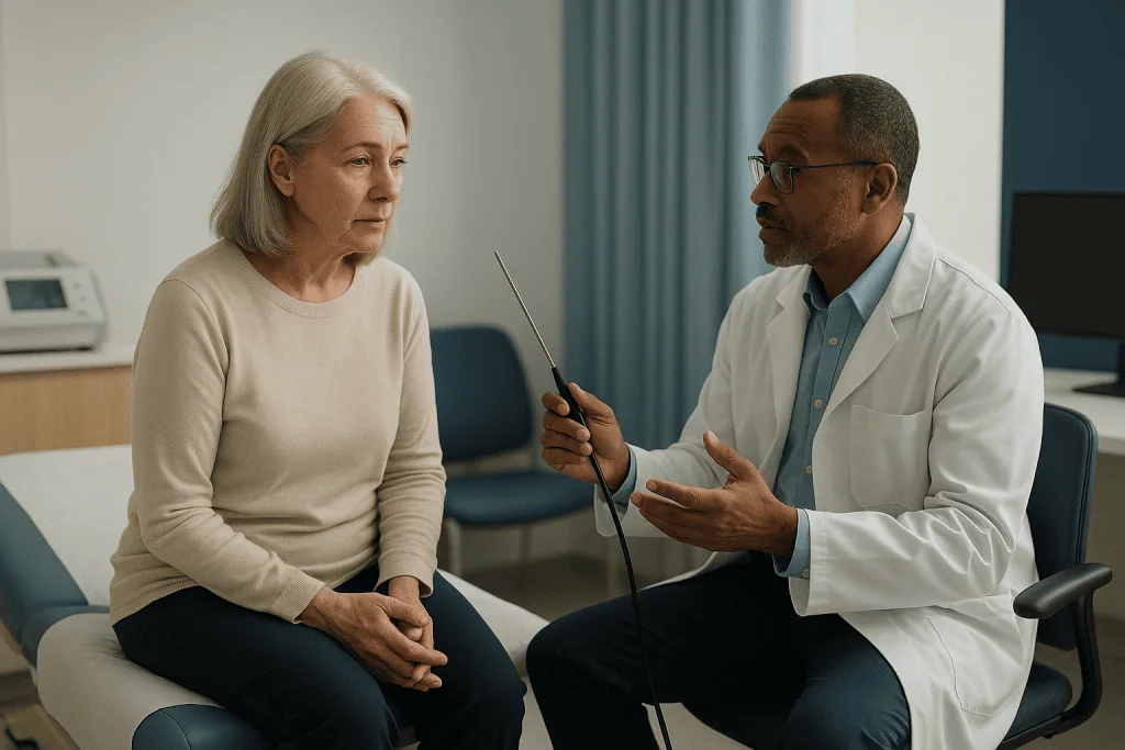
People who have had the test tend to identify two moments. The first is the passage of the scope through the sphincter region, the ring of muscle that holds urine in. For men the urethra is longer and curves past the prostate, so this stage is often felt more, typically as a stinging or pressure sensation lasting a few seconds. For women the urethra is shorter and the moment passes quickly. The second is the filling of the bladder with fluid, which produces a strong, sometimes urgent desire to pass urine. That urge is not injury; it is a stretch signal from a bladder that thinks it is full.
Technique matters more than toughness. Clenching the pelvic floor narrows the urethra and makes the scope’s passage harder. Slow breathing out as the instrument advances, which clinicians often coach, relaxes the muscle. Some departments let you watch the screen, and many patients find that seeing the pink, smooth bladder wall turns dread into curiosity.
The Mayo Clinic notes that a burning sensation during urination and some blood in the urine are common for a day or two afterward. Those after-effects are the part people underestimate, not the exam itself.
Pain that is severe, or that builds rather than fades, is not the normal pattern and should be reported to the team, both during the test and afterward.
Sedation, numbing gel and anesthesia: what is typical and why
The sedation myth runs in two directions at once. One version says you will be “knocked out” for a routine look; the other says you will be strapped down and given nothing. Neither matches standard practice.
For a flexible cystoscopy, the usual approach is local anesthetic gel inserted into the urethra. The gel contains a local anesthetic that temporarily blocks pain signals in the lining, and it doubles as a lubricant. The NIDDK describes this as the standard for an office-based cystoscopy, with the patient awake throughout. Sedation is not routinely added because the test is short and the gel handles most of the discomfort.
For a rigid cystoscopy, or when a biopsy or treatment is planned, general anesthesia (fully asleep) or regional anesthesia such as a spinal (numb from the waist down, awake) is typical. The Mayo Clinic explains that the choice depends on what is being done and on the person’s overall health; an anesthesia team makes that decision, not a leaflet.
What about asking for sedation when it is not offered? That is a legitimate conversation. High anxiety, a previous very painful attempt, a known narrowing of the urethra, or certain pelvic conditions can all change the plan. Some departments can offer light sedation for flexible cystoscopy in selected cases, and others will suggest switching to a rigid cystoscopy under anesthesia instead. The team weighs the small extra risks of sedation against the benefit for you.
Whatever is used, the mechanism is the same: interrupting pain signals for the duration of the exam. Decisions about any medicine before or after the test, including anything you already take, belong to the prescribing clinician.
Can a cystoscopy damage the bladder? What the evidence shows
This is the fear behind the most persistent of the cystoscopy myths, so it deserves a direct answer: lasting damage to the bladder from a diagnostic cystoscopy is rare. The bladder wall is muscular and elastic; it tolerates filling and gentle instrument contact as a matter of design. The scope is designed to look, not to cut, and the fluid used to fill the bladder is at a controlled volume and pressure.
The recognized complications are specific and uncommon. The Mayo Clinic lists infection, bleeding and pain as the main risks. The NHS adds that a urinary tract infection is the most common of these, and that temporary difficulty passing urine or a small amount of bleeding can occur. Perforation, meaning a hole in the bladder or urethral wall, is possible but very rare, and is more associated with therapeutic procedures that remove tissue than with a simple inspection.
Where does the “damage” story come from? Two sources, probably. First, the after-effects feel like injury even when they are not: stinging on urination and pink-tinged urine for a day or two are irritation of the lining, which heals quickly. Second, cystoscopy is frequently performed on people who already have bladder problems, and symptoms that continue after the test get attributed to it.
A related worry is that repeated surveillance cystoscopies, which people with a history of bladder tumors have regularly, will wear the bladder out. Guidance from the NIDDK treats repeat cystoscopy as a standard surveillance tool, which it would not if cumulative harm were a recognized problem.
Rare is not never. That is precisely why the red-flag list later in this article exists.
Who is usually asked to have a cystoscopy, and who is asked to wait
A cystoscopy is a targeted test. It is requested when a direct view of the bladder and urethra will answer a question that urine tests and scans cannot. The Cleveland Clinic and the MedlinePlus Medical Encyclopedia describe the common reasons as investigating blood in the urine, repeated urinary tract infections, persistent pain in the bladder or pelvis, difficulty passing urine, incontinence, suspected bladder stones, and follow-up after treatment of bladder tumors.
Blood in the urine is the headline indication. Visible blood, and in many cases blood found only on testing, prompts a look inside because the bladder lining is where several causes live. A cystoscopy can also assess an enlarged prostate from the inside, check for narrowing of the urethra, and confirm or rule out a fistula, an abnormal connection between the bladder and another organ.
Who is asked to wait? Anyone with a current urinary tract infection is usually postponed until it is treated, because passing an instrument through infected urine raises the risk of spreading bacteria. The NHS notes a urine test is often done beforehand for this reason. People taking medicines that affect clotting are not necessarily excluded, but the team needs to know, especially if a biopsy is planned; any adjustment is the prescriber’s call, never something to change on your own. Pregnancy is discussed case by case. Someone acutely unwell, or with an unstable heart or lung condition, may have the test deferred or moved from the clinic to a supervised setting.
The decision to proceed, delay or choose another test sits with the urology team, who balance the question being asked against your circumstances on the day.
Cystoscopy side effects and the real downsides
Every test has a cost in comfort, and pretending otherwise is its own kind of myth. The downsides of cystoscopy are worth stating plainly, with the evidence attached.
Expected after-effects, per the Mayo Clinic and the NHS, include a burning sensation when passing urine, a need to go more often, and pink or faintly red urine. These commonly last a day or two and settle on their own. Drinking water through the day helps dilute the urine so it stings less, a point both sources make.
Less common problems are the ones that need attention. Urinary tract infection, which the NHS identifies as the most frequent complication, typically announces itself with fever, worsening burning, cloudy or foul-smelling urine, or pain in the back or side in the days afterward. Bleeding heavier than a pink tinge, or clots, is uncommon after a diagnostic look but more possible after a biopsy. A temporary inability to pass urine, more often seen in men with an enlarged prostate, can occur because of swelling and may need a short-term catheter.
Then there are the downsides that are not complications at all: the embarrassment some people feel, the wait for results if a biopsy was taken, and the possibility of a result that is unclear and leads to another test. Those deserve acknowledgment because they are often the parts people actually find hardest.
A rigid cystoscopy under general anesthesia carries the usual anesthesia considerations, discussed with you by the anesthesia team. And for the small group whose scope cannot be passed because of a narrowing, the test may be abandoned and rebooked under anesthesia, which is frustrating but not harmful.
Cystoscopy recovery time: what the next days usually look like
Recovery from a flexible cystoscopy is measured in hours and days, not weeks. The NHS says most people can return to normal activities, including work, the same day or the next, and that it is a good idea to rest for a short while and drink plenty of fluids.
The first few hours are the most noticeable. Passing urine may sting the first two or three times, and the stream may look pinkish. Many people describe it as similar to the start of a mild bladder infection: a sense of urgency and a slight ache low in the pelvis. By the evening, most find it easing; by the second day, most are back to baseline. The Mayo Clinic notes that a warm, damp washcloth over the urethral opening can relieve discomfort, and that drinking water helps flush the bladder.
After a rigid cystoscopy under general anesthesia, the first 24 hours look different. You will need someone to take you home, and the Cleveland Clinic advises against driving, operating machinery or making important decisions until the anesthetic has fully worn off. If tissue was removed, blood-tinged urine can continue for several days and the team will tell you what is within the expected range for your procedure.
Sexual activity, exercise and bathing are usually fine once you feel comfortable after a flexible test; after a biopsy or treatment, the team may ask for a short pause. Results from the inspection itself are typically given at the time, because the urologist has seen the lining directly. Biopsy results take longer, usually a matter of days to a couple of weeks depending on the laboratory, and are reviewed with you at follow-up.
Is there an alternative to having a cystoscopy?
Sometimes, partly, and it depends on the question. That is the accurate answer, and it is more useful than a flat yes or no.
Imaging is the first alternative people reach for. Ultrasound of the bladder and kidneys is painless and is often done alongside cystoscopy, not instead of it, because it is good at showing the kidneys and larger bladder lesions but poor at detecting small or flat changes in the bladder lining. A CT urogram, a scan with contrast dye that outlines the whole urinary tract, is excellent for the kidneys and ureters and is frequently part of the same work-up for blood in the urine. The NIDDK describes cystoscopy as the direct way to see the bladder lining, which is why guidelines keep it in the pathway even when imaging is normal.
Urine cytology, where a laboratory examines cells shed into the urine, can pick up some higher-grade abnormalities but misses many lower-grade ones. Newer urine biomarker tests exist; the evidence so far does not support using them to replace cystoscopy for diagnosis, although research into their role in surveillance continues. That is an honest “not yet,” not a dismissal.
For some symptoms, the alternative is time and treatment. A first simple urinary infection does not usually need a scope. Bladder symptoms that fully resolve after a course of treatment may not either. The judgment about whether direct inspection is needed rests with the urologist, who weighs your age, risk factors and findings.
If anxiety is the barrier, say so. Switching from an awake flexible test to a rigid one under anesthesia is itself an alternative, and it keeps the diagnostic question answered.
What are the signs that a cystoscopy went as planned?
People search for “signs a cystoscopy was successful,” and the question deserves reframing. Success here means two things: the urologist obtained a clear view of what needed to be seen, and you recovered without complication. Neither is about how much it hurt.
On the first point, you will usually know before you leave. Because the inspection is visual, the urologist can often tell you immediately whether the bladder lining looked normal, whether anything needed sampling, and what the next step is. The MedlinePlus Medical Encyclopedia notes that normal findings mean the bladder wall appears smooth and the bladder is of normal size, shape and position. An “abnormal” finding is not a diagnosis; it is a reason for a biopsy result or further test, which the team will explain.
On the second point, the reassuring pattern over the following days is simple. Stinging and urgency ease rather than worsen. Pink urine fades to its usual color within a day or two, as the Mayo Clinic describes. You pass urine without needing to strain. You have no fever and no new pain in the back or side.
What a successful test does not guarantee is a particular answer. A normal cystoscopy in someone with blood in the urine is good news about the bladder, but the team may still look at the kidneys with imaging. An abnormal one does not mean the worst; many findings are benign, and the pathology report settles it.
If you leave without understanding what was seen, ask. A clear explanation on the day is part of the test being done well.
What people often get wrong: cystoscopy myths, corrected
Here are the claims that circulate most, set against the evidence.
- “The scope is as thick as a finger.” A flexible cystoscope is close to pencil-width and bends with the urethra. The wider rigid scope is used under anesthesia, so you do not feel it.
- “You have to be put to sleep.” Flexible cystoscopy is done awake with local anesthetic gel; the NIDDK describes this as the standard office approach. Anesthesia is reserved for rigid procedures or special circumstances.
- “They give you nothing for the pain.” The gel is a local anesthetic; it numbs the lining. Sedation or anesthesia is discussed when the gel alone is unlikely to be enough.
- “It damages the bladder.” Lasting harm is rare. The usual after-effects, stinging and pink urine for a day or two, are irritation of the lining, not injury, per the NHS.
- “Blood in the urine afterward means something went wrong.” A pink tinge is expected. Heavy bleeding or clots are not, and should be reported.
- “It causes incontinence.” The sphincter is passed, not cut. Temporary urgency is common; new lasting incontinence is not a recognized outcome of diagnostic cystoscopy.
- “Men can’t have it awake.” Men do have flexible cystoscopy awake routinely. The longer urethra means a few more seconds of pressure, not a different procedure.
- “A scan would show the same thing.” Imaging complements cystoscopy but cannot reliably see small or flat changes in the lining, which is why the direct look remains in guideline pathways.
One myth runs the other way and is worth correcting too: “It’s nothing, don’t bother reading the leaflet.” The test is minor, but preparation, especially knowing about the urine check beforehand and the fluids afterward, makes the difference between an easy day and an annoying one.
Questions to ask your care team before a cystoscopy
A few minutes of questions before the appointment tend to remove most of the dread. These are the ones that change what you should expect.
- Which type of cystoscopy am I having, flexible or rigid? This single answer determines the anesthesia plan, the time involved and whether you need someone to take you home.
- What question is this test meant to answer? Knowing whether it is a first look for blood in the urine, a check on symptoms, or routine surveillance helps you understand what a “normal” result would and would not mean.
- What will be used for comfort, and can that be changed if I struggle? Ask about the local anesthetic gel, whether sedation is an option in your department, and what happens if the scope cannot be passed.
- Should I do anything about my regular medicines? Tell the team about everything you take, including blood thinners and supplements. Do not stop or alter anything yourself; the prescribing clinician decides.
- Will a urine test be checked first? The NHS notes infection is usually excluded beforehand; confirm how that is done.
- Might a biopsy be taken, and how will I get the result? Ask about the usual waiting time and whether follow-up is in person or by phone.
- What should the next 48 hours look like, and what is outside the normal range? Have them say out loud what amount of blood, pain or fever should prompt a call.
- Can I watch the screen? Many people find it calming; some prefer not to. Either is fine.
- Is there an alternative in my case, and what would we lose by choosing it? A fair question, and a good team will give a fair answer.
Write the answers down. The post-procedure leaflet rarely covers the specifics that apply to you, and a note from the conversation is the version you will actually consult at eleven at night.
When to call your doctor after a cystoscopy
Most people need no follow-up contact beyond the planned results. A short list of red flags separates ordinary irritation from a problem that needs assessment, and the Mayo Clinic and Cleveland Clinic both advise contacting the care team if any of these occur.
- Fever, chills or shaking in the days after the test, which can signal a urinary tract infection spreading beyond the bladder.
- Inability to pass urine despite feeling the need, particularly if the lower abdomen becomes swollen or tense. This is retention and needs same-day attention.
- Heavy bleeding: urine that is dark red rather than pink, blood clots, or bleeding that is increasing rather than fading after the first day or two.
- Pain that worsens rather than settling, especially severe pain in the lower abdomen, back or side.
- Burning on urination that persists beyond a couple of days or that comes with cloudy, foul-smelling urine.
- Nausea, vomiting or feeling generally very unwell after the procedure.
After a rigid cystoscopy under general anesthesia, add the standard post-anesthesia concerns: breathing difficulty, chest pain, confusion, or a wound-related problem if treatment was performed. The discharge instructions will specify who to call and when, and they take precedence over any general list.
When in doubt, call. Urology departments field these questions daily and would much rather hear from someone with a pink tinge and a worry than miss someone with an infection who waited. If you cannot reach the department and symptoms are severe, such as inability to pass urine with a swollen abdomen, high fever with shaking, or heavy bleeding, seek urgent care.
Everything else, the mild stinging, the urgency, the faint discoloration, is the bladder lining doing its ordinary, unhurried repair. Water, rest and a day or two are usually all it asks.
Frequently asked questions
What are the downsides of a cystoscopy?
The main downsides are short-term discomfort and a small risk of complications. Expect stinging when you urinate and pink-tinged urine for a day or two, plus some urgency. Less commonly, a urinary tract infection, heavier bleeding (more likely after a biopsy) or temporary difficulty passing urine can occur. Embarrassment and the wait for biopsy results are real downsides too. Serious injury to the bladder is rare.
What is the most painful part of a cystoscopy?
Most people describe two moments: the scope passing the sphincter muscle near the start, felt as a few seconds of stinging or pressure, and the bladder filling with fluid, which creates a strong urge to urinate. Men often notice the first more because the urethra is longer. Breathing out slowly as the scope advances helps relax the muscle. Severe or building pain is not the normal pattern and should be reported.
Does cystoscopy hurt afterward, and for how long?
Mild burning on urination and some urgency are common for the first day or two and then fade, according to the Mayo Clinic and NHS. Drinking water helps dilute the urine so it stings less. Pain that increases over the following days, or that comes with fever or cloudy urine, suggests infection rather than normal irritation and warrants a call to the care team.
Is there an alternative to having a cystoscopy?
Partly. Ultrasound, CT urography and urine cytology are often used alongside cystoscopy and can answer some questions, especially about the kidneys and ureters. They cannot reliably see small or flat changes in the bladder lining, so cystoscopy remains the direct test for that. If anxiety is the barrier, having the procedure under anesthesia is itself an alternative. The urologist decides which tests your situation needs.
What are the signs that a cystoscopy was successful?
Success means the urologist got a clear view and you recovered without complication. You will usually be told on the day whether the bladder lining looked normal and what happens next. Over the following days, the reassuring pattern is stinging and pink urine easing within a day or two, passing urine without straining, and no fever or new pain in the back or side.
What is the difference between flexible vs rigid cystoscopy?
A flexible cystoscopy uses a thin, bendable scope, is done awake with local anesthetic gel in a clinic, and takes a few minutes; it is mainly for diagnosis and surveillance. A rigid cystoscopy uses a straight, slightly wider instrument under general or regional anesthesia in an operating room, usually when treatment or larger biopsies are planned. The team chooses based on what needs doing, not on how much you can tolerate.
What is the usual cystoscopy recovery time?
After a flexible cystoscopy, most people return to normal activities the same day or the next, per the NHS, with mild burning and urgency settling within a day or two. After a rigid cystoscopy under general anesthesia, you need someone to take you home and should avoid driving for the rest of that day; if tissue was removed, blood-tinged urine may continue for several days within a range the team will describe.
What are the most common cystoscopy side effects?
The most common side effects are a burning sensation when passing urine, needing to go more often, and pink or faintly red urine, all typically lasting a day or two. Urinary tract infection is the most frequent complication and shows up as fever, chills or worsening burning. Temporary difficulty passing urine and heavier bleeding are less common and more likely after a biopsy or treatment.
Can a cystoscopy cause incontinence or permanent bladder damage?
New, lasting incontinence is not a recognized outcome of diagnostic cystoscopy; the sphincter muscle is passed by the scope, not cut. Permanent bladder damage is rare. The stinging and urgency people feel afterward are irritation of the lining, which heals within days. Perforation is a very rare risk, more associated with procedures that remove tissue than with a simple inspection.
Will I be awake during a cystoscopy?
For a flexible cystoscopy, usually yes. Local anesthetic gel numbs the urethra and the test takes only minutes, so sedation is not routinely added. For a rigid cystoscopy, you will typically have general anesthesia or a spinal, because treatment is often performed at the same time. If you are very anxious or have had a painful previous attempt, tell the team; comfort options can be discussed.
References
- NHS: Cystoscopy
- NIH NIDDK: Cystoscopy & Ureteroscopy
- Cleveland Clinic: Cystoscopy
- MedlinePlus Medical Encyclopedia: Cystoscopy
This article is for general information only and is not a substitute for professional medical advice. Please consult a qualified doctor about your individual situation.

More from the Blog

Can a Stricture Return After Urethral Reconstruction? Warning Signs, Checks and Next Options
Yes, a urethral stricture can narrow again after urethroplasty, although open reconstruction holds up far better over time than repeated stretching or endoscopic cutting.…

TURP vs Laser Prostate Surgery: How Urologists Choose When the Prostate Is Very Large
TURP and laser prostate surgery both open a blocked channel through an enlarged prostate, and for average-sized glands their urinary results are broadly similar.…

Bladder Pain Syndrome Treatment in Steps: Lifestyle Changes, Medication and Bladder Therapies
An interstitial cystitis treatment plan is usually built in steps. It starts with self-care such as bladder retraining, stress management, identifying food triggers and…

Do All Bladder Infections Need Antibiotics? Myths and Facts Urologists Explain
Not every bladder infection needs antibiotics, but many do. In otherwise healthy, non-pregnant women with mild cystitis, guidelines allow a short trial of symptom…

When Does a Urinary Infection Need Urgent Care? Fever, Back Pain and Vomiting Explained
A urinary infection needs urgent, same-day care when bladder symptoms are joined by fever, chills, pain in the back or side, nausea or vomiting,…

Varicocele Surgery for International Patients: Stay Length, Flying Home and Remote Follow-Up
People who travel for varicocele surgery are usually asked to stay near the surgical center until the first wound check, often within the first…