

Dental Hygiene Myths: Does Scaling Damage Enamel or Loosen Teeth?
Key Takeaways
- Enamel is the hardest tissue in the body and cannot regenerate once lost, but properly performed scaling removes tartar sitting on top of it rather than the enamel itself.
- Teeth that feel loose or gappy after a deep cleaning are revealing bone loss and gum recession that periodontitis had already caused and that tartar had been masking.
- Post-scaling sensitivity comes from exposed root dentin, not thinned enamel, and typically eases over days to a few weeks as tubules close and gums heal.
- Enamel begins to demineralize below a surface pH of about 5.5, which is why frequent sipping of acidic drinks damages enamel far more than professional cleaning ever could.
- Early demineralization seen as white spots can be reversed with fluoride and reduced acid exposure; enamel that has physically worn away can only be protected or restored.
- Scaling and root planing is the NIH-described first-line treatment for periodontitis, recommended from measured pocket depths and X-rays, with a re-evaluation typically scheduled weeks later.
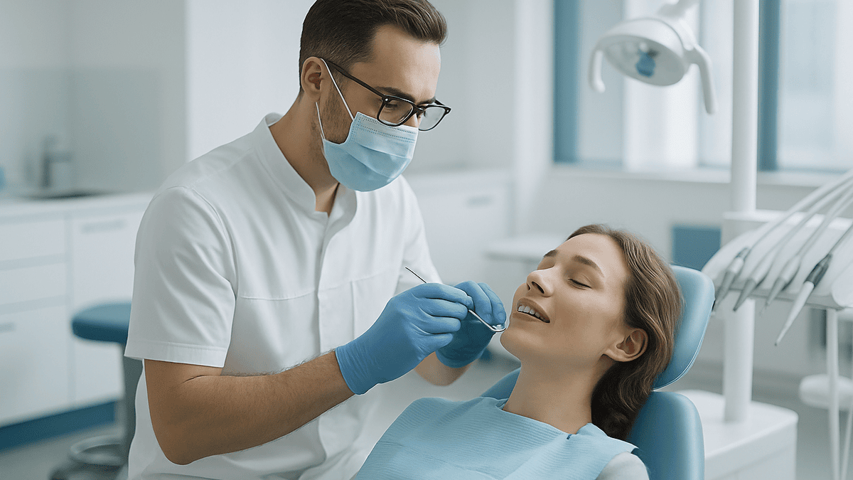
Professional scaling does not damage healthy enamel or loosen teeth when performed correctly. Enamel is the hardest tissue in the body, far harder than the tartar being removed, and dental instruments are designed to work at the gumline and below it. Teeth can feel sensitive or slightly mobile for a short time afterward because tartar that was propping up inflamed gums is gone, not because the tooth itself was weakened.
Maria left the dental office running her tongue along her lower front teeth. They felt strange, almost too clean, with little gaps she was sure had not been there an hour ago. That night, cold water made one tooth zing. By the weekend she had typed the question so many people type after a deep cleaning: does scaling damage enamel? And the follow-up that worried her more: had the hygienist just loosened her teeth?
It is a reasonable fear. Scaling involves metal tips, vibrating instruments and a fair amount of scraping close to the gum. The sensations afterward are real. But the story your mouth tells you in the first few days is not the story of the tooth. It is the story of the gum tissue that had been quietly inflamed for months, sometimes years.
This explainer walks through what scaling actually removes, why enamel is not the vulnerable party, and which sensations are normal versus which ones deserve a phone call.
Does scaling damage enamel? The short, honest answer
No, not when it is done properly on a healthy tooth. That answer needs a little unpacking, because the fear behind the question is not silly. People feel roughness, hear scraping and then notice sensitivity. Putting those three things together, it is natural to conclude that something was taken off the tooth.
Something was taken off. It just was not enamel. Scaling removes plaque, a soft film of bacteria, and tartar, which is plaque that has absorbed minerals from saliva and hardened onto the tooth. The NHS notes that once plaque has hardened into tartar, brushing at home can no longer remove it, which is exactly why a professional has to do the job with instruments (NHS). Tartar sits on top of the enamel and, more importantly, on the root surface below the gumline. The instrument works at that junction.
Enamel itself is the hardest substance in the human body, harder than bone, according to the Cleveland Clinic. Tartar, by comparison, is a chalky mineral crust that a trained clinician can flake or chip away with controlled pressure. The physics favor the tooth. A scaler is designed to catch on the ledge of tartar and pull it free; it is not designed to gouge a smooth, dense crystalline surface, and under normal use it does not.
Where the honest caveat comes in: enamel that is already thin, eroded or cracked is a different surface, and a clinician will adjust technique around it. Root surfaces, which are covered by cementum and dentin rather than enamel, are softer and can be smoothed slightly during root planing. That is intentional. The goal there is to remove bacterial deposits from a rough root so the gum can reattach, not to preserve every micron of a surface that was already contaminated.
So the answer holds: correctly performed scaling does not harm healthy enamel. The rest of this article explains why the aftermath feels the way it does.
What actually happens during scaling and root planing
Picture the tooth as a fence post set into soil. Above ground is the crown, covered in enamel. Below ground is the root, covered in a thin layer called cementum. Where the gum meets the tooth there is a shallow groove, and in a healthy mouth that groove is only a few millimeters deep. When plaque and tartar build up along it, the gum pulls away and the groove deepens into what dentists call a pocket.
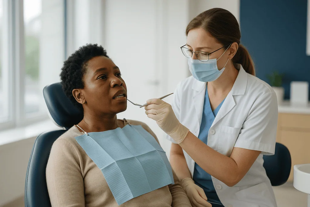
Scaling is the removal of plaque and tartar from the crown and from inside that pocket. Root planing goes one step further, smoothing the root surface so bacteria have fewer rough places to cling to and the gum has a cleaner surface to heal against. The Mayo Clinic describes this pairing as the standard nonsurgical approach to periodontitis, the stage of gum disease where the supporting bone has begun to be lost (Mayo Clinic).
Two kinds of instruments do the work. Ultrasonic scalers use a tip that vibrates very rapidly while a fine water spray flushes debris out of the pocket; the vibration shatters tartar rather than scraping it. Hand scalers and curettes are shaped metal tips the clinician uses to catch ledges of tartar and lift them off, then to plane the root. Most appointments use both.
Deeper pockets are usually treated with local anesthetic, and the Cleveland Clinic notes that a full mouth is often divided across more than one visit so each area gets careful attention. A typical sequence is: numb the area, use the ultrasonic tip to break up bulk deposits, follow with hand instruments to check the root by feel and remove what remains, then rinse. The clinician often runs a fine probe along the root afterward, feeling for any residual roughness the way you might run a fingernail along a painted wall to find a drip.
Why enamel is tougher than tartar, and why the instruments are built around that
Enamel is about 96 percent mineral by weight, arranged in tightly packed rods of hydroxyapatite crystal. The Cleveland Clinic describes it as the hardest tissue in the body and notes that, because it contains no living cells, it cannot repair itself once genuinely lost (Cleveland Clinic). That second fact is why people worry, and it is worth respecting. Enamel is a one-time issue.
The reassuring part is the comparison. Tartar is mineralized plaque: bacteria, proteins and saliva minerals that have set into a porous, brittle crust. It is hard enough to resist a toothbrush but nowhere near as dense or as well organized as enamel. When a hand scaler is set against a ledge of tartar and drawn upward, the tartar fractures at its weakest point, which is usually the interface between deposit and tooth. The instrument is doing what a putty knife does to dried paint on glass: the paint comes off, the glass stays.
Ultrasonic tips work by a different principle but with the same outcome. The tip oscillates tens of thousands of times per second, and that vibration, combined with the tiny implosions created in the water spray, breaks tartar into fragments. Manufacturers and training programs set these tips to be used at a light touch, moving continuously rather than parked in one spot. A tip left pressing on one point at high power for a long time could, in theory, leave a fine mark on any surface. That is why technique matters and why clinicians are trained to keep the tip gliding.
Where the tooth is genuinely softer is below the enamel. Dentin, the layer underneath, and cementum, the root covering, are less mineralized. Root planing does remove a superficial layer of contaminated cementum on purpose. The scale of that removal is microscopic and is accepted in periodontal treatment because the alternative, leaving bacterial toxins embedded in the root, keeps the gum inflamed.
Why teeth can feel loose or gappy after a deep cleaning
This is the sensation that unsettles people most, and it deserves a mechanical explanation rather than reassurance alone.

Consider what a large deposit of tartar was doing before it was removed. It filled the space between teeth. It sat in the deepening pocket like grout, physically occupying the gap where inflamed gum had pulled back. To the tongue, everything felt solid and continuous. Then the tartar is gone. The tongue now finds the true contours of the teeth and the true position of the gum, and the true position is lower than it used to be, because gum disease had already caused recession. The gaps were always there. They were simply hidden.
Mobility works similarly. In periodontitis, the Mayo Clinic explains, the infection destroys the bone and connective tissue that hold teeth in place, and loosening teeth are a sign of the disease itself (Mayo Clinic). A tooth that was already losing bone support may have been splinted somewhat by a collar of tartar bridging it to its neighbors. Remove the collar and the pre-existing looseness becomes noticeable. The cleaning revealed the damage; it did not create it.
There is a second, temporary effect. Inflamed gum tissue is swollen and fragile. In the days after scaling, that swelling begins to subside as the bacterial load drops. The gum tightens back toward the tooth and, in shallower pockets, may reattach. During that transition the teeth can feel slightly different when you bite, and some people describe a vague wobble that settles as healing progresses.
Genuinely new, progressive looseness after scaling is not expected and should be reported. But the common experience, teeth that feel exposed and a little unfamiliar for a week or two, is the mouth recalibrating to a cleaner baseline, not a tooth coming free.
Sensitivity after scaling: what is happening and how long it usually lasts
Cold water, sweet foods, even a breath of winter air through the mouth can produce a sharp, short zing after a deep cleaning. The Cleveland Clinic lists this sensitivity as an expected side effect of scaling and root planing and notes that it typically eases over the following days as the gums heal, though some people notice it for a few weeks (Cleveland Clinic).
The mechanism involves the root, not the enamel. Dentin, the layer under enamel and cementum, is threaded with microscopic tubules that run toward the nerve at the center of the tooth. When a root surface that was buried under tartar and swollen gum is suddenly exposed to the mouth, those tubules are open to temperature and pressure changes. Fluid inside them shifts, and the nerve interprets that shift as pain. Over time the tubules narrow as minerals from saliva deposit inside them, and the gum tissue that recedes less once inflammation resolves covers part of the root again. Both processes reduce sensitivity gradually.
Several things help while that happens. Toothpastes formulated for sensitive teeth work by blocking those tubules or calming the nerve response; your dental team can tell you whether one is appropriate for you and which type. Fluoride, whether in ordinary toothpaste or applied professionally, strengthens the mineral surface. Avoiding very hot, very cold or very acidic drinks for a few days gives the tissue a quieter environment to settle.
Two patterns are worth distinguishing. Sensitivity that is broad, triggered by cold, brief and slowly improving is the expected kind. Sensitivity that is localized to one tooth, spontaneous, throbbing, or waking you at night is a different signal and points toward pulp inflammation or another problem. That kind warrants a call, discussed further in the red-flag section below.
Do dental cleanings damage enamel? Ultrasonic versus hand instruments compared
Researchers have looked at this question directly, because it matters for training and for people with restorations. Laboratory studies in which extracted teeth are scaled and then examined under magnification generally find that properly used instruments leave the enamel intact, while excessive force, a worn tip or a high power setting held stationary can leave superficial marks measured in microns. Those findings are the basis for how clinicians are taught: light pressure, constant motion, correct tip angle.
The practical differences between the two main approaches are summarized here.
| Feature | Ultrasonic scaling | Hand scaling and root planing |
|---|---|---|
| How it removes tartar | Rapid vibration plus water spray fractures deposits | Sharpened tip catches and lifts deposits by controlled pressure |
| Effect on healthy enamel | None with correct power and continuous motion | None with correct angulation and light strokes |
| Where it excels | Bulk tartar removal, flushing pockets, reaching deep or awkward areas | Fine detail, tactile checking of the root, final smoothing |
| Common sensations | Vibration, cold water, occasional cold sensitivity | Scraping sensation, pressure |
| Special considerations | Some pacemakers or implant surfaces need specific tips; clinician will check | Slower for heavy deposits; relies on tip sharpness and operator skill |
Most appointments combine both, using the ultrasonic tip for the heavy lifting and hand instruments to finish. Neither method is inherently the enamel-safe choice; safety comes from technique.
One place where extra care is warranted is around existing dental work. The margins of fillings, crowns and veneers can be chipped or roughened by careless instrumentation, which is why clinicians note restorations before starting and adjust around them. If you have veneers or bonded fillings on front teeth, mention it, even if you assume it is already in your chart.
What are the downsides of teeth scaling? Real risks, stated plainly
Any procedure worth doing has a list of things that can go wrong, and pretending otherwise does not help anyone. Here is the honest inventory for scaling and root planing, framed in the neutral terms your care team would use.
Sensitivity, covered above, is the most common downside and is usually short-lived. Gum tenderness and minor bleeding in the first day or two are expected; the tissue was inflamed to begin with and has just been cleaned thoroughly. Some people notice their gums look slightly lower afterward, and this is the resolution of swelling revealing the recession that had already occurred rather than new tissue loss.
Less common risks include:
- Soreness or a small ulcer where an instrument or the mouth prop contacted soft tissue.
- Jaw ache from keeping the mouth open, particularly during longer appointments.
- Temporary numbness or bruising at an anesthetic injection site.
- Disturbance of a filling margin or, rarely, a chipped restoration.
- A short-lived rise in mobility of teeth that were already compromised, as described earlier.
Because scaling releases bacteria into the bloodstream for a short time, the Mayo Clinic and others note that people with certain heart conditions or recent joint replacements may need a conversation with their physician and dentist beforehand about whether preventive antibiotic cover is appropriate. That decision follows guideline criteria and belongs with the treating clinicians; it is not something to self-assess.
What scaling does not do, on the balance of mainstream evidence, is thin healthy enamel, cause cavities, or make gum disease worse. The comparison that matters is not scaling versus doing nothing to a healthy mouth. It is scaling versus leaving tartar and bacteria in a pocket that is already destroying bone. Against that alternative, the downsides are modest and mostly temporary.
Who scaling is usually for, and who is usually asked to wait
Routine scaling, the kind that happens at a standard check-up, is for almost everyone. Plaque hardens into tartar in a matter of days along the gumline, and no one brushes perfectly. The NHS recommends regular dental visits at an interval your dentist sets, and removal of tartar is a normal part of those visits (NHS).
Scaling and root planing, the deeper procedure done under anesthetic in sections, is specifically for people diagnosed with periodontitis or with pockets deep enough that bacteria cannot be reached by home care. The NIH’s National Institute of Dental and Craniofacial Research describes it as the first-line treatment before any surgical option is considered (NIDCR). Candidates are identified by measuring pocket depths with a probe, reviewing X-rays for bone loss, and noting bleeding on probing. Nobody is offered deep scaling on the basis of a glance.
Some people are asked to wait or to prepare first. Those with poorly controlled diabetes may be advised to work with their physician on glucose control, because healing after periodontal treatment is slower when blood sugar runs high. People taking anticoagulants are not usually excluded, but the dental team may want to coordinate with the prescribing doctor about bleeding risk. Anyone with a recent heart valve procedure, certain congenital heart conditions or a recent joint replacement may need a physician’s input before the appointment. During pregnancy, routine cleaning is generally considered safe and often encouraged, but timing of longer treatments is discussed individually.
Very anxious patients, people with a strong gag reflex, or those with limited mouth opening may need shorter appointments, different positioning or sedation options. None of these are reasons to skip treatment; they are reasons to plan it well. The decision about whether and when to proceed always rests with the treating team, who can see the pocket chart and the X-rays.
What damages tooth enamel the most? The real culprits
If scaling is not the threat, what is? The evidence points to three main mechanisms, and none of them involve a hygienist.
Acid is first and by a wide margin. Enamel begins to lose mineral when the pH at its surface drops below roughly 5.5, a threshold well established in dental research and cited by the Cleveland Clinic in its overview of enamel erosion (Cleveland Clinic). Two sources of acid do the damage. Dietary acid comes from soft drinks, sports and energy drinks, citrus, wine, vinegar and some sparkling waters. Internal acid comes from stomach contents in people with reflux disease or frequent vomiting. Frequency matters more than quantity: sipping an acidic drink across an afternoon keeps the surface below threshold for hours, while drinking the same amount in five minutes allows saliva to neutralize and remineralize afterward.
Bacterial acid is second. The bacteria in plaque ferment sugar and release acid directly onto the enamel surface, which is how cavities form. This is the mechanism the Mayo Clinic describes in its account of tooth decay, and it is why sugar frequency, not just total intake, drives risk (Mayo Clinic).
Mechanical wear is third. Grinding or clenching, especially at night, wears enamel from the biting surfaces. Brushing too hard with a stiff brush, particularly right after an acid exposure when the surface is temporarily softened, abrades enamel at the gumline and contributes to those notched areas that dentists call abfraction or abrasion lesions.
Dry mouth, whether from medicines, medical conditions or dehydration, amplifies every one of these because saliva is the body’s own remineralizing rinse. Genetics play a part too: some people inherit thinner or less well-formed enamel.
Notice what is missing from this list. Professional cleaning does not appear because, in the mainstream evidence, it does not belong there.
Can you reverse enamel damage on teeth?
This is where honesty matters more than comfort. The answer is partly yes and partly no, and the dividing line is whether the enamel has been demineralized or actually lost.
Early damage is demineralization: acid has drawn calcium and phosphate out of the crystal structure, leaving the surface porous but structurally present. This stage can look like a chalky white spot on the tooth. At this stage the process can be reversed, because saliva, and fluoride in particular, can drive minerals back into the weakened crystal lattice. MedlinePlus and the NHS both describe fluoride’s role in helping enamel resist and recover from early acid attack (MedlinePlus, NHS). Reducing acid frequency, letting saliva do its work between exposures, and using fluoride toothpaste as directed give the surface the chance to reharden. Dental teams may also apply professional fluoride treatments or other remineralizing agents; which one, if any, is appropriate is their call.
Established loss is different. Once the enamel has physically worn away, whether from erosion, abrasion or decay that has cavitated, the body cannot grow it back. The Cleveland Clinic states plainly that enamel has no living cells and does not regenerate (Cleveland Clinic). What dentistry can do is protect what remains and restore what is gone. Options range from bonding and fillings to crowns and veneers, chosen according to how much structure is missing and where. Those are restorations, not regrowth, and they carry their own maintenance needs.
The realistic message is this: enamel damage is worth catching early because the early stage is the only one that reverses. Regular check-ups exist partly to spot white spots and thinning before they become cavities or exposed dentin. If you have been told your enamel is thin or eroding, the most useful questions to ask are about the cause, since stopping the cause is what protects the enamel you still have.
What the following days and weeks usually look like
Timelines here are typical ranges drawn from clinical sources, not promises. Healing varies with how much disease was present, how well home care goes and general health.
The first day is often the least comfortable. Numbness wears off over a few hours. Gums feel tender, may bleed slightly when brushed, and the teeth feel unusually smooth and exposed. Cold sensitivity often makes its first appearance. Eating on the treated side can be uncomfortable, so softer, lukewarm foods are easier. Brushing and flossing should continue, gently; the Cleveland Clinic emphasizes that keeping the area clean is what allows healing, so skipping hygiene out of tenderness works against the treatment (Cleveland Clinic).
Over the first week, tenderness usually fades. Bleeding on brushing diminishes as the gum tissue becomes less inflamed. The gums may look pinker and firmer and, in places, slightly lower, revealing recession that had been masked by swelling. Sensitivity typically peaks in this window and then begins to ease.
Across the following weeks, the deeper changes happen. In shallower pockets, the gum can reattach to the freshly cleaned root surface. Mobility that was related to inflammation, rather than to bone loss, tends to settle. Sensitivity continues its slow decline as dentin tubules close and the gum matures.
Most dental teams schedule a re-evaluation somewhere in the range of four to eight weeks after treatment. At that visit, pockets are measured again and compared with the starting chart. Areas that have responded are moved to a maintenance schedule, which for people with a history of periodontitis is often more frequent than the standard recall. Areas that have not responded may be re-treated or referred for a periodontal specialist’s opinion about surgical options. That sequence, and the decisions inside it, belong to the treating team.
What people often get wrong about scaling and enamel
Several beliefs circulate widely enough to deserve a direct correction, each against what the evidence actually shows.
The scraping sound means enamel is being scraped. The sound is tartar fracturing and the instrument gliding over a hard surface. A putty knife scraping paint off a window sounds alarming too; the glass is fine.
My teeth were tight before and loose after, so the cleaning loosened them. As discussed, tartar can bridge and brace teeth that periodontal disease has already loosened. The looseness predated the appointment and was hidden by the deposit that caused it.
Cleanings create gaps between teeth. The gaps are the shape of the gum after inflammation-related swelling resolves and after tartar that filled the space is gone. Nothing was cut away.
If I brush well I never need scaling. Brushing controls plaque, but tartar forms wherever plaque sits for more than a few days, and the NHS is clear that once it has hardened it cannot be brushed off (NHS). Almost everyone accumulates some.
Sensitivity afterward means the enamel is thinner. The sensitivity comes from exposed root dentin, which was never covered by enamel in the first place, and it improves as tubules close and gums heal.
Deep cleaning is a sales tactic. Scaling and root planing is recommended on the basis of measured pocket depths and X-ray evidence of bone loss, and it is described by the NIH as the standard first-line treatment for periodontitis (NIDCR). Anyone unsure about a recommendation is entitled to see their pocket chart and X-rays and to ask what the numbers mean.
Whitening toothpaste will fix eroded enamel. Abrasive whitening products remove surface stain; they do not add mineral. On already-thin enamel they can make things worse. Fluoride and acid control are what protect enamel; restoration is what replaces it.
Questions to ask your care team before and after scaling
A good appointment is a conversation, and the most reassured patients are usually the ones who asked what the numbers meant. These questions are the ones clinicians are glad to hear because they signal engagement rather than mistrust.
Before treatment:
- What are my pocket depths, and which areas are you most concerned about?
- Do my X-rays show bone loss, and if so how much and where?
- Is this a routine cleaning or scaling and root planing, and will I be numbed?
- Will this be done in one visit or split into sections?
- Do you need to speak with my physician first, given my heart history, joint replacement, blood thinner or diabetes?
- I have veneers, bonded fillings or a crown here; how will you work around them?
About enamel specifically:
- Have you noticed any thin, eroded or worn enamel on my teeth, and what do you think is causing it?
- Are there white spots or early lesions that could still remineralize?
- Is a fluoride application or a specific toothpaste worth considering for me?
After treatment:
- What sensations should I expect this week, and which ones would you want to hear about?
- How should I brush and floss the treated area while it is tender?
- When is my re-evaluation, and what will you measure then?
- What maintenance interval do you recommend for me, and why?
- If some areas do not improve, what are the next options?
Write the answers down, or ask for a copy of your periodontal chart. Comparing pocket depths from visit to visit is the clearest way to see whether treatment and home care are working, and it turns an abstract worry about enamel into a concrete, trackable set of numbers.
When to call your doctor or dentist after scaling
Most people need nothing more than gentle hygiene and a little patience after scaling. The signs below are the ones that fall outside the expected pattern and should prompt a call to your dental office, or to a physician or urgent care if the office is closed and symptoms are severe.
- Bleeding that does not slow with gentle pressure after about half an hour, or that restarts heavily hours later.
- Pain that is worsening after the second or third day rather than easing, especially if it is throbbing, spontaneous or wakes you at night.
- Swelling of the gum, face or jaw that is increasing, or a visible lump or pus at the gumline; these can indicate an abscess.
- Fever, chills or feeling generally unwell in the days after treatment.
- A tooth that is becoming progressively looser rather than settling, or a tooth that has shifted position.
- Difficulty swallowing or breathing, or swelling spreading toward the floor of the mouth or the neck. This is an emergency and warrants immediate care.
- Sensitivity that is localized to a single tooth, sharp when biting, and not improving over a couple of weeks, which may signal a crack or a restoration problem.
- Numbness that persists well beyond the expected duration of the local anesthetic.
For people with heart valve disease, certain congenital heart conditions or a recent joint replacement, any fever or unexplained fatigue in the weeks after dental treatment should be discussed with a physician promptly, because of the small risk of bacteria seeding those sites (Mayo Clinic).
None of these signs means something was done wrong. They mean the healing is not following the usual script, and the people best placed to work out why are the clinicians who treated you and who can see the chart, the X-rays and the tissue itself. Every decision about next steps rests with them.
Frequently asked questions
Do dental cleanings damage teeth enamel?
No, routine dental cleanings do not damage healthy enamel when performed with standard technique. The instruments remove plaque and hardened tartar, which are softer than enamel and sit on its surface. Laboratory studies show that only excessive pressure or a stationary high-power ultrasonic tip leaves microscopic marks, which is why clinicians are trained to use light, moving strokes. Restorations such as veneers need extra care, so mention them before treatment.
What are the downsides of teeth scaling?
The most common downsides are temporary: cold sensitivity, gum tenderness and minor bleeding for a few days. Gums may look slightly lower afterward because swelling resolves and reveals existing recession. Less often, a filling margin can be disturbed, the jaw can ache from a long appointment, or an anesthetic site can bruise. People with certain heart or joint conditions may need physician input beforehand. Against untreated periodontitis, these risks are modest and short-lived.
What damages tooth enamel the most?
Acid causes the most enamel damage, whether from frequent acidic drinks and foods, stomach acid from reflux, or the acid that plaque bacteria produce from sugar. Enamel starts losing mineral when its surface pH drops below about 5.5. Mechanical wear from grinding or aggressive brushing adds to the loss, and dry mouth makes every factor worse by removing saliva’s protective effect. Professional cleaning is not among the causes recognized in mainstream evidence.
Can you reverse enamel damage on teeth?
Partly. Early damage, where acid has drawn minerals out but the surface is still present, can be reversed as saliva and fluoride return minerals to the weakened crystal structure. Enamel that has physically worn away cannot grow back because it contains no living cells. At that stage dentistry protects what remains and restores what is missing with bonding, fillings or crowns. Catching white spots early is the only window in which true reversal happens.
Why do my teeth feel loose after scaling?
Because tartar that was bracing already-loosened teeth has been removed, and because inflamed gum tissue is changing as it heals. Periodontitis destroys the bone and ligament that hold teeth, and a collar of tartar can splint neighboring teeth together, hiding that mobility. Once cleaned, the pre-existing looseness becomes noticeable. Mild wobbliness usually settles over the following weeks as inflammation resolves; looseness that is getting worse should be reported to your dentist.
How long does sensitivity last after scaling and root planing?
Sensitivity to cold typically eases over a few days and, for some people, persists for a few weeks before fading, according to the Cleveland Clinic. It arises from exposed root dentin rather than enamel, and it improves as microscopic tubules in the dentin narrow and the gum tissue heals. Toothpastes for sensitive teeth and fluoride can help; your dental team can advise which is suitable. Localized, throbbing or worsening pain is a different pattern and warrants a call.
Is ultrasonic scaling safer for enamel than hand scaling?
Neither method is inherently safer; both leave healthy enamel intact when used correctly. Ultrasonic tips fracture tartar with vibration and water spray and excel at bulk removal and flushing deep pockets. Hand instruments allow fine tactile control and final smoothing of the root. Most appointments combine both. The variables that matter are pressure, tip angle, tip condition and keeping the instrument moving, all of which are matters of clinician technique rather than tool choice.
Why does scaling cause gaps between my teeth?
Scaling does not create gaps; it uncovers them. Tartar builds up in the spaces between teeth and inside deepening gum pockets, physically filling the gaps that gum recession has opened. Swollen, inflamed gum also occupies more space than healthy gum. When tartar is removed and swelling subsides, the true shape of the teeth and the true gumline become apparent. The recession was caused by the disease, and the cleaning made it visible.
How often is scaling needed?
For most people, tartar is removed at routine check-ups at whatever interval the dentist sets based on individual risk, which the NHS notes can vary from person to person. People treated for periodontitis are usually placed on a more frequent maintenance schedule after their re-evaluation, because pockets that have been deep tend to reaccumulate deposits faster. The right interval is set by the treating team using pocket measurements and bleeding scores rather than a fixed rule.
Can scaling be done if I have thin or eroded enamel?
Usually yes, with adjusted technique. A clinician who knows enamel is thin will use lighter pressure, avoid resting an ultrasonic tip on the affected areas, and may recommend a fluoride application afterward to support the surface. Tell your dental team about any diagnosed erosion, reflux, grinding or sensitivity before the appointment. Leaving tartar in place on eroded teeth exposes them to more bacterial acid, so cleaning remains beneficial; the decision on approach rests with your dentist.
References
- Cleveland Clinic: Tooth Enamel
- NHS: How to keep your teeth clean
- NIH National Institute of Dental and Craniofacial Research: Periodontal (Gum) Disease
- MedlinePlus: Periodontitis
This article is for general information only and is not a substitute for professional medical advice. Please consult a qualified doctor about your individual situation.

More from the Blog

Impacted Tooth Treatment in Adults vs Teens: Why Timing Changes the Approach
An impacted tooth is one blocked from fully coming through the gum. In teens, roots are still forming and bone is more adaptable, so…

Keeping Up Orofacial Myology Exercises Between Visits: Why Daily Practice Shapes the Result
Daily myofunctional exercises matter because orofacial myology works through motor learning: the tongue, lips and jaw muscles only adopt a new resting posture and…

Bleeding Gums in Pregnancy and With Diabetes: Why Gum Care Is Planned Differently
Bleeding gums in pregnancy are usually pregnancy gingivitis: hormonal changes make gums react more strongly to plaque, so they swell and bleed with brushing.…

How Oral and Maxillofacial Surgeons Train, and Questions to Ask Before Your Procedure
Oral and maxillofacial surgeons first complete dental school, then a hospital-based surgical residency that typically runs four to six years and includes rotations in…

Recovery After Gummy Smile Treatment: Swelling, Sensitivity and the Reshaped Gum Line
Recovery after gummy smile treatment depends on the procedure. Gum contouring and crown lengthening usually bring mild swelling, tenderness and temporary tooth sensitivity that…
Is Sensitivity After Teeth Whitening Normal? What Helps and When to Call the Dentist
Yes. Temporary sensitivity is one of the most common side effects of peroxide-based teeth whitening. It usually appears during or shortly after treatment as…