

How Echocardiography and Cardiac Imaging Locate a Clot Before Cardiac Thrombosis Treatment
Key Takeaways
- A clot inside a heart chamber usually appears on echocardiography as a solid mass with a defined edge sitting in an area where blood is stagnant, most often the left atrial appendage or the scarred apex of the left ventricle.
- A standard chest echocardiogram frequently cannot see into the left atrial appendage, which is why a transesophageal scan or cardiac CT is routinely used before cardioversion or ablation in atrial fibrillation.
- Ultrasound contrast, a microbubble injection that makes blood glow white on screen, markedly improves detection of clots layered against a motionless ventricular wall.
- Cardiac MRI can distinguish a clot from a tumor because a clot has no blood supply and stays dark on delayed contrast images while most tumors light up.
- Anticoagulants do not dissolve an existing clot; they prevent growth and new clots while the body's own enzymes clear it, which is why repeat imaging is arranged over weeks to months.
- A heart attack is a clot blocking a coronary artery on the heart's surface, whereas a chamber thrombus is a clot inside the pumping cavity that threatens to travel to the brain or elsewhere.
Echocardiography uses sound waves to build moving pictures of the heart, and a clot usually appears as a distinct mass inside a chamber or on a valve. A standard chest echocardiogram is the first step; a transesophageal echocardiogram sees the left atrial appendage far more clearly, and cardiac MRI or CT can settle uncertain cases. Where the clot sits guides the treatment plan your care team then discusses with you.
The follow-up letter said the test was routine, but the word in the referral was not: thrombus. Three weeks after a heart attack, a retired schoolteacher found himself at his kitchen table at midnight, typing “echocardiogram heart clot” into his phone and reading forum posts written by strangers with entirely different hearts.
His question was reasonable. If a clot is sitting inside a pumping chamber, how does anyone see it, and how do they know it is not something else? The answer turns out to be a small detective story in which sound waves, a probe passed behind the heart, and occasionally a magnet the size of a small car each play a part.
What follows explains what these scans actually show, why the clot’s address matters as much as its existence, and what usually happens between the scan report and the first conversation about treatment.
Can an echocardiogram detect a blood clot in the heart?
Yes, and it is usually the first tool a cardiologist reaches for. An echocardiogram is an ultrasound scan of the heart: a handheld probe sends high-frequency sound waves through the chest, and the echoes that bounce back are assembled into a live moving image. A thrombus, the medical word for a blood clot that forms inside a vessel or heart chamber, reflects sound differently from flowing blood, so it tends to show up as a solid-looking mass with a visible edge sitting where blood should be moving freely.
The scan does more than spot a lump. It measures how well each chamber squeezes, whether any segment of the heart wall has stopped moving, and how the valves open and close. Doppler, a technique that measures the speed and direction of blood flow from the shift in returning sound waves, adds color maps showing where blood swirls or stalls. Stalled blood is exactly where clots like to form, so a sluggish corner of the heart raises suspicion even before a mass is seen.
The test has honest limits. Ribs, lung tissue and body habitus can blur the picture from the chest wall. Small clots, and clots tucked into the left atrial appendage, are easy to miss on a standard scan. That is why a cardiologist reading a report will often describe a finding as “consistent with thrombus” rather than declaring it outright, and why a second imaging method is sometimes requested (Mayo Clinic, NHS). The echocardiogram is the opening move, not the whole game.
Why the clot's location changes everything before treatment
Two people can both be told “there is a clot in your heart” and face quite different situations, because the heart has four rooms and several ledges where clots settle, each with its own cause and its own risk.

The left atrium, particularly its small side pouch called the appendage, is the classic site in atrial fibrillation, an irregular rhythm in which the upper chambers quiver rather than contract. Blood pools, and a clot that forms there can travel straight to the brain.
The left ventricle, the main pumping chamber, collects clots after a heart attack has left part of its wall scarred and motionless. These sit at the tip of the chamber, the apex, and can shed fragments into the body’s arterial circulation.
Valves and devices form a third category. Artificial valves, pacemaker leads and long-term catheters give blood a foreign surface to stick to, and a clot on a mechanical valve can interfere with the way its leaflets swing.
The right side of the heart is a different story again. A clot found in the right atrium or ventricle has usually traveled up from a leg vein and is passing through on its way to the lungs. That is not a heart problem in origin; it is a clot in transit, and the concern shifts to pulmonary embolism, a blockage of a lung artery.
Each address implies a different underlying cause, a different embolic route and a different treatment conversation. Imaging that says “clot present” without pinning down the location leaves the treating team with half the information they need, which is why the scan protocols below are built around finding not just whether, but where.
Transthoracic echocardiogram: what actually happens in the room
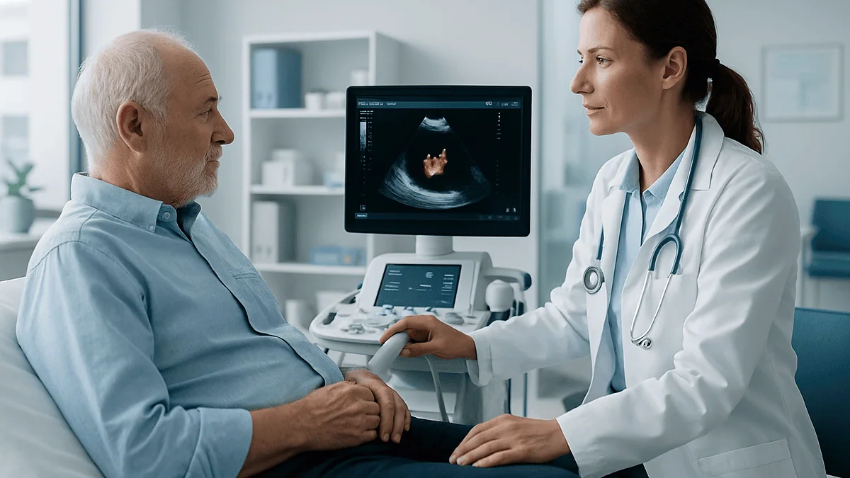
A transthoracic echocardiogram, meaning one performed through the chest wall, is the standard version most people receive. You undress from the waist up, lie on a couch, and a sonographer places three or four sticky electrodes on your skin to record the heart’s electrical rhythm alongside the pictures. You will spend most of the scan lying on your left side with your left arm behind your head, which lets the heart swing forward against the ribs.
Cool gel goes on the chest, and the probe is pressed firmly between the ribs, under the ribcage and at the base of the neck. Firm pressure is normal and sometimes briefly uncomfortable, but the test itself does not hurt and involves no radiation. You may be asked to breathe out and hold still for a few seconds at a time while a clip is recorded. The NHS notes the scan usually takes between 15 minutes and an hour, and there is no need to fast or stop usual medicines unless your team says otherwise (NHS).
When the question is specifically about a clot at the apex of the left ventricle, the sonographer may inject a small amount of ultrasound contrast into an arm vein. This is a suspension of tiny gas-filled microbubbles that make blood glow bright white on screen; a clot stays dark against the glow, so its outline becomes far easier to see. The microbubbles are cleared by breathing them out within minutes.
The sonographer records the images but does not usually give results on the spot. A cardiologist reviews the full study, measures the chambers, grades wall motion and writes a report that goes to the doctor who requested it (Mayo Clinic). That report, rather than anything said in the scan room, is what shapes the next step.
Transesophageal echocardiogram for clot: why the probe goes down the throat
The esophagus, the swallowing tube, runs directly behind the left atrium with only a thin wall of tissue between them. A transesophageal echocardiogram, usually shortened to TEE, takes advantage of that geography by placing a slim ultrasound probe at the tip of a flexible tube and passing it down the throat, so the heart is imaged from behind with no ribs or lungs in the way. The resulting pictures of the left atrium, the appendage, the valves and any artificial valve are far sharper than anything obtainable from the chest.

Preparation matters more than for the chest scan. Cleveland Clinic advises fasting for several hours beforehand so the stomach is empty, because the procedure involves sedation and suppresses the gag reflex. A numbing spray is applied to the back of the throat, a sedative is given through a vein, and a small bite guard protects the probe and your teeth. Most people remember little of the probe being placed. The probe itself is usually in position for a matter of minutes, though preparation and recovery from sedation stretch the whole visit to well over an hour, and you will need someone to take you home and to avoid driving for the rest of the day (Cleveland Clinic).
A sore throat for a day or two is common. Serious complications are rare but real: injury to the esophagus, breathing difficulty from sedation, or a reaction to the medicines used. People with known esophageal disease, swallowing disorders or recent upper gastrointestinal bleeding are usually steered toward CT instead.
The trade is a more involved test for a much clearer answer. When the clinical question is “is there a clot in the appendage before we reset this rhythm,” that clarity is precisely what the team needs.
Left atrial appendage thrombus: the hiding place in atrial fibrillation
Picture a small windsock attached to the side of the left atrium. That is the left atrial appendage, a ridged pouch that empties briskly when the atrium contracts normally. In atrial fibrillation it stops contracting and the pouch fills with slow, swirling blood. A clot forming there is called a left atrial appendage thrombus, and it is the reason atrial fibrillation is so closely linked with stroke: the NHS lists stroke as the main complication of the rhythm, precisely because clots formed in the quivering atrium can travel to the brain (NHS).
On a transthoracic scan the appendage is often no more than a smudge. Through the esophagus it fills the screen. The cardiologist looks for three things. First, an actual mass, which may be a rounded blob or a flat layer lining the pouch. Second, “smoke”: a hazy swirling pattern of clumped red cells, known as spontaneous echo contrast, which signals blood stagnant enough to clot. Third, how fast blood exits the appendage on Doppler; a slow emptying velocity means higher risk even when no clot is yet visible.
This scan sits at a key decision point. Before cardioversion, the electrical or medicine-based reset of the rhythm, or before an ablation procedure that burns or freezes the tissue driving the arrhythmia, teams want to know the appendage is clear. Restoring a strong atrial contraction while a clot is sitting in the pouch can fling it into circulation. If a clot is found, the planned procedure is postponed, anticoagulation is continued or adjusted by the prescribing clinician, and the scan is repeated later to confirm the pouch has cleared. A clear scan, on the other hand, is a snapshot, not a permanent certificate.
Left ventricular thrombus after a heart attack
A heart attack happens when a coronary artery is blocked and the muscle it feeds is starved of oxygen. The damaged segment stops contracting, a state called akinesis, and if the damage is at the front and tip of the left ventricle the apex can balloon slightly with each beat. Blood that should be swept out with every contraction instead eddies in the dead zone.
Clinicians describe clot formation in terms of three ingredients: stagnant flow, an injured surface, and blood that is more prone to clotting, all of which are present after a large heart attack. The result is a left ventricular thrombus, most often a layered deposit hugging the motionless wall at the apex.
On echocardiography the reader looks for a mass with a clear boundary that sits next to, and only next to, an area of wall that does not move. That pairing is important: a clot rarely forms on healthy, vigorously contracting muscle, so a suspicious shadow beside a normal wall is more likely an artifact. Ultrasound contrast, the microbubble injection described earlier, markedly improves the ability to separate a real clot from muscle trabeculations, the natural ridges inside the chamber.
The report will usually describe the clot’s shape. A flat, laminar clot that lines the wall behaves differently from a rounded clot that protrudes into the cavity, and a clot that wobbles with each beat is watched more closely than one that sits still, because mobile material is more likely to break away. These descriptions are not verdicts; they are inputs for the treating team, alongside the size of the heart attack, kidney function and bleeding history, when the conversation turns to anticoagulation and how soon to re-image.
Is it a clot, or something else? How readers tell the difference
Not every mass inside the heart is a clot, and telling them apart is one of the more careful judgments in cardiac imaging.
A myxoma is a benign tumor that classically hangs from a stalk on the wall dividing the two atria and flops toward the valve with each beat. Its attachment point and its slightly speckled texture distinguish it from a clot, which tends to sit where blood is stagnant rather than dangling into a fast-flowing chamber.
A vegetation is an infected growth on a valve leaflet, the hallmark of endocarditis, an infection of the heart’s inner lining. Vegetations look shaggy and irregular and usually come with fever and abnormal blood tests, whereas clots on valves are more often seen on artificial valves and tend to be smoother.
Normal anatomy causes its share of false alarms. The appendage has ridges of muscle that can mimic small clots. A fold of tissue between the appendage and the nearby pulmonary vein is famous for being mistaken for a mass. Fine, lacy remnants in the right atrium are common and harmless.
Then there are artifacts, images created by the physics of ultrasound rather than by anything real: reverberations off a metal valve, clutter close to the probe, or side-lobe echoes that paint a ghostly line across a chamber. Experienced readers change the probe angle, adjust settings and look for the mass in at least two views before believing it.
When doubt remains, the team has two further strategies. One is time: a clot on anticoagulation typically shrinks over weeks, a tumor does not. The other is cardiac MRI, which can characterize the tissue directly, as the next section explains.
Beyond ultrasound: where cardiac CT and MRI fit in
Echocardiography answers most questions, but two other technologies step in when the picture is unclear or a TEE is unsafe.
Cardiac CT uses X-rays and an iodine-based contrast dye injected into a vein. Timed correctly, the dye fills every chamber, and a clot shows as a dark filling defect where dye cannot reach. CT is fast, needs no sedation and images the appendage well, which makes it a common alternative before ablation. Its drawbacks are radiation, the need for reasonable kidney function to handle the dye, and the fact that slow-mixing blood in a sluggish appendage can itself look dark, producing a false positive that sometimes requires a delayed second scan.
Cardiac MRI uses a powerful magnet and radio waves rather than radiation, often with a gadolinium-based contrast agent. Its strength is tissue characterization: a clot has no blood supply, so on delayed images it stays black while surrounding muscle and most tumors take up contrast. MRI is generally considered the most sensitive method for small clots layered against the ventricular wall. It takes longer, is noisy and enclosed, and some older implanted devices and severe kidney impairment limit its use.
| Method | How it sees | Strongest at | Main limits |
|---|---|---|---|
| Transthoracic echo | Sound waves through chest | First look; wall motion; ventricular clots with contrast | Poor view of appendage; image quality varies |
| Transesophageal echo | Sound waves from esophagus | Appendage; valves; artificial valves | Sedation; fasting; rare esophageal injury |
| Cardiac CT | X-rays with iodine dye | Appendage when TEE unsafe; anatomy before procedures | Radiation; dye and kidneys; slow-flow false positives |
| Cardiac MRI | Magnet with gadolinium | Small ventricular clots; clot versus tumor | Time; enclosed space; some devices |
No single test wins every contest. The treating team chooses based on where the clot is suspected, what else needs answering, and what is safe for the individual in front of them.
Who is usually sent for an echocardiogram heart clot check, and who is asked to wait
The request for a clot-focused scan almost always follows one of a handful of clinical situations rather than arising out of the blue.
People recovering from a large heart attack, especially one affecting the front wall, are commonly scanned because that is where ventricular clots form. Anyone who has had a stroke or a transient ischemic attack, a brief stroke-like episode that resolves, without an obvious cause in the neck or brain arteries will often have the heart examined as a possible source. People with atrial fibrillation who are due for cardioversion or ablation, and who have not been on uninterrupted anticoagulation beforehand, are routinely offered a TEE or CT first. New symptoms in someone with an artificial valve, or a valve that suddenly shows a higher pressure gradient on a routine echo, prompt a closer look for clot on the leaflets. A weakened heart with a low ejection fraction, the percentage of blood pumped out with each beat, and someone who has had a clot lodge in an arm, leg or organ artery round out the usual list (American Heart Association, MedlinePlus).
Some people are asked to wait or are routed differently. Those already established on stable anticoagulation with a recent clear scan may not need repeat imaging before a procedure; their team weighs the small chance of a new clot against the burden of another test. Someone who is acutely unwell will usually get a quick bedside scan to answer the urgent question and a detailed study later, once stable. Individuals with esophageal disease, swallowing problems or recent upper gastrointestinal bleeding are generally offered CT rather than TEE.
None of these are rigid rules. They are the patterns behind most referrals, and the final call rests with the team that knows the whole history.
What the following days and weeks usually look like
The scan report reaches the requesting doctor, and the path splits.
If a clot is found, the first conversation is usually about anticoagulants, the class of medicines that slow the blood’s clotting system. A common misunderstanding is worth correcting early: these medicines do not dissolve a clot the way a solvent dissolves glue. They stop the clot from growing and prevent new ones from forming, which gives the body’s own clot-clearing enzymes time to break down what is already there. That process unfolds over weeks to months rather than days, which is why teams usually arrange repeat imaging after an interval to see whether the clot has shrunk, changed shape or disappeared. Whether the medicine is started, switched or continued, and for how long, is a decision for the prescribing clinician, who balances embolic risk against bleeding risk for each person.
Planned procedures shift. A cardioversion or ablation is postponed until a later scan shows the appendage is clear. Some people with a ventricular clot are asked to avoid heavy straining or contact sports until re-imaging, though gentle activity is generally encouraged. Everyone on anticoagulation is taught to watch for bleeding and to tell dentists, surgeons and pharmacists what they are taking.
If no clot is found, the planned procedure typically goes ahead as scheduled. The team will still remind you that a clear scan describes one moment in time; the conditions that prompted the scan, whether atrial fibrillation or a scarred ventricle, remain, and ongoing prevention is discussed on its own merits.
After a TEE specifically, expect a scratchy throat, a mild sedative hangover and a day without driving (Cleveland Clinic). Most people are back to their usual routine the following day.
How serious is a blood clot in the heart? An honest reading of the evidence
It is serious, and it is also treatable, and both halves of that sentence deserve equal weight.
The danger from a clot inside a heart chamber is rarely the clot itself, which does not usually block the heart’s own pumping. The danger is embolization: a fragment breaks free and lodges downstream. From the left side of the heart that means the brain, and stroke is the outcome imaging and treatment are principally trying to prevent (NHS, American Heart Association). Fragments can also reach the arteries of the arms, legs, gut or kidneys. From the right side of the heart, the destination is the lungs.
Seriousness therefore depends on where the clot is, how large it is, whether it is mobile or firmly layered, what caused it, and how quickly it is recognized. A flat, stable clot lining a scarred ventricle in someone promptly started on anticoagulation is a different proposition from a large mobile mass in an untreated appendage. This is why the internet search “what are the odds of surviving a blood clot in the heart” cannot produce a single honest number: the figures scattered across forums and articles are drawn from very different groups of patients, imaged at different stages, treated in different ways.
What the evidence does support is that clots inside the heart are a recognized, well-studied complication with established pathways. Anticoagulation reduces the chance of embolism and allows many clots to resolve over time. Repeat imaging confirms progress. When a clot threatens a mechanical valve’s function or is very large and mobile, teams may discuss more intensive options, including clot-dissolving medicines given in hospital or surgery, each with its own risks that are weighed individually.
The appropriate response to the finding is neither panic nor dismissal. It is a plan, made with the people who can see your whole picture.
What people often get wrong about echocardiograms and clots
Several ideas circulate widely enough to be worth addressing head on.
“Needing an echocardiogram means something is badly wrong.” An echocardiogram is one of the most common tests in cardiology, requested for murmurs, breathlessness, blood pressure follow-up, chemotherapy monitoring and routine checks after a heart attack. Being asked to have one is a sign of a thorough team, not a hidden verdict (Mayo Clinic).
“If the echo was clear, there is definitely no clot.” A standard chest scan can miss clots in the appendage and small ventricular clots. A report that says “no thrombus seen” is accurate as far as the images allow; it is not the same as a guarantee, which is why teams add contrast, TEE or MRI when the stakes are high.
“A clot in the heart is the same as a heart attack.” They are different events. A heart attack is a clot blocking a coronary artery on the outside surface of the heart, starving the muscle. A chamber thrombus is a clot inside the pumping cavity, threatening to travel elsewhere. One can lead to the other, but they are not interchangeable.
“Blood thinners dissolve the clot.” They prevent growth and new clots; the body does the dissolving, slowly.
“The scan is clear, so I can stop the medicine.” A clear scan changes the conversation, not the prescription. Only the prescribing clinician should adjust anticoagulation, because the condition that caused the clot usually persists.
“TEE is a kind of surgery.” It is a scan under light sedation with a probe in the swallowing tube, over in minutes, with no incision.
“Red flags on an echocardiogram mean an emergency.” A report full of technical language is not a list of alarms; it is a description for clinicians, discussed in the next section.
Questions to ask your care team about the scan and what it found
Scan reports are written for clinicians, and the most useful thing you can do is turn the report into a conversation. Bring a written list; the appointment often moves quickly.
- Exactly where is the clot, and what do you think caused it to form there?
- How confident are you that it is a clot rather than an artifact, a normal structure or something else, and would another imaging test change that confidence?
- Is the clot described as flat and layered or as protruding and mobile, and how does that affect the plan?
- What does the rest of the report say about my heart’s pumping strength and my valves?
- If anticoagulation is being discussed, what is the intended goal, how will you know it is working, and when will imaging be repeated?
- Which of my other medicines, supplements or activities should I ask about before changing anything?
- If I had a procedure planned, when will it be reconsidered, and what would need to be true on the next scan for it to go ahead?
- What bleeding signs should I watch for, and who do I contact outside office hours?
Two habits make these conversations more productive. Ask for a copy of the report itself, not just a summary, so you can follow the terminology over time. And when a percentage or a timeline is quoted, ask where it comes from; a figure drawn from a large guideline population may or may not apply to your particular heart, and a good clinician will say so.
You are entitled to understand the reasoning behind the plan. You are not expected to make the plan alone, and the team should never leave you feeling that you must.
When to call your doctor: red-flag signs after a heart clot is found
Some symptoms mean calling emergency services immediately rather than waiting for a clinic line. Sudden drooping of one side of the face, weakness or numbness in an arm or leg, slurred or confused speech, or sudden loss of vision are signs of a possible stroke, and every minute matters. Sudden severe chest pain, sudden breathlessness, coughing up blood or fainting can signal a clot in the lungs or a new heart attack. A limb that suddenly turns cold, pale, painful or numb, or sudden severe abdominal pain with vomiting, can mean a clot has lodged in an artery to that limb or to the gut.
After a transesophageal echocardiogram, seek urgent care for severe or worsening chest or throat pain, difficulty or pain on swallowing that is getting worse, vomiting blood, fever, or breathing difficulty. A mildly scratchy throat for a day or two is expected; the symptoms above are not (Cleveland Clinic).
If you are taking anticoagulants, contact your team promptly for bleeding that will not stop after firm pressure, black or tarry stools, blood in the urine, large unexplained bruises, coughing or vomiting blood, or any significant fall or blow to the head, even if you feel fine afterwards.
Same-week, non-emergency calls are appropriate if you are unsure how to take a medicine around a dental or surgical appointment, if you develop new palpitations or breathlessness that is gradually worsening, or if you have questions about the report that were not answered.
A useful rule: any symptom that is sudden, severe or on one side of the body is an emergency until a clinician says otherwise. Your team would far rather hear from you about a false alarm than not hear about a real one.
Frequently asked questions
How serious is it to have a blood clot in your heart?
It is a serious finding because a fragment can break off and travel to the brain, lungs, limbs or organs, but it is also a recognized complication with established treatment pathways. Seriousness depends on where the clot sits, how large and mobile it is, what caused it and how quickly anticoagulation begins. Your treating team weighs these factors together rather than relying on any single number.
What are the odds of surviving a blood clot in the heart?
No single honest figure exists, because published numbers come from very different groups of patients with clots in different locations, imaged at different stages and treated in different ways. Many clots shrink or resolve on anticoagulation with repeat imaging confirming progress. Ask your own team how the location, size and cause of your clot shape the outlook, since general statistics may not reflect your situation.
Is it serious if you need an echocardiogram?
Not by itself. An echocardiogram is one of the most commonly requested heart tests and is used for murmurs, breathlessness, blood pressure follow-up and routine checks after a heart attack. Being asked to have one reflects a thorough team wanting a clear picture. The scan is painless, uses no radiation and usually takes between 15 minutes and an hour.
What are red flags on an echocardiogram?
Readers pay particular attention to a mass sitting beside a wall segment that does not move, hazy swirling blood in the left atrial appendage, very slow flow out of the appendage, a mobile mass that wobbles with each beat, or a sudden rise in pressure across an artificial valve. These are descriptions for clinicians, not verdicts, and each is interpreted alongside your history and often confirmed with a second imaging method.
Can an echocardiogram detect a blood clot every time?
No. A standard chest scan can miss small clots and often cannot see clearly into the left atrial appendage, where atrial fibrillation clots form. Adding ultrasound contrast, performing a transesophageal echocardiogram or using cardiac CT or MRI closes most of that gap. A report reading “no thrombus seen” is accurate for the images obtained, which is why teams choose the method according to how high the stakes are.
What is a left atrial appendage thrombus?
It is a clot that forms in the small windsock-shaped pouch attached to the left atrium. In atrial fibrillation the atrium quivers instead of contracting, blood pools in the pouch and can clot, and a fragment can travel to the brain. This is the main reason atrial fibrillation raises stroke risk, and it is why the appendage is checked closely before rhythm-resetting procedures.
Does a transesophageal echocardiogram for clot hurt?
Most people find it uncomfortable rather than painful. The throat is numbed with a spray, a sedative is given through a vein, and the probe is usually in place for only a few minutes; many people remember little of it. A scratchy throat for a day or two afterwards is common. Severe throat or chest pain, trouble swallowing, fever or vomiting blood are not expected and need urgent attention.
Do I need to fast before a heart clot scan?
It depends on the type. A standard transthoracic echocardiogram needs no fasting and no change to usual medicines unless your team says otherwise. A transesophageal echocardiogram does require fasting for several hours beforehand because sedation is used and the stomach must be empty. CT and MRI with contrast have their own instructions, which your team will provide in advance.
Can I drive home after a transesophageal echocardiogram?
No. The sedation used during the procedure slows reaction times for the rest of the day, so you will need someone to take you home and should avoid driving, operating machinery or making important decisions until the following day. Most people return to their normal routine within a day, apart from a mildly sore throat.
What happens if a clot is found before my cardioversion or ablation?
The procedure is postponed, because restoring a strong atrial contraction while a clot sits in the appendage could fling it into circulation. Your prescribing clinician will continue or adjust anticoagulation, and the scan is repeated after an interval to confirm the appendage has cleared before the procedure is rescheduled. This delay is a safety step, not a sign the procedure is off the table.
References
This article is for general information only and is not a substitute for professional medical advice. Please consult a qualified doctor about your individual situation.

More from the Blog

Sternum Pain: Where the Breastbone Sits, Costochondritis and Other Causes and When to Seek Care
Sternum pain most often comes from the chest wall itself: costochondritis, an inflammation of the cartilage that joins the ribs to the breastbone, or…

Questions to Ask Your Cardiologist After an Angina Diagnosis
After an angina diagnosis, the most useful questions to ask your cardiologist cover five areas: which type of angina you have (stable, unstable, microvascular…

Life Expectancy After Cardiac Ablation: What Long-Term Studies Show and Who Benefits Most
Life expectancy after cardiac ablation is shaped mainly by the condition being treated and by overall heart health, not by the procedure itself. For…

Recovery After Congenital Heart Surgery: Intensive Care, Wound Care and the First Two Months
Congenital heart surgery recovery time varies with the defect and the child's age, but a typical path after open-heart repair is 2–4 days in…

Caring for Someone With Cardiorenal Syndrome: Tracking Weight, Swelling and Appointments
Caring for someone with cardiorenal syndrome means helping their heart and kidneys stay in balance from one day to the next: weighing them each…

Living With Cardiac Sarcoidosis: Repeat Imaging, Rhythm Monitoring and Daily Routines
Living with cardiac sarcoidosis usually means a long partnership with a heart team: repeat scans such as cardiac MRI and FDG-PET to check whether…