

Peri-Implantitis Warning Signs: Bleeding, Pus and Implant Mobility That Mean a Prompt Visit
Key Takeaways
- Bleeding on gentle probing is the defining sign of peri-implant mucositis, the reversible stage before bone is lost, according to the 2017 World Workshop consensus.
- A systematic review indexed on PubMed found peri-implant mucositis in about 43 percent and peri-implantitis in about 22 percent of implant patients across the studies analyzed.
- Peri-implantitis progresses in a non-linear, accelerating pattern and generally faster than gum disease around natural teeth.
- Movement of the implant post itself signals loss of osseointegration and is treated as implant failure, not as a diagnostic criterion of peri-implantitis.
- Pain is typically a late feature because implants lack a nerve-rich pulp, so bleeding, discharge and exposed threads usually appear first.
- Bone lost to peri-implantitis does not regrow on its own, which is why professional assessment within days of recurring bleeding matters more than waiting for pain.
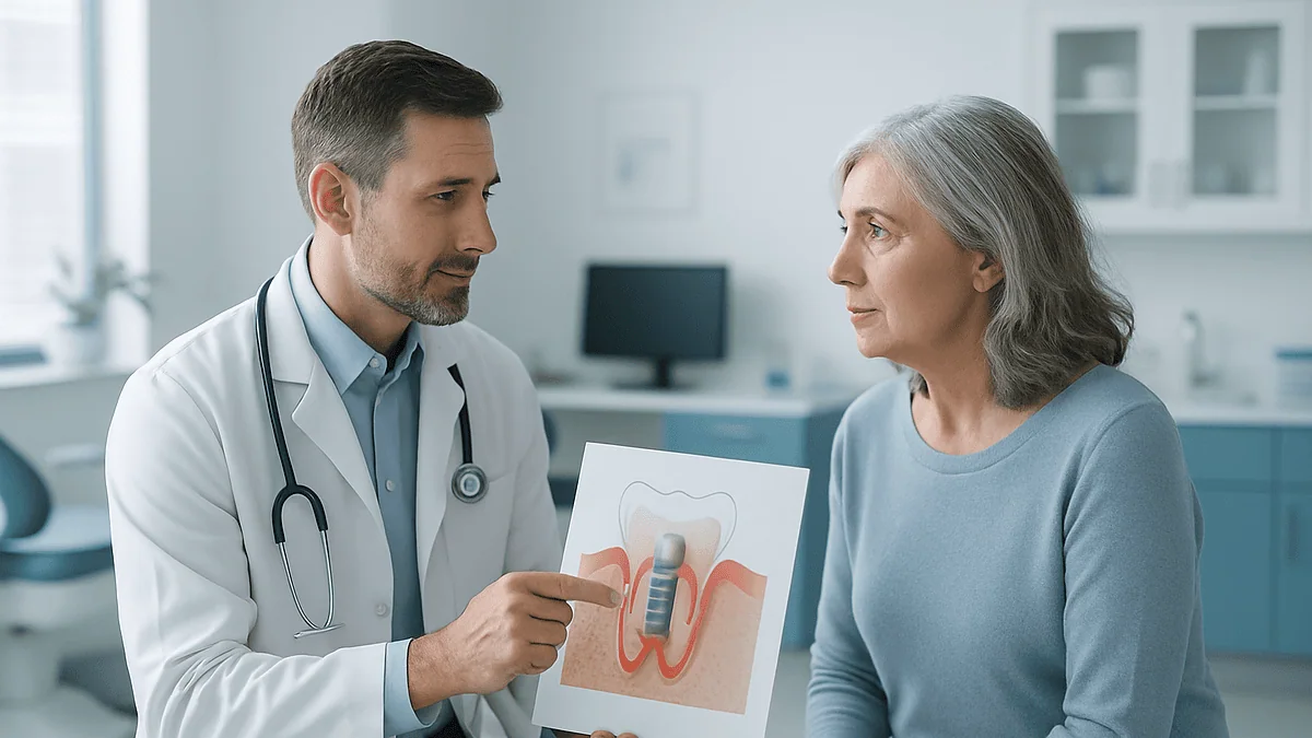
Peri-implantitis warning signs include gums around a dental implant that bleed when brushed or probed, redness or swelling, pus or a persistent bad taste, a gum line that is pulling back to expose implant threads, and any looseness of the implant or crown. Bleeding may be reversible mucositis; pus, bone loss and mobility mean prompt professional assessment is needed.
The floss comes out pink. Not dramatically, not the red of a cut lip, just a faint blush on the thread as it slides past the back molar that was replaced with an implant six years ago. The tooth itself feels fine. It chews fine. It has never ached. So the floss goes in the bin and the day carries on.
That small, easily dismissed moment is exactly where most peri-implantitis warning signs begin. Dental implants do not decay the way natural teeth do, and that is part of the problem: people stop watching them. The bone and gum that hold an implant in place can still become inflamed and, over time, quietly recede.
This explainer walks through what bleeding, pus and implant mobility each actually mean, how quickly the condition tends to move, who is most exposed, and what a dental team can and cannot do once bone has been lost. The decision about your own implant always rests with the team looking after it.
What peri-implantitis actually is, in plain language
A dental implant is a titanium or ceramic post placed into the jawbone to hold a replacement tooth. Over a few months the bone grows tightly against its surface, a process called osseointegration, which is simply bone fusing to metal with no gum ligament in between. Mayo Clinic describes this healing window as taking several months before a crown can be loaded.
Peri-implantitis is inflammation of the soft tissue around an implant that has progressed to loss of the supporting bone. It is best understood as a two-stage story. The first stage, peri-implant mucositis, is inflammation confined to the gum: red, swollen, bleeding tissue with the bone underneath still intact. The 2017 World Workshop consensus, summarized in the Schwarz review on PubMed, defines mucositis as reversible and peri-implantitis as the stage where bone has begun to disappear.
The mechanism is bacterial. Plaque, a sticky film of bacteria, collects at the junction between the implant crown and the gum just as it does around a natural tooth. The body responds with inflammation. Around a natural tooth, a cushioning ligament and a rich blood supply help contain that response. Around an implant, the gum attaches directly to the surface with fewer defensive fibers and less circulation, so once bacteria get a foothold below the gum line the inflammatory front tends to reach bone sooner.
Bone that has been lost around an implant does not regrow on its own. That single fact explains why dental teams treat bleeding around an implant more seriously than many patients expect, and why the goal is always to catch the process while it is still mucositis.
Peri-implantitis warning signs: bleeding that keeps coming back
Bleeding is the earliest and most common signal, and it is also the one people most often talk themselves out of. Healthy implant tissue should not bleed when you brush along the gum margin, floss around the crown, or when a dentist gently runs a probe around it. The NHS makes the same point about natural gums: bleeding during brushing is a sign of gum disease, not a sign of brushing too hard.

Around an implant the pattern matters more than a single episode. A one-off spot of blood after a new floss threader or an aggressive interdental brush can be irritation. Bleeding that returns each time you clean the same site for a week or more suggests plaque is sitting below the gum line where your brush cannot reach it.
Watch for what accompanies the bleeding. Redness that is deeper than the surrounding pink gum, a shiny or puffy look to the tissue collar around the crown, and tenderness when you press the gum with a fingertip all point toward mucositis. At this stage the bone is usually still intact, and the Schwarz review describes mucositis as reversible with thorough professional cleaning and improved home care.
Bleeding on probing is also the single most useful clinical sign for a dentist. In the 2017 case definitions cited in that review, bleeding or suppuration on gentle probing is required for both mucositis and peri-implantitis diagnoses. In other words, the thing you noticed on your floss is the same thing your dentist will be checking for with an instrument. That overlap is why reporting it promptly, rather than waiting for pain, changes the likely outcome. Implants rarely hurt until late; bleeding speaks first.
Pus, swelling and a bad taste: what discharge around an implant means
Pus is a thick, yellow-white fluid made of dead white blood cells and bacteria. Its appearance around an implant, whether as a visible bead at the gum line or as a persistent salty, metallic or foul taste that no amount of rinsing clears, marks a shift from irritation to active infection.
Clinicians call this suppuration, and it carries more weight than bleeding. The 2017 World Workshop case definition, as summarized in the Schwarz review on PubMed, lists bleeding and/or suppuration on probing alongside increased probing depth and radiographic bone loss as the features of peri-implantitis. Pus is not required for the diagnosis, but when it is present it strongly suggests the pocket around the implant has deepened enough to trap bacteria in a low-oxygen space where they thrive.
Swelling behaves differently around implants than around infected natural teeth. There is no pulp inside an implant to die and form an abscess at the root tip, so the swelling tends to sit right at the gum collar rather than higher up in the cheek. A gum that looks like a small raised cushion around one implant while its neighbors look flat is a pattern worth describing to your dentist exactly that way.
Two other changes often travel with pus. Bad breath localized to one area, which a partner or family member may notice before you do, and a gum margin that appears to be shrinking away from the crown so that a darker band of metal or the ridged threads of the implant become visible. Exposed threads mean bone has already receded below them.
None of these signs tells you how much bone remains. Only a probe and an X-ray can do that, which is why discharge is a prompt to be seen within days, not a problem to manage with rinses at home.
Implant mobility: why a loose implant is a different emergency
Mobility needs careful separation into two very different problems, because they lead to very different conversations with your dentist.

The first is a loose crown or a loose connecting screw. Most implant crowns are attached to the post by an internal screw, sometimes through a small connector called an abutment. Screws can loosen with years of chewing. When they do, the crown may wobble, click or rotate slightly while the implant beneath remains perfectly solid in the bone. This is mechanical, usually straightforward to address, and not peri-implantitis, though a loose crown can create a gap where plaque collects and inflammation follows.
The second is movement of the implant post itself. A natural tooth has a tiny amount of physiological give because of its ligament. An integrated implant has none. Any perceptible movement of the post means the bone-to-metal bond has failed, either because it never fully formed in the months after placement or because inflammation has destroyed enough surrounding bone that the remaining contact cannot hold it. The Schwarz review is explicit that mobility is a sign of complete loss of osseointegration, not a diagnostic criterion of peri-implantitis; by the time the post moves, the disease has typically run most of its course.
You usually cannot tell the two apart yourself. A crown that spins or lifts is most likely a screw issue. A whole unit that rocks when you press it, especially with tenderness, swelling or pus, is far more concerning. Either way, stop chewing on it and arrange to be seen promptly.
Seek care urgently if mobility comes with facial swelling, fever, difficulty swallowing, or spreading pain along the jaw. Those signs suggest infection is no longer confined to the implant site and warrant same-day attention, not a routine appointment.
Mucositis, peri-implantitis and a failing implant at a glance
The three stages overlap and blur into one another, but seeing them side by side clarifies why the same symptom can be minor in one column and serious in the next. The definitions below follow the 2017 World Workshop consensus as summarized in the Schwarz review on PubMed; the reversibility judgments and typical clinical findings are drawn from the same source.
| Feature | Peri-implant mucositis | Peri-implantitis | Failing or failed implant |
|---|---|---|---|
| Where the problem sits | Gum only | Gum plus supporting bone | Bone-to-implant bond lost |
| Bleeding on gentle probing | Yes | Yes, often with pus | Often, may be less obvious |
| Bone loss on X-ray | None beyond initial remodeling | Progressive, visible | Extensive, may surround the post |
| Probing depth | May be unchanged | Increased compared with baseline | Deep, may reach the tip |
| Implant movement | No | No | Yes |
| Pain | Usually none | Often none until advanced | Variable |
| Reversible? | Yes, with cleaning and home care | Progression can be controlled; lost bone does not regrow spontaneously | No; removal is usually discussed |
The row worth memorizing is pain. Across all three columns, discomfort is an unreliable messenger. Patients frequently arrive at the failing-implant stage having felt nothing more than occasional bleeding, because implants lack the nerve-rich pulp that makes a natural tooth ache when infected.
The other point the table makes quietly is that probing depth only means something against a baseline. A measurement taken when the crown was first fitted lets a dentist see change years later. If you do not know whether that baseline exists in your records, it is a reasonable question to ask at your next visit.
How quickly does peri-implantitis progress?
Faster than gum disease around natural teeth, and not in a straight line. That is the honest summary of the evidence, and it should shape how promptly bleeding gets reported.
The Schwarz review on PubMed describes peri-implantitis as progressing in a non-linear, accelerating pattern. Bone loss may be slow or barely detectable for a period and then speed up, so an implant that looked stable on one X-ray can show meaningful change on the next. The same review notes that, in the studies available, onset often occurred relatively early after the crown was placed, within the first few years of function, rather than being purely a problem of very old implants.
Why the acceleration? Two mechanisms are usually proposed. First, the pocket around an inflamed implant deepens as bone recedes, and a deeper pocket shelters more bacteria in a low-oxygen environment, which drives more inflammation, which costs more bone. Second, as bone is lost, the rough, threaded portion of the implant becomes exposed inside the pocket. Those threads and surface textures were designed to help bone grip; once exposed to plaque they become almost impossible to clean thoroughly, either at home or in the chair.
How this translates to an individual is genuinely uncertain. Progression varies with smoking status, blood sugar control, history of gum disease around natural teeth, the design and position of the implant, and how well the gum around it can be cleaned. Nobody can tell you from a single bleeding episode how many months you have. What the evidence does support is that the window during which the problem is still reversible mucositis is finite, and that waiting for a routine check that is many months away is a gamble the acceleration curve does not favor.
Can a dental implant get infected years later?
Yes. An implant that has functioned uneventfully for a decade is not immune, and this surprises people more than almost any other aspect of implant care.
The reasoning is simple once the mechanism is clear. Peri-implantitis is driven by plaque bacteria at the gum-implant junction, and plaque forms every day for as long as the implant is in the mouth. Nothing about successful early healing removes that exposure. What changes over the years is the set of circumstances around the implant: gums naturally recede with age, dexterity for flossing may decline, a new medical condition or medication may dry the mouth, a partner who used to remind you about check-ups may no longer be there, or a neighboring tooth may be lost and change the way food packs around the crown.
The scale of the problem is not small. A systematic review of prevalence studies by Derks and Tomasi, indexed on PubMed, calculated a weighted mean prevalence of peri-implant mucositis of about 43 percent of patients and of peri-implantitis of about 22 percent, with wide variation between studies depending on how disease was defined and how long implants had been in place. Those figures describe people who had implants, not people who had been careless with them.
Late-onset infection tends to be caught later, too, precisely because vigilance fades. The implant has become just another tooth, its check-ups are treated as optional, and the bleeding that would have prompted a call in year one is shrugged off in year nine.
The practical consequence is that lifelong maintenance is part of having an implant, not an add-on. Cleveland Clinic’s overview of dental implants frames them as requiring the same daily care and regular professional attention as natural teeth. Treating that as a permanent arrangement rather than a post-surgery phase is the single most protective mindset shift a patient can make.
Who is most at risk, and who is usually asked to wait before an implant
Risk is not evenly spread, and the same factors that raise the chance of peri-implantitis are often the ones that lead a dental team to postpone or reconsider implant placement in the first place.
A history of periodontitis around natural teeth stands out. Periodontitis is the advanced form of gum disease in which the bone supporting teeth is destroyed, and Mayo Clinic describes it as a common cause of tooth loss. People who lost teeth to periodontitis carry the same bacterial profile and the same tissue response into implant treatment, and the Schwarz review identifies prior periodontitis as a consistent risk indicator for peri-implantitis.
Smoking is the second major factor. Tobacco narrows blood vessels in the gum, blunts the immune response and slows healing; the NHS lists smoking among the main contributors to gum disease. Poorly controlled diabetes works through related mechanisms, impairing the body’s ability to fight infection and repair tissue. Both conditions are commonly discussed before surgery, and patients are often asked to stabilize blood sugar or make progress on stopping smoking before placement proceeds.
Plaque control matters independently of everything else. An implant crown that is shaped or positioned so that floss cannot pass around it, or a patient whose dexterity, vision or health limits daily cleaning, is at higher risk regardless of medical history.
Who is usually asked to wait? Mayo Clinic notes that implant candidates need adequate bone, healthy oral tissues, and the ability to commit to several months of healing. Active gum disease is generally treated first. Recent radiation to the jaw, certain medications that affect bone turnover, and uncontrolled systemic illness each prompt further discussion. None of these is an automatic bar; each is a reason for the treating team to weigh timing and alternatives such as a bridge or denture with the patient.
How dentists diagnose peri-implantitis and why 'it looks fine' isn't enough
A healthy-looking implant on a quick glance can be losing bone underneath. Diagnosis rests on three measurements, none of which you can make at home.
The first is gentle probing. A thin, blunt instrument is walked around the gum collar of the implant to check for bleeding, pus and the depth of the pocket between gum and implant. Around a natural tooth, probing depth has a fairly consistent healthy range. Around an implant, the healthy depth varies with how deeply the post was placed and how thick the gum is, so the 2017 World Workshop consensus, as described in the Schwarz review, emphasizes comparing current readings against a baseline recorded when the crown was fitted. An increase over time is the signal, not any single number.
The second is radiography. A dental X-ray shows the level of bone against the implant threads. Some bone remodeling in the first year after placement is expected and is not disease. Bone loss beyond that, especially a saucer-shaped defect around the implant neck, points to peri-implantitis. When no baseline X-ray exists, the same consensus proposes thresholds for probing depth and radiographic bone loss that can support a diagnosis, which is why dental teams sometimes ask patients to obtain records from wherever the implant was placed.
The third is the clinical picture: redness, swelling, tenderness, discharge, exposed threads and any mobility of the crown or post.
Bringing these together, the dentist assigns one of the stages in the table above. What they are really deciding is whether bone is still intact, in which case cleaning and home-care coaching may be all that is needed, or whether the process has moved into bone and a more involved conversation begins. Asking to see the X-ray and to have the probing depths explained is entirely reasonable and helps you follow the reasoning.
Can you fix peri-implantitis? What treatment involves
The answer people want is a clean yes. The evidence-honest answer is that peri-implantitis can usually be controlled, its progression can often be stopped, and inflamed tissue can be brought back to health, but bone that has already been lost does not reliably return, and no single approach is proven superior. The Schwarz review on PubMed is candid that treatment outcomes vary and that recurrence is common without ongoing maintenance.
Treatment is layered. The first layer is always mechanical: professional removal of plaque and hardened deposits from the implant surface, using instruments designed not to scratch it, along with adjustment of any crown contours that trap food, and detailed coaching on how to clean the site at home. For mucositis, this layer alone is often sufficient.
Where bone has been lost, non-surgical cleaning reaches its limits because exposed threads inside a deep pocket cannot be adequately decontaminated blind. The next layer is surgical access: the gum is lifted so the implant surface can be seen, cleaned and treated directly. Depending on the shape of the bone defect, the surgeon may either reshape the bone and reposition the gum lower to make the site cleanable, or attempt to rebuild the defect with bone graft material. Which is chosen depends on the defect’s anatomy and is a judgment for the treating team.
Antimicrobials are sometimes used alongside these steps. Antiseptic rinses aim to reduce bacterial load on the surface; systemic antibiotics, when prescribed, target the bacteria driving inflammation. Their role is supportive rather than primary, and whether, which and for how long are decisions for the prescribing clinician based on the individual case.
When bone loss is extensive or the implant is mobile, removal is discussed. That is not a failure of treatment so much as recognition that the implant can no longer be maintained in health, and it opens a conversation about what, if anything, replaces it.
Early signs of dental implant failure that are easy to miss
Failure is a word that covers two different timelines, and the early signs differ for each.
Early failure happens in the months after placement, before or shortly after the crown is fitted, when bone does not fuse to the implant. Mayo Clinic notes that this healing phase takes several months and that if the bone does not integrate the implant is removed, the site allowed to heal, and a second attempt sometimes considered. Signs during this window include persistent dull ache at the site well after normal surgical soreness has faded, a sense that the healing cap or temporary crown feels different from day to day, and any movement of the post itself. Because there is no crown to load yet, these are easy to attribute to ordinary healing.
Late failure is the endpoint of peri-implantitis, and its early signs are the ones this article has traced: bleeding that recurs, a puffy gum collar, a bad taste from one area, threads becoming visible as the gum pulls back, and a crown that seems to sit slightly differently against its neighbors. Food catching where it never used to is another quiet signal, because it usually means the gum contour has changed.
Two signs deserve particular attention because they are so often misread. The first is a gum that looks perfectly pink but bleeds readily when probed; the surface can look healthy while the pocket beneath is inflamed. The second is the absence of pain. People reason that a problem serious enough to threaten an implant would surely hurt. Implants have no pulp and few nerve endings in the surrounding tissue, so pain is typically a late feature, if it appears at all.
The practical rule is that any change at an implant site that persists beyond a week or two is worth a call, however trivial it feels. Dental teams would far rather examine a healthy implant than meet a failing one.
What the following days and weeks usually look like after treatment
Recovery depends heavily on how far the disease had progressed and which layer of treatment was needed, so any timeline is a typical range rather than a promise.
After non-surgical cleaning for mucositis, most people notice the gum becoming less tender and less prone to bleeding over the following couple of weeks as inflammation subsides and home cleaning improves. The tissue may feel slightly sensitive to cold at first where deposits were removed. Dentists commonly arrange a review within weeks to re-probe the site and confirm the bleeding has settled; if it has not, that itself is information and may prompt closer investigation of the crown shape or a deeper pocket.
After surgical treatment, expect a recovery closer to that of minor gum surgery. Mayo Clinic’s description of dental implant surgery recovery, which involves similar tissue handling, lists swelling of the gums and face, bruising, minor bleeding and pain at the site as typical for the first few days, easing over one to two weeks. Stitches may be dissolvable or removed at a follow-up visit. Soft foods and gentle cleaning of the area, as instructed, are the norm while the gum heals. Because the goal of surgery is often to make the implant surface cleanable, the gum may end up sitting lower than before, exposing more of the crown or a small band of metal; this is expected and is the trade-off for a site you can keep clean.
Where grafting was attempted, the team will typically wait several months before an X-ray can show whether bone has formed, and will be cautious about interpreting early images.
Whichever route was taken, the weeks after treatment establish a new maintenance rhythm. Schwarz and colleagues stress that supportive care at regular intervals is central to preventing recurrence, so the closing conversation of treatment is usually about how often you will be seen going forward.
What people often get wrong about peri-implantitis
The myths around implant infection are remarkably consistent, and each one delays care in a predictable way.
“Implants can’t get gum disease because they’re not real teeth.” Implants cannot decay, which is true and often over-generalized. The gum and bone around them are living tissue, exposed to plaque every day, and they respond to bacteria much as they do around natural teeth, in some ways less resiliently.
“If it doesn’t hurt, it’s fine.” This is the single most costly misunderstanding. Pain is a late sign, when it appears at all. Bleeding, discharge and exposed threads all arrive first and all tend to be painless.
“Bleeding means I’m brushing too hard, so I should brush more gently.” The NHS is direct that bleeding gums signal gum disease rather than over-brushing. Backing off cleaning at an inflamed implant site leaves more plaque in place and usually makes the bleeding worse.
“A mouthwash will sort it out.” Rinses reach the surface of the gum. Peri-implantitis lives in a pocket below the gum line, on a roughened implant surface, where rinsing has little effect. Rinses can be a supporting measure if a dentist suggests one, not a substitute for professional cleaning.
“Once the implant has healed, the risky period is over.” The Derks and Tomasi prevalence review on PubMed found peri-implantitis in roughly a fifth of implant patients across the studies analyzed, many of them years into function. The risk period is the lifetime of the implant.
“If it’s infected, it will just have to come out.” Not necessarily. Early disease is often controlled without surgery, and even established bone loss can frequently be stabilized. Removal is one option on a spectrum, reserved for implants that can no longer be maintained.
The through-line is that implants ask for the same watchfulness as teeth, with a lower threshold for asking a professional to look.
Questions to ask your care team about an implant that bleeds
A short list carried into the appointment tends to produce a clearer conversation than trying to remember everything in the chair. These are the questions that most reliably surface the information you need.
- Is this mucositis or peri-implantitis, and how do you know? Ask which findings, bleeding, pocket depth, X-ray changes, led to that judgment.
- Do you have baseline probing depths and an X-ray from when the crown was fitted? If not, can they be obtained, and what will you use for comparison instead?
- How much bone has been lost, and can you show me on the image? Seeing the defect makes the stage tangible.
- What is making this site hard to clean? Crown shape, position, a gap at the margin and gum thickness can each be part of the answer, and some are adjustable.
- What treatment are you proposing first, and what would prompt moving to the next step? Understanding the sequence helps you recognize when a review is showing progress and when it is not.
- If surgery is discussed, will the aim be to rebuild bone or to reshape the site so it can be cleaned, and how will the gum look afterward?
- How often should I be seen for maintenance now, and what exactly will be checked each time?
- Which home tools suit this specific implant, and can someone demonstrate them on my mouth rather than a model?
- Is anything in my general health or medication list affecting healing here, and should my doctor be involved?
- At what point would you consider the implant unmaintainable, and what would the options be then?
You are entitled to unhurried answers. If the plan is not clear after the visit, asking for a brief written summary of the diagnosis and next steps is reasonable and increasingly routine.
When to call your doctor
Most peri-implantitis warning signs justify a prompt but routine appointment; a few require same-day or emergency attention. Knowing which is which removes the guesswork.
Arrange to be seen within days, not weeks, if you notice any of the following at an implant site: bleeding when brushing or flossing that persists for more than a week or two; a gum collar that looks redder, puffier or shinier than around neighboring teeth; a persistent bad taste or localized bad breath coming from one area; visible pus or a yellow-white bead at the gum line; a gum margin that seems to be shrinking so that a dark metal band or implant threads have become visible; food catching where it never did before; or a crown that clicks, rotates or feels different against its neighbors.
Seek urgent, same-day care, contacting your dental team or, out of hours, an urgent dental or medical service, if any of these red flags appear:
- Movement of the implant post itself, not just the crown
- Swelling that is spreading into the cheek, under the jaw or toward the eye
- Fever, chills or feeling generally unwell alongside a swollen or discharging implant site
- Difficulty swallowing, difficulty opening the mouth, or any change in breathing
- Severe, worsening pain that is not controlled by the measures your dentist has previously advised
- Numbness or tingling of the lip, chin or tongue that is new
Those red flags suggest infection may be moving beyond the implant into surrounding tissue spaces, and the NHS and Cleveland Clinic both treat spreading facial swelling with fever as a reason for emergency assessment rather than waiting.
Whatever the level of urgency, the assessment, staging and treatment plan belong to the dental team who can examine, probe and image the site. This article can help you recognize what you are seeing and describe it accurately; it cannot tell you how much bone remains or what should be done about it.
Frequently asked questions
How quickly does peri-implantitis progress?
Faster than gum disease around natural teeth, and in an accelerating rather than steady pattern. The Schwarz review of the 2017 World Workshop consensus describes bone loss that can be slow at first and then speed up as the pocket deepens and rough implant threads become exposed. Individual pace varies with smoking, diabetes control, previous gum disease and how cleanable the site is, so no one can predict a timeline from a single bleeding episode.
Can peri-implantitis be treated, or can you fix it?
It can usually be controlled and its progression stopped, but bone already lost does not reliably return. Early mucositis often resolves with professional cleaning and improved home care. Established bone loss may need surgery to access and decontaminate the implant surface, sometimes with reshaping or grafting. Outcomes vary between people, no single method is proven superior, and lifelong maintenance is needed to prevent recurrence. The treating team decides the approach.
Can a dental implant get infected years later?
Yes. Plaque forms at the gum-implant junction every day for as long as the implant is in place, so infection can begin at any point, including a decade or more after placement. A prevalence review on PubMed found peri-implantitis in roughly a fifth of implant patients, many well into function. Late infections are often caught later because vigilance fades, which is why regular professional checks remain important indefinitely.
What are the early signs of dental implant failure?
Recurring bleeding when brushing or flossing around the implant, a puffy or reddened gum collar, a persistent bad taste from one area, pus, and implant threads becoming visible as the gum recedes are the usual early signals of late failure. Early failure in the months after placement may show as persistent ache or movement of the post. Pain is unreliable and often absent until late, so painless changes still warrant a check.
Is bleeding around an implant always peri-implantitis?
No. A single episode after a new interdental tool can be simple irritation, and bleeding with intact bone is mucositis, the reversible earlier stage. Peri-implantitis is diagnosed only when probing depth has increased and an X-ray shows bone loss beyond normal early remodeling. Bleeding that recurs over a week or more is the signal to have the site probed and imaged so the stage can be established.
Why doesn't peri-implantitis hurt?
Implants have no pulp, the nerve-rich core that makes an infected natural tooth ache, and the gum attached to an implant contains fewer nerve endings than the ligament around a tooth. Inflammation and bone loss can therefore advance with little or no discomfort. Pain, when it appears, is typically a late sign. This is why dental teams rely on probing and X-rays rather than symptoms to detect the condition.
What is the difference between a loose crown and a loose implant?
A loose crown usually means the internal screw holding it to the implant has loosened; the crown may click or rotate while the post beneath is solid. A loose implant means the post itself moves because the bone-to-metal bond has failed, which is treated as implant failure. They can be hard to tell apart at home, so any movement should be assessed promptly and the implant not chewed on meanwhile.
Will mouthwash or antibiotics clear up peri-implantitis?
Not on their own. The infection sits in a pocket below the gum line on a roughened implant surface that rinses cannot reach, and antibiotics cannot remove the plaque and deposits driving it. Mechanical cleaning by a dental professional is the primary treatment; antiseptics or antibiotics are occasionally used as supporting measures, and whether they are appropriate in a given case is a decision for the prescribing clinician.
Who is most at risk of peri-implantitis?
People with a history of periodontitis around natural teeth, smokers, and those with poorly controlled diabetes are consistently identified as higher risk in the consensus literature. Implants that are positioned or shaped so floss cannot pass around them, and patients whose dexterity or health limits daily cleaning, are also more exposed. Many of these factors are the same reasons a dental team may ask someone to wait before placing an implant.
How often should an implant be checked once it is healthy?
The consensus literature emphasizes regular supportive care as central to preventing peri-implantitis, but the exact interval is individual and set by the treating team based on risk factors, previous disease and how easily the site can be cleaned. Each visit typically includes gentle probing for bleeding and pocket depth, occasional X-rays to monitor bone level, and cleaning of the implant surface with instruments that do not scratch it.
References
This article is for general information only and is not a substitute for professional medical advice. Please consult a qualified doctor about your individual situation.

More from the Blog
Getting Used to a New Dental Bridge: Sensitivity, Speech and Bite Adjustments in Early Weeks
The dental bridge adjustment period is the stretch of days to a few weeks in which a newly cemented bridge stops feeling foreign. Mild…

How Long Do Inlays and Onlays Last? Wear, Bonding Strength and When Replacement Is Discussed
Inlays and onlays usually last many years, and patient-facing sources place comparable restorations in roughly the five-to-fifteen-year range that Cleveland Clinic gives for crowns,…
3-on-6 Dental Implants: How This Full-Arch Alternative Differs From All-on-4
3-on-6 dental implants replace a full arch of teeth using six upright implants that support three separate ceramic bridges, while All-on-4 uses four implants,…

Who Needs Maxillofacial Prosthetics? Obturators and Facial Prostheses After Surgery Explained
Maxillofacial prosthetics treat openings and missing structures in the mouth, jaws and face left by cancer surgery, injury, infection or birth differences. An obturator…
Dental Bone Graft Warning Signs: Exposed Membrane, Persistent Swelling and When to Call
Dental bone graft failure signs include swelling or pain that worsens after the third day instead of easing, a membrane or grayish graft material…
What Happens in Dental Implant Surgery: Flap or Flapless, Drilling and Seating the Post
In dental implant surgery, the area is numbed, the jawbone is reached by lifting a small gum flap or through a small opening (flapless),…