

Retinal Scans: What an Eye Photo Reveals About Diabetes, Blood Pressure and the Brain

Key Takeaways
- Roughly one in four American adults with diabetes has some diabetic retinopathy, and the early stages cause no symptoms, which is why photographs rather than vision complaints drive screening.
- Retinal screening photography is backed by decades of program data and a prospective trial of autonomous software that detected referable retinopathy with about 87 percent sensitivity and 90 percent specificity.
- England's screening program now invites people with two consecutive clear results every two years, a risk-based interval introduced from 2023 rather than a reduction in screening.
- Narrowed retinal arterioles predicted future hypertension and stroke in large observational cohorts, but a normal-looking retina never rules out high blood pressure.
- A thinner retinal nerve fiber layer on OCT is associated with cognitive decline at the group level, yet no eye scan is validated to diagnose or predict Alzheimer's in an individual.
- Authorized AI readers are restricted to detecting diabetic retinopathy on specific cameras, so a clean automated result says nothing about glaucoma, macular degeneration or the retinal periphery.
A retinal scan is a photograph or light-based scan of the back of the eye. It can show early blood-vessel damage from diabetes and high blood pressure before symptoms appear, and screening programs built on it are well proven for diabetic retinopathy. Links to dementia and heart risk come from observational research and are not yet diagnostic tests, so any result should be interpreted by an eye care professional.
The clip runs about forty seconds. A woman rests her chin on a camera that looks like a desk lamp, a flash blinks, and a caption promises the photo can reveal Alzheimer’s a decade before memory slips. Versions of it have racked up millions of views, and as of early 2026 the retinal scan has become one of the most searched health tests in the United States, helped along by camera systems that read images automatically and are appearing in primary care offices and retail pharmacies.
The surge arrives at an awkward moment for the evidence. Part of what the videos claim is settled science that has protected sight for decades. Part of it is promising research that no clinician can yet act on. The gap between the two is where most of the confusion lives.
So here is the sorting job: what an image of the back of the eye genuinely tells us about diabetes, blood pressure and the brain, how strong each piece of evidence is, and what to do with a result.
What does a retinal scan show?
Start with the word itself. The retina is the thin, light-sensing tissue lining the inside back wall of the eye; it turns light into nerve signals the brain reads as sight. A retinal scan is any imaging test that records that tissue through the pupil, and in practice it means one of two things. Fundus photography is a color photograph of the back of the eye (the fundus) taken with a specialized camera. Optical coherence tomography, or OCT, uses reflected light waves to build a cross-section of the retina’s layers, rather like an ultrasound that uses light instead of sound.
What the image contains is remarkable for a tissue barely half a millimeter thick. The pale disc where the optic nerve enters the eye sits toward one side. The macula, the darker central zone responsible for reading and face recognition, sits near the middle. Fanning out from the disc are arteries and veins small enough that a retinal scan is the only place in the body where a clinician can look directly at living blood vessels without an incision.
Disease leaves recognizable marks on that landscape. Microaneurysms are pinpoint bulges in capillary walls and are often the earliest sign of diabetic damage. Hemorrhages appear as dots, blots or flame shapes depending on the layer involved. Hard exudates are waxy yellow deposits of leaked fat and protein. Cotton-wool spots are fluffy white patches where nerve fibers have swollen because their blood supply was cut off. Arteries that look narrowed, or that pinch a vein where they cross, point toward long-standing high blood pressure.
Equally important is what the test does not do. It measures no blood sugar, records no blood pressure number and names no disease on its own. It shows damage, or its absence, and a trained grader or clinician decides what that pattern means for you.
Why the retina is a window on the body's smallest blood vessels
Think of the capillaries in the retina as about one tenth the width of a human hair. Vessels that fine are the first to suffer when blood sugar runs high for years or when pressure inside the arteries stays elevated, and the same type of vessel supplies the kidneys, the nerves in the feet and the deep white matter of the brain. Damage visible in the eye is therefore a reasonable proxy for damage happening in places no camera can reach.
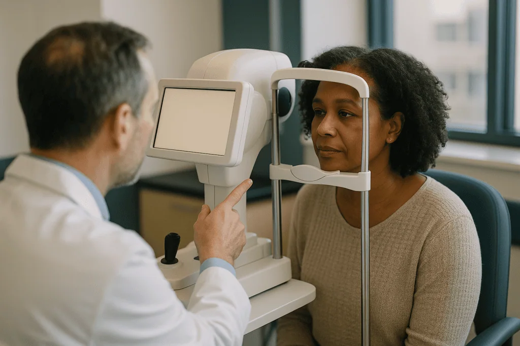
Each disease writes in its own hand. Chronically high glucose weakens the supporting cells that wrap capillary walls, the vessels bulge and leak, and in advanced disease the starved retina grows fragile new vessels that bleed easily. High blood pressure thickens and narrows arteriole walls instead. Under the camera, a hardened artery crossing a vein compresses it like a foot on a garden hose, a sign called arteriovenous nicking. The artery’s own reflection brightens and takes on a coppery or silvery look as the wall stiffens.
The brain connection is anatomical, not metaphorical. During development the retina grows outward from the same tissue that forms the brain, so it is, strictly speaking, central nervous system. The innermost retinal layer is made of the long fibers of ganglion cells, and those fibers bundle together to form the optic nerve. When nerve cells elsewhere in the brain shrink, researchers have asked whether this accessible slice of nerve tissue shrinks too, and OCT can measure its thickness to within a few micrometers.
None of this means an eye photo is a full-body check. It means the retina reports on two systems with unusual honesty: the small vessels and the nerve tissue they feed.
What changed recently
Three developments explain why the retinal scan has moved from eye clinics into the broader conversation.
The first is automation. In 2018 the United States Food and Drug Administration authorized the first software allowed to read retinal photographs for diabetic retinopathy without a specialist reviewing the image, which meant a camera in a primary care office could flag who needed an eye appointment. Similar systems have followed, and that is why a person with diabetes may now be offered a scan during a routine visit rather than being told to book one elsewhere.
The second is a shift in how often low-risk people are screened. The National Health Service in England, which runs one of the longest-standing population screening programs for diabetic eye disease, now invites people whose previous two screenings showed no retinopathy every two years instead of every year, a change rolled out from 2023. The change rests on data showing that progression to sight-threatening disease within two years is rare in that group, and it reflects a mature program adjusting intervals to risk rather than a retreat from screening.
The third is scale of need. The Centers for Disease Control and Prevention estimates that roughly one in four American adults with diabetes has some retinopathy, and diabetic retinopathy remains a leading cause of blindness among working-age adults. Those figures, drawn from 2021 data published in 2023, sit behind the push to make screening easier to reach.
Layered on top is a fast-growing research field sometimes called oculomics, which uses machine learning to look for signs of systemic disease in eye images. Those studies generate headlines, and a fair share of viral claims, but as the next sections explain they have not yet changed what any clinician should do with your photo.
Retinal scan for diabetes: what diabetic eye screening looks for
Diabetic retinopathy is the term for damage to the retina’s blood vessels caused by diabetes, and it is the reason the retinal scan exists as a mass screening tool. The disease moves through recognizable stages. In the early, non-proliferative stage, graders look for microaneurysms, small hemorrhages and exudates. As more capillaries close, cotton-wool spots and beaded veins appear. In the proliferative stage the retina sprouts new, abnormal vessels that can bleed into the eye or pull the retina loose. Separately, fluid can collect at the macula, a condition called diabetic macular edema, which blurs central vision and is best seen on OCT because swelling shows as thickness rather than color.
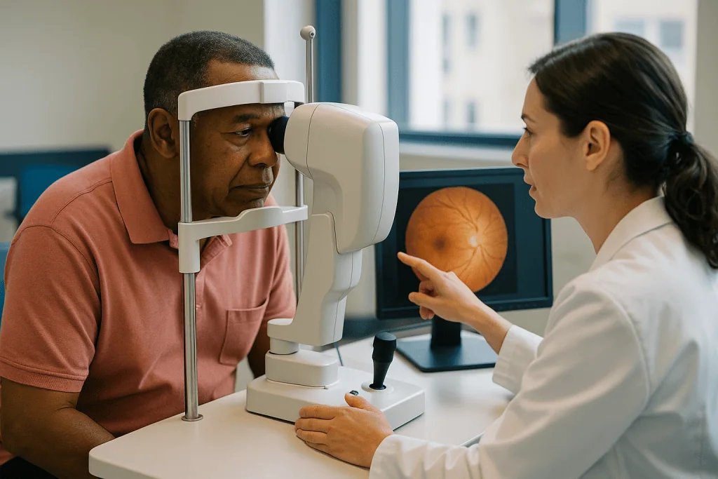
Screening works here because of a long silent window. Early retinopathy produces no symptoms; a person can have changes that merit treatment while reading fine print comfortably. Laser treatment and injected medicines that block a growth signal called VEGF can protect sight when started in time, and the National Eye Institute states that early detection and treatment can reduce the risk of blindness from diabetic retinopathy substantially. Waiting for blurred vision means waiting for damage that is harder to reverse.
Guidance from the NHS, the Mayo Clinic and the National Eye Institute agrees on the broad shape of a program: everyone with diabetes should have regular retinal examination, beginning at or soon after diagnosis for type 2 diabetes and within a few years of diagnosis for type 1, with closer monitoring during pregnancy for anyone with pre-existing diabetes. Exact timing is set by the clinician who knows your blood sugar history, blood pressure, kidney function and previous results.
One practical point: a retinal scan complements, and does not replace, the daily work of managing glucose and blood pressure. The photo tells you how the vessels are faring; the management decides what the next photo looks like.
Can an eye photo reveal high blood pressure?
It can reveal the footprints of high blood pressure, which is not quite the same thing. Hypertensive retinopathy is the name for retinal changes caused by elevated blood pressure, and clinicians have graded it since the 1930s. Mild changes include generalized narrowing of the arterioles and the arteriovenous nicking described earlier. Moderate changes add hemorrhages, cotton-wool spots and hard exudates, the same vocabulary as diabetes but in a different distribution. Severe changes, with swelling of the optic nerve head (papilledema), signal a hypertensive emergency in which pressure has risen high enough to threaten the brain and kidneys.
Here the evidence is observational but consistent and large. Population cohorts that photographed thousands of retinas and then followed participants for years found that narrower retinal arterioles predicted who went on to develop hypertension and were associated with higher risk of stroke, independent of the blood pressure reading taken at the start. That is why a clinician may examine the fundus when blood pressure is newly discovered to be very high, or when a severe headache accompanies it: the eye can show how long and how hard the vessels have been under strain.
Two caveats keep this honest. First, many people with well-documented hypertension have unremarkable retinas, so a clean photo never rules the condition out; a cuff on the arm remains the test. Second, machine-learning models that estimate systolic blood pressure from a fundus image have, in published work, missed the true value by an average of around eleven millimeters of mercury, which is far too imprecise to replace measurement. The photo adds context to a known diagnosis. It does not make one.
Eye scan for dementia: what the brain research actually shows
Because the inner retina is nerve tissue continuous with the brain, researchers have measured it in people with Alzheimer’s disease, Parkinson’s disease and multiple sclerosis. The most repeated finding is that the retinal nerve fiber layer, the band of ganglion-cell fibers headed for the optic nerve, is thinner on OCT in people with Alzheimer’s than in age-matched controls. A large British cohort published in 2018 extended that observation to people without dementia: those with the thinnest layers performed worse on cognitive tests and were more likely to show decline over the following years.
Beyond thickness, small studies have reported deposits of amyloid, the protein that accumulates in Alzheimer’s, in post-mortem retinas, and have attempted to image it in living eyes using special dyes. Others have used OCT angiography, a scan that maps blood flow without any injection, and found sparser capillary networks around the macula in people with early cognitive impairment. In multiple sclerosis the picture is clearer still, because inflammation of the optic nerve directly thins the fiber layer and OCT is routinely used in research to track it.
Now the hard part. Every one of these dementia findings comes from group comparisons. The thinning seen in Alzheimer’s overlaps heavily with thinning caused by glaucoma, ordinary aging, high myopia and vascular disease, so a single person’s measurement cannot be assigned to one cause. No OCT or photograph is validated to diagnose Alzheimer’s or to predict who will develop it, no professional guideline recommends scanning the eye for that purpose, and no clinician should offer a dementia verdict from one. The right description of this field is promising and unfinished. If a scan prompts concern about memory, the path forward is a conversation with a physician, not another scan.
What the evidence actually says
Grading evidence is the step most viral claims skip, so here it is laid out plainly, from strongest to weakest.
- Strong, with program-level and prospective trial data: retinal photography to screen for diabetic retinopathy. Decades of structured screening in England coincided with diabetic eye disease losing its place as the leading cause of certified blindness in working-age adults there. The first autonomous reading software was authorized after a prospective study of about 900 people with diabetes in primary care, where it detected more-than-mild retinopathy with roughly 87 percent sensitivity (the share of true cases it caught) and 90 percent specificity (the share of healthy eyes it correctly cleared).
- Moderate, from large observational cohorts: the link between retinal arteriolar narrowing and future hypertension and stroke, and the association between a thinner retinal nerve fiber layer and later cognitive decline. These studies followed thousands of people for years, which rules out many biases but cannot prove cause or show that acting on the finding changes outcomes.
- Preliminary, from retrospective datasets: deep-learning models that estimate age, sex, smoking status, blood pressure or cardiovascular risk from a fundus photo. The best-known 2018 study predicted age within about three years on average and identified smokers with modest accuracy, impressive as computer science, not yet tested as a way to improve anyone’s health.
- Expert opinion only: using a retinal scan to look for dementia or heart disease in a healthy person with no diabetes and no symptoms. No guideline body recommends it.
The pattern is consistent. Where the scan looks for the disease it was designed to find, in the people at known risk, it performs well and saves sight. Where it is asked to infer distant conditions from subtle patterns, the science is real but the clinical use is not yet there.
Retinal scan vs dilated eye exam: which do you need?
People often assume a camera has replaced the eye doctor peering in with a bright light. The two overlap, but each sees something the other misses.
| Test | What it captures | Dilating drops | Best at | Main limits |
|---|---|---|---|---|
| Fundus photograph | Color image of the central retina, optic disc and vessels | Sometimes | Screening for diabetic and hypertensive changes; keeping a permanent record to compare year to year | Standard cameras miss the far edges of the retina; cataract or small pupils can make images ungradable |
| OCT scan | Cross-sectional map of retinal layers and thickness | Usually not | Detecting macular swelling, measuring nerve fiber layer, following glaucoma and macular degeneration | Shows structure, not color or bleeding pattern; cannot see the peripheral retina |
| Dilated examination | Live, three-dimensional view by a clinician | Yes | Examining the periphery, the lens and the vitreous gel; judging subtle depth and movement | No permanent image unless photographed; blurred near vision and light sensitivity for several hours |
In diabetic eye screening the photograph has proven itself as the workhorse because it is fast, reproducible and can be graded remotely. OCT is increasingly added because macular swelling, a common cause of lost reading vision in diabetes, changes thickness before it changes appearance. The dilated examination remains the reference standard whenever a photograph is unclear, whenever symptoms suggest a problem at the retinal edge such as a tear, and in the comprehensive eye exams that adults are advised to have periodically regardless of diabetes.
The honest answer to the heading, then, is that for most people with diabetes the retinal scan is the right screening tool, and anyone with a flagged result, a poor image or new symptoms needs the full examination. Your eye care professional will say which applies.
How AI reads a retinal photograph, and where it falls short
The software behind an automated retinal scan is a deep-learning model: a program trained on tens of thousands of photographs that human graders had already labeled, until it learned to recognize the texture of microaneurysms and hemorrhages on its own. It does not understand diabetes. It recognizes patterns, and on the one task it was built for, telling referable retinopathy from non-referable, it does so about as well as trained human graders.
Its shortcomings are specific. A sizable share of images, in some settings one in five, come back as ungradable because of cataract, small pupils or a blink, and those people still need an in-person examination. Models trained mostly on one population can perform worse on retinas with different pigmentation or on cameras they have not seen, which is why regulators restrict each system to the camera and task it was tested with. The authorized systems detect diabetic retinopathy and nothing else; a clean automated result says nothing about glaucoma, macular degeneration or a retinal tear at the edge of the image.
The most important limit is conceptual. An automated result is a sorting decision, not a diagnosis. A positive screen means a clinician should look more closely, and a proportion of those referrals will turn out to be fine. A negative screen means no referable disease was seen today, not that the eye is healthy in every respect. The same logic applies to the experimental models in the news that estimate heart risk or brain age from a photograph: a probability score generated by a pattern-matcher is a research output until a trial shows that acting on it helps people, and no such trial has yet reported.
Used for what it was validated to do, the software widens the net. It does not replace the person who decides what to do with the catch.
Who should have a retinal scan, and how often
The clearest answer is for people living with diabetes, where every major body agrees screening should be routine. The NHS invites everyone with diabetes from age twelve, with annual photographs for most and two-yearly intervals for those whose previous two results were clear. In the United States, guidance summarized by the Mayo Clinic and the National Eye Institute advises an examination at diagnosis for type 2 diabetes, within a few years of diagnosis for type 1, then yearly or as the examiner recommends. Pregnancy in someone with pre-existing diabetes calls for more frequent checks because retinopathy can progress quickly during those months.
For high blood pressure the picture is different. There is no population screening program, and a fundus check is something a clinician may do when blood pressure is severely elevated, newly diagnosed at a high level, or accompanied by symptoms such as headache or visual change. If you have long-standing hypertension and no diabetes, the retina is typically examined as part of a general eye exam rather than on a fixed schedule.
For adults without either condition, the relevant advice is about comprehensive eye examinations, which include a look at the retina. The Mayo Clinic suggests an exam every five to ten years in the twenties and thirties, every two to four years from forty to fifty-four, every one to three years from fifty-five to sixty-four, and every one to two years after sixty-five, with closer follow-up for anyone with a family history of eye disease or a personal history of eye problems.
What none of these schedules include is a retinal scan to look for dementia or heart disease in an otherwise well person. If that is the reason you are considering one, the more useful appointment is with your primary care physician, who can assess those risks with tools that have been tested for the job.
What happens during a retinal scan
The whole thing takes less time than parking. You sit at a tabletop camera, rest your chin and forehead against padded supports, and fix your gaze on a small light inside the lens. The operator aligns the camera with your pupil, and a flash fires. Most people see a bright afterimage for a few seconds. Both eyes are photographed, usually from more than one angle, so the central retina and optic disc appear in full.
Whether you receive dilating drops depends on the camera and the program. Many modern cameras can image through an undilated pupil in a dimmed room, which is why pharmacy and primary care screening often needs no drops. Where drops are used, they widen the pupil over about fifteen to twenty minutes and leave near vision blurred and eyes light-sensitive for several hours. Sunglasses help, and arranging for someone else to drive is wise. The drops can sting briefly on instillation; that is expected and passes within a minute or so.
An OCT scan, when added, feels like a second photograph without the flash. You look at a target while the machine sweeps a faint light across the retina for a few seconds. Nothing touches the eye in either test.
Afterward, the images go to a grader, a clinician or an authorized software system. In structured screening programs you typically receive a letter or message within a few weeks stating whether retinopathy was seen, at what level, and whether an appointment with an eye specialist is needed. In an eye clinic the clinician often reviews the images with you on the screen during the same visit, which is worth asking for: seeing your own vessels makes an abstract risk concrete.
Understanding your results: grades, referrals and false alarms
Screening reports use grading systems that can look cryptic. Programs modeled on the English one report retinopathy as R0 (none), R1 (background changes such as a few microaneurysms), R2 (more advanced non-proliferative changes) and R3 (proliferative disease with new vessels), alongside a separate maculopathy code, M0 or M1, indicating whether changes threaten the macula. Clinics in the United States more often describe results in words: no retinopathy, mild, moderate or severe non-proliferative, proliferative, with or without macular edema. The underlying logic is the same, and the question the grade answers is whether an ophthalmologist needs to see you and how soon.
A background grade is common and, on its own, is not a treatment decision. It is information that your clinician folds into the bigger picture of glucose, blood pressure and cholesterol control, and it usually means a repeat photograph in a year. A referable grade means a specialist examination, where decisions about laser or injected medicines are made face to face and belong entirely to that specialist.
Expect some noise. Every screening test produces false positives, where a referral finds nothing serious, and false negatives, where early disease is missed on one photograph and caught on the next. Ungradable images are not a bad result; they are a reason for a dilated exam. Incidental findings happen too: a photo taken for diabetes may show signs suggesting glaucoma or macular degeneration, and those follow their own pathways.
One thing a result should never prompt is a change to your own medicines. A reassuring photograph does not mean blood pressure or glucose treatment can be relaxed, and a worrying one is not a signal to adjust anything on your own. Take the report to the clinician who prescribes for you and let it inform the next conversation.
Common myths about retinal scans
The viral claims deserve direct answers, one at a time.
“An eye photo can diagnose Alzheimer’s years early.” Research has found group differences in retinal thickness and blood flow between people with and without Alzheimer’s, and that is genuinely interesting. No test built on those differences has been validated for individuals, and no guideline recommends scanning the retina for dementia. A scan cannot tell you that you have, or will develop, Alzheimer’s.
“If my vision is fine, my eyes are fine.” Diabetic retinopathy is silent in its early and even moderate stages, which is the entire reason screening exists. Good vision today is not evidence of healthy vessels.
“A normal retinal scan means my blood pressure and heart are healthy.” Many people with hypertension have unremarkable retinas, and estimates of heart risk from photographs remain experimental. The cuff, the blood test and the conversation with your physician measure those things; the photo adds context.
“The camera flash damages the eye.” Fundus cameras use a brief flash within established safety limits, and millions of screening photographs are taken every year without harm. The afterimage fades within seconds.
“The AI result is the diagnosis.” Automated software sorts images into referable and non-referable. It flags people for examination; it does not examine them.
“A retinal scan replaces my regular eye exam.” A screening photograph looks for specific diseases in the central retina. It does not check your glasses prescription, eye pressure, the lens or the far periphery, and it cannot detect a retinal tear at the edge of the image. The comprehensive exam still has its place.
“Once the scan is clear, I can skip next year.” Intervals are set by risk and by program rules, not by a single good result. Follow the schedule your clinician or screening service gives you.
When to see a doctor
Routine screening is for people without symptoms. Certain changes should not wait for the next scheduled photograph and warrant same-day medical attention or an emergency department:
- Sudden loss of vision in one or both eyes, even if it recovers within minutes.
- A dark curtain or shadow spreading across part of your visual field.
- A sudden shower of new floaters, flashes of light, or both, which can signal a retinal tear or detachment.
- Blurred or double vision together with a severe headache, confusion, chest pain or weakness on one side of the body, which can indicate a hypertensive emergency or a stroke.
- Eye pain with redness, nausea and halos around lights.
Less urgent but still prompt reasons to book an appointment include straight lines that look wavy or bent, a persistent blurred or missing patch in central vision, difficulty reading that new glasses do not fix, and any screening letter that advises specialist follow-up. If you have diabetes and have not had retinal imaging in the past year, that alone is a reason to call.
Bring to the appointment whatever the scan produced: the report, the grade, the images if you have them, and a list of your current medicines. The decisions that follow a retinal finding, whether that is a change in how often you are screened, a referral for laser or injection treatment, or an adjustment to blood pressure or diabetes medicines, belong to the clinicians who prescribe for you and examine you. A retinal scan is a strong reason to have those conversations. It is never a substitute for them, and it should never be used to start, stop or change a treatment on your own.
Frequently asked questions
What does a retinal scan show that a normal eye test does not?
A retinal scan records the back of the eye in detail, showing the optic nerve, macula and blood vessels, whereas a standard vision test measures how well you read letters. The photograph or OCT can reveal microaneurysms, hemorrhages, exudates and swelling that cause no symptoms yet. It does not check your glasses prescription or eye pressure, so the two tests complement rather than replace each other.
How is diabetic eye screening different from a regular eye exam?
Diabetic eye screening is a targeted photograph of the central retina, graded for one disease using set criteria, and repeated on a fixed schedule. A comprehensive eye exam checks vision, eye pressure, the lens and the whole retina including its edges, usually with dilation. People with diabetes benefit from both: screening catches retinopathy early, and periodic full exams cover everything else.
Can a retinal scan detect high blood pressure I did not know about?
Sometimes it reveals changes that suggest long-standing or severe high blood pressure, such as narrowed arterioles, arteriovenous nicking, hemorrhages or swelling of the optic nerve. Many people with hypertension have normal retinas, though, so the scan cannot rule it out, and software estimates of blood pressure from photographs are too imprecise for clinical use. A cuff measurement remains the diagnostic test.
Is an eye scan for dementia available, and does it work?
No eye scan is approved or recommended for diagnosing or predicting dementia. Research using OCT has found that people with Alzheimer’s tend to have thinner inner retinal layers and sparser capillaries as a group, but those changes overlap with glaucoma, aging and other conditions. If you are worried about memory, a physician can assess it with validated tools; a retinal image cannot.
Retinal scan vs dilated eye exam: do I still need the drops?
Many modern cameras image through an undilated pupil, so screening often needs no drops. A dilated exam is still required when the photograph is unclear, when you have symptoms such as new floaters or flashes, and as part of a comprehensive eye exam, because it lets the clinician see the far edges of the retina, the lens and the vitreous gel directly.
How accurate is AI at reading a retinal scan?
For the task it was authorized to perform, detecting more-than-mild diabetic retinopathy, the first autonomous system caught about 87 percent of true cases and correctly cleared about 90 percent of unaffected eyes in a prospective primary care study. Accuracy drops if images are poor or the camera differs from the one it was validated on, and the result is a referral decision, not a diagnosis.
How often should someone with diabetes have retinal imaging?
Most guidance calls for an examination at diagnosis for type 2 diabetes, within a few years of diagnosis for type 1, and then yearly, with some programs extending to two years after consecutive clear results. Pregnancy with pre-existing diabetes usually means more frequent checks. The exact interval is set by your clinician or screening service based on your results and risk factors.
Does a retinal scan hurt or carry any risks?
The scan itself is painless; nothing touches the eye, and the flash leaves only a brief afterimage. If dilating drops are used they may sting for a moment and leave near vision blurred and eyes light-sensitive for several hours, so bring sunglasses and avoid driving until it wears off. Serious reactions to the drops are rare, and the camera flash is within established safety limits.
What does a background retinopathy result mean?
It means the grader saw early changes, typically a few microaneurysms or small hemorrhages, that do not currently threaten vision and do not need treatment. It is a prompt to keep blood sugar and blood pressure well managed and to attend the next scheduled photograph, usually in a year. Any decision about your medicines in light of the result belongs to your prescribing clinician.
Can a healthy person without diabetes benefit from a retinal scan?
A retinal view is part of the comprehensive eye exams adults are advised to have periodically, and those can pick up glaucoma, macular degeneration or incidental vascular changes. Using a scan specifically to screen for heart disease or dementia in a well person is not recommended by any guideline body, because the research models have not been tested in trials showing they improve health.
References
- NHS: Diabetic eye screening
- NIH National Eye Institute: Diabetic retinopathy
- MedlinePlus: Diabetic eye problems
This article is for general information only and is not a substitute for professional medical advice. Please consult a qualified doctor about your individual situation.

More from the Blog

The BRCA Gene Test: Who Should Consider It and What Results Change
The BRCA gene test is a blood or saliva test that looks for inherited changes in the BRCA1 and BRCA2 genes, which can substantially…
Prostate MRI: How It Changed Prostate Cancer Diagnosis
Prostate MRI, a detailed magnetic scan of the prostate gland, is now widely used before biopsy when a PSA test or examination raises concern.…
Ovarian Cancer Screening and CA-125: Why There Is Still No Routine Test, and Who Is Monitored
No routine ovarian cancer screening test is recommended for women at average risk. Two large randomized trials, PLCO and UKCTOCS, found that annual CA-125…
3D Mammograms and Breast Ultrasound: How Modern Breast Screening Layers Up
A 3D mammogram, or digital breast tomosynthesis, captures X-ray images in thin slices so radiologists can see through overlapping tissue. Studies suggest it detects…

APOE4: What Carrying the Alzheimer’s Risk Gene Means, and Does Not Mean
APOE4 is the most common gene variant linked to late-onset Alzheimer's disease. One copy roughly doubles to triples lifetime risk compared with the most…

Vitamin D Test: What Level Is Actually Deficient and Who Needs Testing
A vitamin D test measures 25-hydroxyvitamin D in blood. Most US laboratories and the NIH call a level below 12 ng/mL (30 nmol/L) deficient…