

Tympanoplasty Step by Step: How Surgeons Graft a Perforated Eardrum Under the Microscope

Key Takeaways
- A chronic eardrum perforation stays open because the outer skin grows around the rim and seals the edge, so the surgeon must scrape that margin raw before any graft can take.
- The graft is usually temporalis fascia from above the ear or tragal cartilage from the flap in front of the canal, both harvested within centimeters of the incision.
- In the common underlay technique the graft sits beneath the remaining drum, propped from below by absorbable sponge in the middle ear and held from above by packing in the canal.
- Hearing is normally worse for the first two to four weeks after surgery because packing and swelling block sound, and a fair hearing test waits until the ear is open and dry.
- The Wullstein type on your operation note describes how many middle ear bones were rebuilt; a type I repair grafts the drum alone and has the most predictable hearing outcome.
- The chorda tympani, the taste nerve to the front of the tongue, runs just behind the drum, which is why a one-sided metallic taste is a recognized and usually temporary effect.
Tympanoplasty repairs a perforated eardrum with a small graft of the patient's own tissue, most often thin fascia from above the ear or cartilage from the flap in front of the ear canal. Working through the canal or a cut behind the ear, the surgeon freshens the hole's edges, tucks the graft beneath the remaining drum, supports it with absorbable packing and lets the ear heal over the following weeks.
The audiologist slides the headphones off and says the words you half expected: the hole has not closed. Two years after a bad ear infection, a small gap in the eardrum is still letting water in, letting sound leak out, and turning every shower into a careful tilt of the head. The surgeon mentions a graft. You nod, and only in the parking lot do you realize you have no picture of what that means.
This is the tympanoplasty procedure step by step, written for exactly that moment. It follows the operation from the first hearing test to the day the packing dissolves, and it names each part in plain language: where the graft comes from, how it is tucked into place, why the ear feels blocked afterward, and what the evidence honestly says about hearing.
Some of it is reassuring. Some of it is simply practical. All of it is meant to make the conversation with your own surgeon shorter, calmer and more useful.
What is a tympanoplasty, and why does an eardrum need a graft?
The eardrum, or tympanic membrane, is a shallow cone of tissue roughly the size of a pencil eraser and about a tenth of a millimeter thick. It has three layers: an outer skin, a middle fibrous sheet that gives it tension, and an inner lining continuous with the middle ear. When a perforation is fresh, the skin layer usually grows across the gap by itself; the NHS and Mayo Clinic both describe most perforations healing without treatment within a few weeks to a couple of months.
Chronic holes behave differently. The outer skin can curl around the rim and grow onto the inner surface, sealing the edge so that neither side has anything to grow toward. The result is a stable, open window into the middle ear. Sound energy escapes through it instead of vibrating the membrane, water and bacteria find their way in, and the ear may discharge again and again.
Tympanoplasty is the operation that closes that window and, where needed, restores the chain of three tiny bones behind it. The word breaks down simply: tympano for the eardrum, plasty for reshaping. When only the membrane is repaired, many surgeons use the narrower term myringoplasty; when the middle ear bones are also inspected or rebuilt, tympanoplasty is the fuller name.
The graft is not a patch glued over the surface. It is a scaffold. The surgeon places a thin sheet of the patient’s own tissue where the fibrous layer should be, and over the following weeks the remaining eardrum skin migrates across it, blood vessels grow in, and the scaffold becomes living membrane. That biology is why dryness and gentle pressure matter so much in recovery, and why the surgeon spends more effort preparing the edges than placing the graft itself.
Who is tympanoplasty usually for, and who is asked to wait?
Surgeons generally consider the operation when a perforation has stayed open long enough that spontaneous closure is unlikely, and when it is causing something that matters to the person: recurrent discharge, a measurable conductive hearing loss, water intolerance, or the wish to swim and shower freely. Cleveland Clinic lists chronic infection, trauma and a hole that has not healed after months as common reasons. A perforation that lingers after an ear tube has fallen out belongs in the same group.

Not everyone with a hole is booked for surgery. Several situations usually lead to a pause rather than a date:
- An actively infected, wet ear. Most teams treat the discharge first, because a graft placed onto inflamed tissue has a harder time surviving.
- Young children. The Eustachian tube, the passage that equalizes middle-ear pressure, matures through childhood; many surgeons prefer to wait until a child is older and the opposite ear has been healthy for a season. The decision is highly individual.
- A recent cold or sinus infection, which can be a reason to postpone a scheduled operation by a few weeks.
- Medical conditions that make anesthesia riskier until they are better controlled.
Some people are offered a simpler first step. A small, dry, central perforation may be trialed with an office procedure in which the edges are freshened and a paper or gel patch is laid across the hole to guide healing. It is not a substitute for a graft when the hole is large, but it can avoid an operating room for the right person.
Equally, a hole that causes no symptoms in an adult who is happy to keep water out may simply be observed. Surgery treats a problem; it is not required just because a perforation exists. Your treating team weighs hearing tests, the state of the other ear and your own priorities before recommending anything.
Tympanoplasty procedure step by step: anesthesia and reaching the eardrum
Most tympanoplasties are done under general anesthesia, meaning you are fully asleep, though some adults with a hole that can be reached through the ear canal are offered local anesthesia with sedation. Johns Hopkins and Cleveland Clinic both describe the operation as commonly an outpatient procedure, with most people going home the same day. Once you are asleep, the team cleans the ear and the skin around it and injects a numbing solution containing a small amount of a blood-vessel-constricting agent, which keeps the field clear enough to see through a microscope.
The surgeon then chooses a route. There are three classic ones, and the choice depends on the size and position of the hole, the width of the ear canal, and the surgeon’s training.
- Transcanal: everything is done through the natural opening of the ear, with no external cut. This suits smaller or central holes and is the standard approach for endoscopic ear surgery, in which a slim camera replaces the microscope’s line of sight.
- Endaural: a short incision at the notch just above the ear canal opening widens the view without going behind the ear.
- Postauricular: a curved incision in the crease behind the ear folds the outer ear forward and gives the widest access. It is favored for large holes, holes at the front edge, and cases where the middle ear bones must be rebuilt.
Whichever route, the next step is the same. The surgeon makes a semicircular cut in the skin of the deep ear canal and lifts that skin, together with the rim of the eardrum, forward as a hinged flap. This is the tympanomeatal flap, and raising it is the moment the middle ear is properly exposed. Under magnification the surgeon can now see the hole from behind, check the three tiny bones, and confirm nothing else needs attention before the graft is brought in.
Tympanoplasty procedure step by step: harvesting the graft
Before the flap is raised, or sometimes just after, the surgeon takes the tissue that will become the new eardrum. The three tissues used most often all come from within a few centimeters of the ear, so no second surgical site is needed.
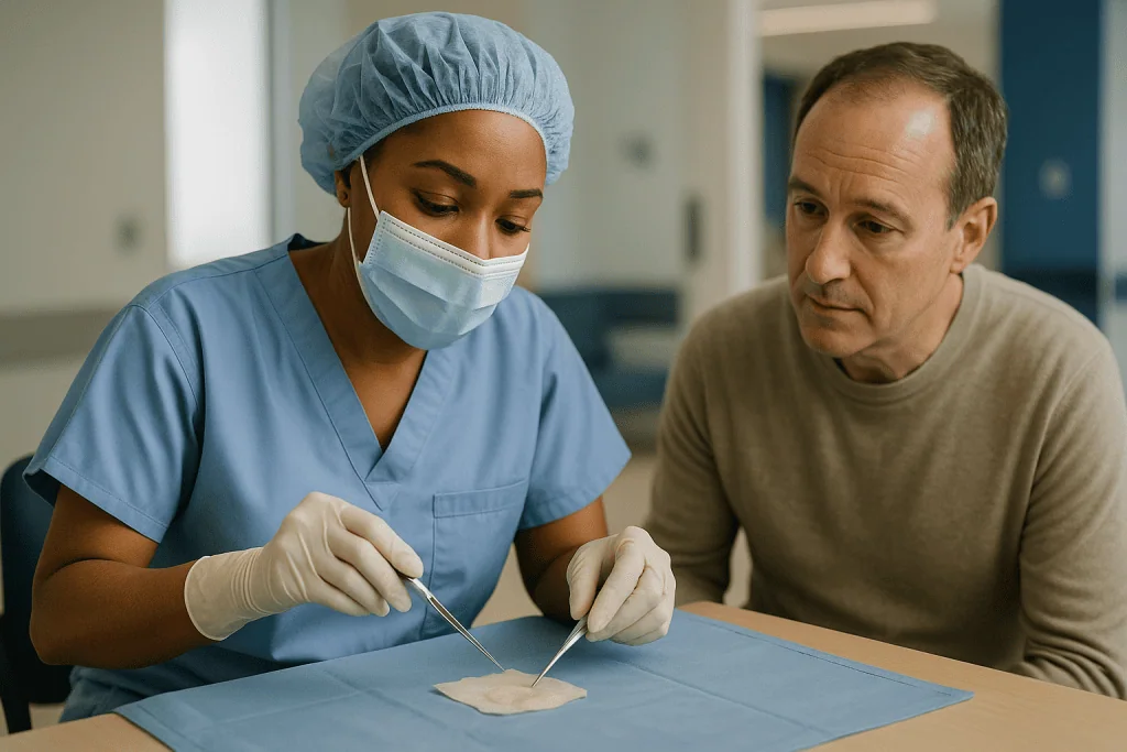
Temporalis fascia is the thin, pearly sheet of connective tissue covering the chewing muscle above and behind the ear. Through the postauricular incision, or a small cut in the hairline, the surgeon lifts a piece a little larger than the hole, scrapes off any muscle fibers and lets it dry until it becomes a firm, translucent film that handles like tissue paper. Tragal cartilage comes from the tragus, the small firm flap in front of the ear canal; a sliver is removed with its covering membrane, the perichondrium, through an incision hidden on the tragus’s inner side. A fat plug from the earlobe serves the smallest perforations.
| Graft tissue | Where it comes from | Character | Typically chosen when |
|---|---|---|---|
| Temporalis fascia | Above the ear, over the chewing muscle | Thin, pliable, close to the natural drum | Most standard perforations |
| Tragal cartilage with perichondrium | Front flap of the ear canal | Stiffer, resists retraction | Large holes, poor Eustachian tube function, revision surgery |
| Perichondrium alone | Same site as cartilage | Thin like fascia, small piece | Small to medium holes via the canal |
| Earlobe fat | Back of the earlobe | Soft plug rather than sheet | Pinhole perforations |
None of these is universally best. Cartilage vibrates slightly less freely but is far more resistant to being sucked inward when middle-ear pressure turns negative, so surgeons often reserve it for ears with a history of retraction or failed repair. Fascia remains the everyday choice for a straightforward hole. Synthetic and donor materials exist, but the patient’s own tissue is the mainstream standard described by Johns Hopkins and Cleveland Clinic.
Placing the graft under the microscope: underlay, overlay and packing
With the flap raised and the graft ready, the surgeon turns to the hole itself. The edges are the whole story here. A chronic perforation is lined with skin that has grown around its rim, and a graft cannot fuse with skin. So the surgeon uses a fine hook to scratch away a millimeter or so of the margin all the way around, creating a fresh, raw edge that will bleed slightly and, more importantly, will grow.
The graft is then positioned in one of two ways. In the underlay technique, the more common of the two, the graft slides beneath the remaining eardrum and rests against its inner surface, supported from below by small pieces of absorbable sponge packed into the middle ear so it cannot sag. In the overlay technique, the surgeon first peels the outer skin off the fibrous layer of the remnant, lays the graft on top of that fibrous layer, and then returns the skin over the graft. Overlay gives a larger contact area and suits very large or front-edge holes, but it demands more dissection and carries a small risk of trapping skin, which is why most surgeons default to underlay.
Once the graft is seated, the surgeon folds the tympanomeatal flap back down over it. The rim of the old drum and the canal skin now overlap the graft’s edges like a lid on a jar. Absorbable sponge, sometimes moistened with an antibiotic solution, or strips of dressing are packed into the outer ear canal to hold everything against the middle ear wall while healing starts. If a behind-the-ear incision was made, it is closed with stitches and a padded dressing is wrapped around the head.
From the outside, very little has changed. Inside, a scaffold now spans the gap, and the next several weeks decide whether the ear accepts it.
Types of tympanoplasty: what the type numbers actually mean
If your operation note says type I or type III, the surgeon is using a classification from the 1950s that describes how much of the middle ear’s sound-conducting chain had to be rebuilt. It is useful shorthand because hearing expectations differ across the types.
Sound normally travels from the eardrum to the malleus, incus and stapes, the three tiny bones that lever vibrations into the inner ear. When those bones are intact and mobile, repairing the drum alone restores the system. When infection or cholesteatoma, a growth of trapped skin, has eroded a bone, the surgeon must bridge the gap.
- Type I: the eardrum is grafted; all three bones are present and working. This is the simple perforation repair most people mean by tympanoplasty.
- Type II: the malleus is partly eroded; the graft is laid onto the remaining malleus or the incus.
- Type III: malleus and incus are missing or removed; the graft or a small prosthesis rests directly on the head of the stapes.
- Type IV: only the stapes footplate remains; the graft is placed so that sound reaches it directly while a small air pocket protects the round window.
- Type V: the footplate is fixed and a new window is created into the inner ear. This is rarely performed today.
Two practical points follow. First, the type is often confirmed during surgery rather than before, because the bones cannot always be fully assessed through a hole in a scarred drum; consent forms therefore sometimes list several possibilities. Second, when bones are rebuilt, the surgeon may use a shaped piece of your own cartilage or a small prosthesis, and hearing results become less predictable than in a type I repair. If your diagnosis includes cholesteatoma, the eardrum graft may be one part of a broader operation that also opens the mastoid bone behind the ear, and your surgeon will explain that separately.
Is tympanoplasty a big surgery?
People ask this to mean two different things: how serious is the risk, and how disruptive is the recovery. The answers diverge.
In terms of the body’s response, tympanoplasty is a small, contained operation. It involves no body cavity, minimal blood loss and, in the standard type I case, no bone drilling. Cleveland Clinic and Johns Hopkins describe it as usually outpatient, with operating time on the order of one to a few hours depending on the approach and whether the middle ear bones are addressed. Most people are eating and walking within hours of waking. Complications that would count as serious, such as injury to the facial nerve, are rare and are covered under risks below.
In terms of precision, it is demanding. The eardrum is thinner than a sheet of paper, the space behind it is only a few millimeters deep, and the facial nerve, the taste nerve to the tongue and the inner ear all sit within a centimeter of the working area. This is why the operation is done under a microscope or an endoscope, why it can take longer than the small incision suggests, and why surgeons train for years before doing it alone.
In terms of disruption, expect a middle path. The ear will feel plugged for weeks because of packing, there will be a period of restrictions on water, straining and travel, and hearing cannot be judged until the packing is gone. Based on the recovery ranges those hospital pages give, many adults return to desk work within about a week and to unrestricted activity over one to two months, though your surgeon’s advice for your ear takes precedence over any general figure.
So: minor in risk, major in precision, moderate in patience. Framing it that way tends to settle the question.
How painful is tympanoplasty recovery?
For most people, less than they feared. The ear canal and eardrum carry relatively few pain fibers compared with the skin of the fingertips, and patient information from Cleveland Clinic and Johns Hopkins describes discomfort after tympanoplasty as mild to moderate and manageable with the pain relief recommended by the surgical team. What it does feel like is unfamiliar.
The first sensation is fullness, as though the ear were full of water or you were descending in an airplane. That is the packing, and it lasts as long as the packing does. The second is a dull ache around the ear, most noticeable for the first two or three days and usually worst when a behind-the-ear incision was made. Chewing may tug on the incision or on the tragal cartilage site, so softer foods for a couple of days are common advice.
Several sensations alarm people but are usually expected:
- Clicking, popping or crackling as the sponge softens and shifts.
- Hearing your own pulse, because the packed ear reflects sound inward.
- A small amount of blood-tinged or yellowish fluid on the dressing in the first days.
- Numbness of the top of the ear after a postauricular incision, from small skin nerves that were cut; sensation typically returns over months.
- A metallic or altered taste on one side of the tongue, discussed under risks.
Pain that steadily increases after the third day, rather than easing, is not part of the normal pattern, and neither is pain accompanied by fever or foul-smelling discharge. Those belong in the section on when to call your doctor.
Any medicine you are given, whether a simple analgesic, ear drops or an antibiotic, has a purpose, a duration and a stopping point that only your prescribing clinician should set. If something is not controlling the ache, say so rather than adjusting it yourself.
What not to do after tympanoplasty
The graft’s enemies are water, pressure and infection. Nearly every instruction you receive protects against one of them. Specifics vary by surgeon, and their written sheet overrides anything here, but the reasoning is worth understanding so the rules feel less arbitrary.
Do not let water into the ear. A wet graft becomes a culture plate. Most teams ask for a cotton ball coated with petroleum jelly in the outer ear during showers and a ban on swimming or submerging the head until they have looked in the ear and cleared you. Cleveland Clinic advises keeping the ear dry until the surgeon says otherwise.
Do not blow your nose. Pressure travels from the back of the nose up the Eustachian tube and can lift the graft off its bed. Sniff gently and wipe. If you must sneeze, open your mouth so the pressure escapes forward.
Do not strain, lift heavy objects or bend with your head below your heart during the early weeks. Each raises pressure in the middle ear and the veins around it.
Do not fly, dive or travel to high altitude until your surgeon has cleared you. Cabin pressure changes act on the drum exactly as nose-blowing does. Ask for a specific clearance rather than assuming a number of weeks.
Do not put anything into the ear canal: no cotton swabs, no earbuds pressed deep, no attempts to remove packing that seems loose. Fragments of sponge appearing on the pillow are normal; pulling them is not.
Do not stop or start any medicine, including ear drops, on your own. If drops have been prescribed, the timing and technique the team showed you matter more than anything a general article can say.
Sleeping with the operated ear up and the head slightly raised for the first week is a small habit many surgeons recommend to reduce swelling and drainage. It costs nothing and is easy to forget.
Tympanoplasty recovery time: what the first days and weeks usually look like
Recovery follows the biology of the graft rather than the calendar of the incision. Skin heals in days; a graft becomes a living membrane over weeks. The ranges below reflect typical patterns described in patient information from Cleveland Clinic, Johns Hopkins and the NHS, not promises for any individual ear.
The first 24–48 hours. You go home with a head dressing or a cotton ball in the ear, a follow-up appointment and written instructions. Drowsiness from anesthesia fades within a day. Light spotting onto the dressing is common; the dressing usually comes off the next day.
Days 3–7. Aching eases. Many adults return to work that does not involve exertion. The ear remains blocked, and hearing on that side is worse than before surgery, which surprises people who were not warned. Stitches behind the ear, if any, are checked toward the end of this period.
Weeks 2–4. The packing begins to dissolve or is removed in clinic. Sounds may start to filter through, often with a distorted, echoing quality. Water precautions and the no-nose-blowing rule remain in force.
Weeks 4–8. The surgeon examines the drum, usually under a microscope in the office, and can see whether the graft has taken. Many restrictions are lifted at this point if healing looks secure, though swimming often waits longer.
Weeks 8–12 and beyond. A formal hearing test is repeated once the ear is dry and open. Cleveland Clinic’s patient information describes full recovery, including hearing settling, as commonly taking around two to three months.
Children follow a similar arc but need more help with water precautions and more reassurance about the blocked sensation. For any age, a slower timeline is not in itself a sign of failure; a persistently wet or painful ear is the signal to be checked sooner.
How long does it take for hearing to recover after tympanoplasty?
Later than most people expect, and for an honest reason. Immediately after surgery the ear canal is packed, the middle ear is filled with sponge, and a fresh graft is swollen with fluid. Every one of those blocks sound. Hearing after tympanoplasty is therefore usually worse for the first two to four weeks than it was before, and it cannot be assessed until the packing has gone and the graft has thinned. Cleveland Clinic notes that hearing may not reach its settled level for two to three months.
What the operation can and cannot do also needs stating plainly. A perforation causes a conductive hearing loss: sound leaks through the hole instead of moving the drum, and the bones behind it lose some of their lever. Closing the hole addresses that component. It does nothing for a sensorineural loss, the inner-ear or nerve type that shows on a hearing test as a drop in bone-conduction thresholds. Your pre-operative audiogram separates the two, and your surgeon should be able to point to the gap between the air and bone lines and say how much of it a healed graft might narrow.
Several factors make hearing outcomes less predictable:
- Erosion of the middle ear bones requiring a type II to IV reconstruction.
- A thick cartilage graft, which seals reliably but moves slightly less freely than fascia.
- Scar tissue or fluid in the middle ear that persists after healing.
- Poor Eustachian tube function, which can leave the new drum retracted.
None of these is a reason to expect a poor result, but they explain why surgeons speak of aims rather than guarantees, and why this article quotes no single success percentage. The relevant number for you is the one your treating team gives after seeing your ear, your audiogram and the state of your other ear. If hearing has not improved once the drum is healed, a repeat hearing test and, occasionally, a discussion of a hearing aid or further surgery are the usual next steps.
Risks of tympanoplasty and the alternatives your team may discuss
Every consent conversation covers the same core list. Understanding the anatomy behind each risk makes it less abstract.
- Graft failure or re-perforation. The graft may not take, or may take and later break down after an infection. This is the most common way the operation disappoints. A second repair is usually possible.
- Infection. A wet, painful ear with foul discharge in the weeks after surgery needs prompt review; treatment is decided by the team.
- Taste disturbance. The chorda tympani, the nerve carrying taste from the front of the tongue, crosses the middle ear just behind the drum and is sometimes stretched or cut. A metallic or salty taste on one side is common early and usually fades over months.
- Tinnitus or dizziness. Ringing and brief unsteadiness are usual in the first days; persistent new symptoms should be reported.
- Hearing worse than before. Uncommon, and a permanent inner-ear loss is rare, but it is a real possibility whenever the middle ear is entered.
- Facial weakness. The facial nerve runs through the middle ear in a bony canal. Injury is rare in standard drum repair and is monitored during complex cases.
- Numbness or a subtle change in ear shape after a behind-the-ear incision, and trapped skin forming a cholesteatoma years later, both uncommon.
Alternatives exist, and a good team names them. Watchful waiting with strict water precautions is reasonable for a dry, symptom-free hole in an adult. An office patch procedure can be tried for small central perforations. A hearing aid addresses the hearing component without surgery, though it does nothing for water intolerance or discharge. For a child, waiting for the Eustachian tube to mature is itself an option. The MedlinePlus and Mayo Clinic pages on ruptured eardrum both frame surgery as the step taken when healing has not happened and the hole is causing problems, not as the automatic response to a perforation.
What people often get wrong about eardrum repair
Myths gather around small operations as readily as large ones. These are the ones ENT clinics hear most.
The graft is glued or stitched on. It is neither. It is placed against freshened tissue and held by packing while your own cells grow across it. That is why pressure and moisture rules matter, and why nothing looks attached when you glance in a mirror.
A hole that has been there for years cannot be fixed. The age of the perforation is not the deciding factor; the state of the middle ear and Eustachian tube is. Long-standing dry holes are repaired routinely.
Hearing should be better as soon as you wake up. The opposite is true for the first weeks. Judging the operation before the packing has cleared is like judging a painting before the varnish dries.
Ear drops will heal a chronic perforation. Drops can treat infection and dry a discharging ear, which may be essential before surgery, but they do not close a hole whose edges are covered in skin. The NHS notes that antibiotics may be advised when infection is present or likely; they are not a repair.
Flying will burst the new eardrum. A healed graft tolerates normal cabin pressure changes like any eardrum. The vulnerable window is the early weeks before it has attached, which is why clearance to fly is individual rather than a fixed rule.
Cartilage grafts always mean worse hearing. Cartilage is stiffer, but when cut thin its effect on sound transmission is small, and its resistance to retraction is why surgeons choose it for high-risk ears. The trade-off is a judgment call, not a downgrade.
Endoscopic means better. Endoscopic and microscopic techniques are different tools for reaching the same place. Which one your surgeon uses reflects the hole’s position and their training, and neither should be presented to you as a guarantee.
Questions to ask your care team
A consultation goes better when you arrive with questions that produce specific answers. These are the ones that tend to change decisions or set expectations.
- Which type of tympanoplasty are you planning, and how likely is it that you will need to reconstruct the middle ear bones once you can see inside?
- Where will the graft come from, and why that tissue for my ear?
- Will you go through the ear canal or behind the ear, and what does that mean for the incision and for numbness afterward?
- On my hearing test, how much of the loss is conductive and how much is inner ear, and what could hearing realistically be after healing?
- Is my ear dry enough to operate on now, or do you want to treat discharge first?
- What happens if I choose not to have surgery, or if I wait a year?
- Which packing will you use, and will it dissolve or need removing in clinic?
- Exactly what water, nose-blowing and lifting rules apply, and until which appointment?
- When will you clear me to fly?
- What signs in the first two weeks would you want a phone call about the same day?
- If the graft does not take, what would the next step be?
- Who do I contact after hours, and how?
Bring your most recent audiogram if you have a copy, a list of every medicine and supplement you take, and a note of any previous ear operations or tube insertions. If the patient is a child, ask how the team explains the procedure to children of that age and what you can do at home to make water precautions easier to keep. Write the answers down during the visit; the blocked-ear weeks are a poor time to rely on memory.
When to call your doctor
Most recovery is quiet, and most worries are settled by a phone call to the clinic. A few signs, though, should prompt same-day contact with the surgical team or, when they occur suddenly and severely, emergency care.
- Bleeding that soaks through the dressing or continues beyond the first day, rather than light spotting.
- Foul-smelling, thick or increasing discharge from the ear, especially with fever.
- Pain that grows steadily after the first few days instead of easing, or pain that returns after it had settled.
- New weakness or drooping on one side of the face, including difficulty closing the eye or a crooked smile.
- Severe spinning dizziness, vomiting or an inability to walk steadily.
- A sudden drop in hearing on either side, or a loud new ringing that does not fade.
- Stiff neck, severe headache, confusion or a high fever, which are rare but can signal infection spreading beyond the ear.
- Any allergic-type reaction to a prescribed medicine, such as rash, swelling of the lips or face, or trouble breathing; the last of these is an emergency.
Some milder concerns still merit a call within a day or two: water accidentally getting into the ear, a hard sneeze or nose-blow followed by a popping sensation and a change in hearing, packing that seems to have fallen out early, or an incision that looks red and spreading. None of these necessarily means the graft has failed, but the team may want to look sooner than planned.
Keep the after-hours number from your discharge paperwork somewhere visible. Describing the symptom, when it started and what you have already been prescribed lets the clinician decide quickly whether you need to be seen. That decision, like every other in this article, belongs to the people who have examined your ear.
Frequently asked questions
Is tympanoplasty a big surgery?
It is a small operation in terms of risk and a demanding one in terms of precision. Cleveland Clinic and Johns Hopkins describe it as usually outpatient, with no body cavity opened and minimal blood loss, but the surgeon works under a microscope within a centimeter of the facial nerve and inner ear. Recovery is more about patience with packing and restrictions than about pain or physical limitation.
How painful is tympanoplasty recovery?
Discomfort is usually mild to moderate and eases over the first few days, according to hospital patient information. Most people describe fullness and pressure rather than sharp pain, plus a dull ache around any incision behind the ear. Clicking, hearing your pulse and slight numbness of the ear are expected. Pain that keeps increasing after the third day, or pain with fever, should be reported to the surgical team.
What should you not do after tympanoplasty?
Keep water out of the ear, do not blow your nose, sneeze with your mouth open, avoid heavy lifting and straining, do not fly or dive until cleared, and put nothing into the ear canal. Each rule protects the graft from moisture or pressure while it attaches. Do not stop, start or adjust any prescribed medicine, including ear drops, without your clinician’s instruction.
How long does it take for hearing to recover after tympanoplasty?
Hearing is typically worse for the first two to four weeks because packing and swelling block sound, then improves as the packing dissolves. Cleveland Clinic notes it may take two to three months for hearing to settle. The operation addresses the conductive part of the loss caused by the hole; any inner-ear component identified on your pre-operative hearing test will not change.
What is the tympanoplasty recovery time before I can swim?
Swimming usually waits until the surgeon has examined the healed drum in clinic and confirmed the graft has taken, which is often several weeks to a few months after surgery. There is no fixed date that applies to everyone; the clearance depends on what the surgeon sees under the microscope. Showering with a protected ear is usually allowed much earlier, following the team’s instructions.
What are the types of tympanoplasty?
The classification runs from type I to type V and describes how much of the middle ear’s bone chain was rebuilt. Type I grafts the eardrum alone with intact bones. Types II and III bridge missing malleus or incus onto the remaining bones or stapes. Type IV places the graft against the stapes footplate, and type V, rarely used today, creates a new window into the inner ear.
Will I be awake during tympanoplasty?
Most people are fully asleep under general anesthesia, particularly children and anyone having a behind-the-ear approach or bone reconstruction. Some adults with a hole reachable through the ear canal may be offered local anesthesia with sedation, in which the ear is numbed and you are relaxed but not unconscious. The anesthesia team decides with you based on your health and the planned approach.
Where does the graft for a tympanoplasty come from?
Almost always from your own body, near the ear. Temporalis fascia is a thin connective tissue sheet over the chewing muscle above the ear; tragal cartilage and its covering, perichondrium, come from the small flap in front of the canal; earlobe fat serves pinhole perforations. Using your own tissue avoids rejection, and no second surgical site is needed.
Can a tympanoplasty fail, and what happens then?
Yes. The graft may not take, or may take and later break down after an infection. This is the most common way the operation falls short. If a hole persists once the ear has healed, the surgeon reassesses the middle ear and Eustachian tube function and may propose a repeat repair, often with cartilage, or continued water precautions. That decision rests with your treating team.
Why is my hearing worse right after tympanoplasty?
Because the ear is deliberately blocked. Absorbable sponge fills the middle ear to support the graft, more packing fills the canal, and the graft itself is swollen with fluid. All three stop sound reaching the inner ear. As the packing dissolves over the following weeks, sound returns, often with a hollow or echoing quality at first, before hearing is formally retested once the ear is dry.
References
This article is for general information only and is not a substitute for professional medical advice. Please consult a qualified doctor about your individual situation.

More from the Blog

After Professional Earwax Removal: Temporary Sensitivity and Symptoms Worth a Call
After professional earwax removal, most people notice a few brief effects: the ear may feel oddly open or ticklish, ordinary sounds can seem loud,…

Preparing Your Child for Adenoid Surgery: Fasting, Comfort Items and What to Say Beforehand
Preparing a child for adenoidectomy usually means three things: following the written fasting instructions exactly (solid food stops earliest, clear fluids last), packing familiar…

After a Repositioning Maneuver for Vertigo: Head Movements to Avoid in the Days That Follow
After an Epley maneuver, most current guidance no longer requires strict head restrictions, and the major ENT guideline says postural limits do not clearly…

Who Needs SMR Rather Than a Simpler Septum Repair? What the ENT Evaluation Shows
Submucous resection, or SMR, is usually reserved for a septum that a conservative septoplasty cannot straighten: thick bony spurs, deviations involving both cartilage and…

Who May Be a Candidate for Implantable Hearing Aids When Regular Hearing Aids Fall Short?
Implantable hearing aids are usually considered for people whose hearing loss is moderate to profound and who still struggle to understand speech with well-fitted…

Preparing for a Tonsillectomy: Bleeding-Risk Checks, Medicines to Pause and Fasting Rules
Preparing for a tonsillectomy usually means three things: telling the surgical team about any personal or family history of easy bleeding, following their instructions…