

Video-EEG, MRI and Neuropsychological Testing: How the Epilepsy Surgery Target Is Defined
Key Takeaways
- The epilepsy surgery evaluation looks for concordance: a target is defined only when video-EEG, MRI and neuropsychological findings point to the same region.
- Drug-resistant epilepsy is defined by the International League Against Epilepsy as failure of two appropriately chosen and tolerated antiseizure medicine schedules, which is when a surgical evaluation becomes relevant.
- Video-EEG monitoring may last from several hours to several days because it must capture typical seizures, and any supervised medicine reduction to provoke them happens only inside the hospital under the team's plan.
- A routine EEG can be normal in someone who has epilepsy, and an MRI can be normal in someone with a surgically treatable focus, which is why neither test is used alone.
- Neuropsychological testing is not pass or fail; it lateralizes dysfunction, estimates risk to memory and language, and provides a baseline for honest comparison after surgery.
- A seizure lasting longer than 5 minutes, or seizures without recovery in between, is an emergency that warrants an immediate call for emergency help.
Before epilepsy surgery, specialists combine several tests because no single one can both locate where seizures begin and show what nearby brain tissue does. Video-EEG monitoring records seizures as they happen to pinpoint their electrical origin, a high-resolution epilepsy-protocol MRI looks for a structural cause, and neuropsychological testing maps memory and language. When these lines of evidence point to the same spot, a surgical target can be defined; when they disagree, more testing follows.
The suitcase is packed with a week of loose-necked shirts, a phone charger with an extra-long cable, and a notebook with two columns: seizures the family has seen, and seizures the family suspects. Nobody packs like this for a holiday. People pack like this for video EEG monitoring before epilepsy surgery, a hospital stay in which the goal is, strangely, to have the very seizures everyone has spent years trying to prevent.
That reversal unsettles almost everyone. For a decade the message was control them, record them, avoid the triggers. Now a neurologist is saying: come in, let us watch, and if the seizures are shy we may need to coax them out.
What follows is an honest walk through how an epilepsy team turns a cloud of symptoms into a precise map of the brain, test by test, and why the recording week is only one piece of a much larger puzzle.
Why can't an MRI alone show where seizures start?
An MRI is a photograph of structure. A seizure is an event in time. Those are different things, and the gap between them is the whole reason epilepsy surgery evaluation exists.
Doctors use a handful of terms for the territory they are hunting. The epileptogenic zone is the patch of brain that, if removed or disconnected, would stop the seizures. The lesion is whatever abnormality the scan shows, such as a scar or a malformation. The irritative zone is where abnormal spikes appear between seizures. These areas often overlap, but not always. A scar can sit quietly for life while seizures arise two centimeters away. Conversely, a scan can look flawless while electrical recordings show a focus firing from the same temporal lobe every time.
So each test answers a different question. Video-EEG asks: where does the electrical storm begin, and in what order does it spread? MRI asks: is there tissue that looks abnormal? Neuropsychological testing asks: what does the brain around the suspected target currently do for you, and how much of that function sits on one side? Only when the answers converge does a team feel confident naming a target.
The scale of the need is real. The World Health Organization estimates around 50 million people live with epilepsy worldwide and that up to 70 percent could live seizure-free with proper diagnosis and medicines. The arithmetic leaves a substantial minority whose seizures continue despite treatment, and that is the group for whom this detailed, multi-test mapping becomes relevant. The rest of this article explains how each layer of evidence is gathered, and how the team weighs them against one another before anyone discusses an operation.
Who is usually offered an epilepsy surgery evaluation, and who is asked to wait?
The doorway into evaluation is a definition rather than a feeling. The International League Against Epilepsy, in a consensus proposal published in Epilepsia, defined drug-resistant epilepsy as the failure of two appropriately chosen, appropriately used and tolerated antiseizure medicine schedules to produce sustained freedom from seizures. The word “appropriately” matters: a medicine stopped early because of a rash, or one chosen for the wrong seizure type, does not count against the person.
Candidates for a surgical workup typically share a few features. Their seizures are focal, meaning they begin in one region rather than across the whole brain at once. The seizures interfere with daily life, driving, work, school or safety. And their diagnosis has been confirmed as epilepsy rather than another cause of episodes.
Who is usually asked to wait, or redirected? Several groups:
- People whose events have not yet been captured on EEG, because some episodes that resemble seizures are not epileptic and surgery would not help.
- People with generalized epilepsy syndromes, where seizures start everywhere simultaneously and there is no single focus to target.
- People who have not yet had a fair trial of two suitable medicines.
- People with unstable medical or psychiatric conditions that need attention first, so that testing and recovery are safe.
None of these is a verdict. A person asked to wait may return after a medicine trial is completed or an event is finally recorded. Mayo Clinic describes surgery as an option when medicines have not controlled seizures and the source can be identified; both halves of that sentence have to be true. The judgment of whether they are true belongs to the treating epilepsy team, who see the full record and can weigh factors no article can.
What is video EEG monitoring before epilepsy surgery, and what actually happens?
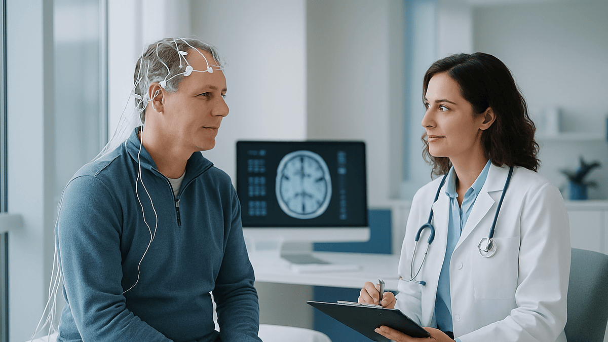
An EEG (electroencephalogram) is a recording of the brain’s electrical activity through small metal discs glued to the scalp. Video-EEG pairs that recording with continuous camera footage so that every movement, pause or word can be lined up with the brain waves at the same instant.
The stay begins with a technologist measuring the head and placing electrodes, usually two dozen or more, with a conductive paste and a gauze cap. The wires gather into a bundle that plugs into a bedside unit, which limits movement to the room and sometimes a short tethered walk. A camera, often with infrared for the dark, watches the bed. A push button lets the person or a companion mark the moment they sense something starting.
Two kinds of data accumulate. Interictal recordings, taken between seizures, show whether sharp waves or spikes cluster over one region. Ictal recordings, taken during a seizure, show where the rhythm first changes and how it spreads. MedlinePlus notes that a routine EEG can be normal in people with epilepsy, which is exactly why teams want the longer, seizure-capturing version before surgery rather than a single outpatient tracing.
The video half carries its own weight. The order of events, such as a hand posturing before the head turns, or speech continuing while one arm jerks, can indicate which hemisphere and which lobe is involved before anyone reads a tracing. Reviewers scrub through hours of footage frame by frame alongside the waveforms.
Family members are often asked to stay. They recognize the subtle early signs, press the event button, and confirm whether what the camera caught resembles the seizures at home. That confirmation is not a formality; a monitoring stay that records three events the family does not recognize has answered a different question than the one being asked.
How long does video EEG monitoring take?
Longer than people expect, and for a reason that has nothing to do with scheduling. The recording needs to capture a representative sample of the person’s typical seizures, and seizures do not arrive on request.
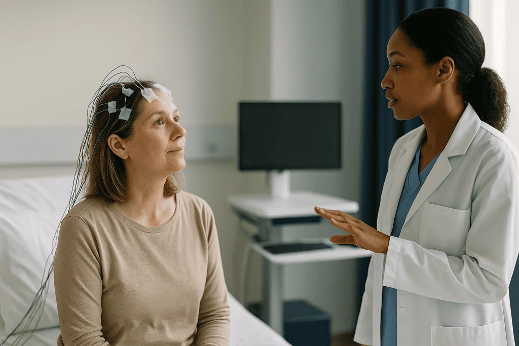
Mayo Clinic describes video-EEG for epilepsy evaluation as lasting from several hours to several days in the hospital. The spread reflects how often seizures happen. Someone with daily episodes may give the team what it needs quickly; someone with monthly episodes may need more time or a change of approach. The team is usually looking for more than one event, because a single seizure can start in an unusual place or be obscured by movement artifact, the electrical noise created when muscles contract or wires shift.
Several factors stretch or shorten a stay:
- How frequent the seizures normally are, and whether they cluster at particular times of day or around sleep.
- Whether the team chooses to lower medicines under supervision to make seizures more likely (see the next section).
- How clearly the recorded events point to one region. Concordant, clean recordings end a stay sooner than ambiguous ones.
- Whether both wake and sleep need to be sampled, since some seizure types appear mainly in sleep.
It helps to treat the stay as open-ended within a range rather than a fixed appointment. Bring entertainment, work that does not require a laptop balanced on the chest, and comfortable clothes that button at the front so the wire bundle does not need to come over the head. Showers are usually not possible while the electrodes are on, and the paste is stubborn, so plan for a thorough hair wash afterward.
Ask your team the specific question: how many typical events do you want to record, and what will you do if they do not come? A clear answer sets expectations and reduces the frustration of a quiet first few days.
What happens to your medicines during the monitoring stay?
This is the part that surprises families most, so it deserves plain language. In a monitored hospital setting, the epilepsy team may deliberately reduce antiseizure medicines to increase the chance that seizures occur while the cameras and electrodes are recording. The decision, the pace and the safety plan belong entirely to the team; it is done with nursing staff nearby, intravenous access often in place, and rescue plans ready.
Why would anyone do this on purpose? Because a stay that records no seizures is a stay that has not answered the surgical question. Medicines that work well enough to suppress seizures for a week in hospital, while still failing to control them at home, can make the recording uninformative. Supervised reduction trades a short period of higher seizure risk, inside a safety net, for the information needed to decide about a permanent intervention.
Other provocation methods are gentler and are standard parts of EEG practice. MedlinePlus lists sleep deprivation, flashing lights (photic stimulation) and deep, rapid breathing (hyperventilation) as techniques used to bring out abnormal activity during recordings. Staff may ask a person to stay up late, or to breathe fast for a few minutes while the tracing runs.
Two cautions follow directly from this. First, nobody should reduce or skip medicines before admission in the hope of speeding things up. Unsupervised withdrawal raises the risk of prolonged or clustered seizures without any of the hospital’s safeguards, and the team cannot interpret recordings against an unknown medicine background. Second, the schedule for returning to the usual regimen after monitoring is set by the prescribing clinician and is not a detail to improvise at home.
If a medicine change is planned during the stay, ask what warning signs the nurses will watch for, and what the plan is if a seizure runs long. Knowing the plan exists is a large part of feeling safe inside it.
What does an epilepsy-protocol MRI look for?
Not every MRI is the same MRI. An epilepsy-protocol MRI is a scan tuned specifically to find the small, subtle abnormalities that cause focal seizures, with thin slices through the temporal lobes and particular sequences that highlight scar tissue and malformed cortex. A scan done years ago in an emergency department to rule out a bleed may simply not have been looking in the right way.
The findings radiologists hunt for include:
- Hippocampal sclerosis, scarring and shrinkage of the hippocampus, the seahorse-shaped memory structure in the inner temporal lobe. It is one of the most common findings in adults evaluated for temporal lobe surgery.
- Focal cortical dysplasia, a patch of brain where the layering of neurons formed abnormally before birth. These can be tiny and easy to miss.
- Cavernous malformations, clusters of leaky blood vessels that leave iron deposits in surrounding tissue.
- Low-grade tumors and old injuries from infection, trauma or stroke.
A key sequence is FLAIR (fluid-attenuated inversion recovery), which darkens normal fluid so that abnormal brightness in tissue stands out. Volumetric sequences allow the two hippocampi to be compared side by side, since a difference in size can be as telling as a difference in signal.
Two practical points matter for patients. First, experienced epilepsy neuroradiologists often re-read outside scans, and a “normal” report can change when someone knows which lobe the EEG has implicated. Second, a scan that remains normal after expert review, often called MRI-negative, does not end the conversation. It makes the other tests more important and raises the likelihood that intracranial recording will be discussed, because the team now has to define the target by function rather than by a visible landmark.
The MRI is powerful, but it is one witness. The team wants its testimony to match what the EEG saw.
Neuropsychological testing for epilepsy surgery: what it measures and why it matters
Neuropsychological testing is a structured set of tasks, done with a specialist psychologist over several hours, that measures memory, language, attention, problem solving and processing speed. There are no needles and no scanner. There is a quiet room, a table, word lists to remember, pictures to name, patterns to copy, and a great deal of patient encouragement.
People sometimes assume it is a test of whether they are “smart enough” for surgery. It is not. It serves three distinct purposes in the surgical evaluation.
First, lateralization: the pattern of strengths and weaknesses can point to which hemisphere is struggling. In most right-handed people, the left temporal lobe handles verbal memory and the right handles visual-spatial memory. A person whose word-list recall is far weaker than their recall of shapes gives the team a clue that fits, or fails to fit, with the EEG and MRI.
Second, risk assessment. If the proposed target sits in the hemisphere that holds language or dominant memory, the team needs to know how much function that tissue is currently carrying. A hippocampus that is scarred and already performing poorly poses a different risk than one that is working hard. Mayo Clinic lists memory and language problems among the possible risks of epilepsy surgery, and this testing is how the team estimates that risk for a particular person rather than quoting a general average.
Third, a baseline. The same battery is usually repeated after surgery, so that any change can be measured honestly instead of guessed.
Two companion tests sometimes follow. The Wada test briefly puts one hemisphere to sleep with a short-acting anesthetic delivered through a catheter in the groin, so the awake hemisphere can be tested alone for language and memory. Functional MRI maps language areas noninvasively by watching blood flow while a person performs tasks in the scanner. Which, if either, is used depends on the center and the question being asked.
PET, SPECT, MEG and functional MRI: how the extra scans fill the gaps
When the three core tests agree, the team may stop there. When they half-agree, or when the MRI is normal, additional imaging adds independent votes. Each measures something different, which is the point: a consistent answer from unrelated methods is more persuasive than a loud answer from one.
| Test | What it measures | What it adds to the puzzle | What it cannot do |
|---|---|---|---|
| Video-EEG (scalp) | Electrical activity over time, with video | Shows where seizures begin and spread; confirms events are epileptic | Blind to deep or small sources; needs seizures to occur |
| Epilepsy-protocol MRI | Brain structure in fine detail | Finds scars, malformations, vascular lesions | Cannot say whether a lesion causes the seizures |
| Neuropsychological testing | Memory, language, attention | Lateralizes dysfunction; estimates risk; sets a baseline | Does not locate a focus by itself |
| PET (interictal) | Glucose metabolism between seizures | Underactive regions often match the focus | Low-metabolism area is often larger than the true target |
| Ictal SPECT | Blood flow injected at seizure onset | Highlights the region most active during the seizure | Timing is critical; late injection misleads |
| MEG | Magnetic fields from neural currents | Localizes spikes with good spatial precision | Usually captures interictal spikes, not seizures |
| Functional MRI | Blood-flow changes during tasks | Maps language and motor areas to protect | Does not show where seizures start |
A few definitions. PET (positron emission tomography) uses a small amount of radioactive sugar to show which regions are using less energy between seizures; an underperforming lobe is often the culprit. SPECT (single-photon emission computed tomography) uses a tracer injected during a seizure, sometimes by a nurse waiting at the bedside during video-EEG, to capture where blood flow surges at onset. MEG (magnetoencephalography) records the faint magnetic fields produced by neurons, which pass through the skull less distorted than electrical signals.
None of these is ordered routinely. Each is chosen to answer a specific unresolved question, and the team will usually explain what they hope it will settle.
When scalp recordings aren't enough: intracranial EEG explained
Scalp electrodes sit on the outside of a skull that blurs and weakens the signals passing through it. For many people that is enough. For others, particularly those with normal MRI scans, deep-seated suspected sources, or recordings that implicate two neighboring regions, the team may propose recording from inside the head.
Intracranial EEG means placing electrodes directly on or into the brain through a neurosurgical procedure, then returning to the monitoring unit for a second recording period. Two main approaches exist. Stereo-EEG uses thin depth electrodes inserted through small holes in the skull along planned trajectories, sampling deep structures as well as the surface. Subdural grids and strips are flat sheets of electrodes placed on the brain surface after a larger opening, giving dense coverage of one area.
Why accept a surgical procedure just to record? Because the resolution changes completely. Scalp EEG might say “left temporal region.” Intracranial EEG can say which gyrus, and in what order the storm travels. It also allows stimulation mapping, in which tiny currents are passed through electrode contacts to see what each spot does, such as moving a finger or interrupting speech. That map is how surgeons learn which tissue must be left untouched.
It carries real risks, described neutrally: bleeding, infection, swelling, and the general risks of anesthesia. The electrodes are removed at the end, either as a separate step or at the same operation as the definitive surgery if the findings support one. Mayo Clinic describes intracranial monitoring as a step used when noninvasive tests cannot pinpoint the source.
The honest framing is this: intracranial EEG is not a sign that things are going badly. It is a sign that the team is refusing to guess. A target defined by direct recording and stimulation mapping is held to a higher standard than one inferred from the surface, and the extra caution is on the patient’s behalf.
How the team puts it all together: the surgical conference
At some point, usually weeks after monitoring, every piece of evidence is laid out in a single room. Epileptologists, neurosurgeons, neuroradiologists, neuropsychologists, nurses and often social workers review the video clips, tracings, scans and test scores together. This multidisciplinary conference is where a target is either defined or declared not yet definable.
The central word is concordance. If the seizures on video-EEG begin over the right temporal region, the MRI shows right hippocampal sclerosis, PET shows right temporal underactivity, and neuropsychological testing shows relatively weaker visual-spatial memory, the evidence is concordant. A surgical target can be named with reasonable confidence, and the discussion turns to what operation fits and what functions sit nearby.
If the evidence is discordant, the conference does not force an answer. It may recommend intracranial EEG, another scan, a repeat monitoring stay, or a different path entirely. A team that admits uncertainty is doing its job.
The options discussed are broader than most people assume. Resection removes the identified tissue. Laser interstitial thermal therapy heats and destroys a small target through a thin fiber under MRI guidance. Disconnection procedures cut pathways so a seizure cannot spread. Neurostimulation devices, such as vagus nerve, responsive or deep brain stimulators, deliver electrical pulses to reduce seizures without removing tissue, and are often considered when no safe single target exists. Each has distinct trade-offs that the team weighs for the individual.
On outcomes, the evidence base is anchored by a randomized trial of temporal lobe surgery published in the New England Journal of Medicine, in which roughly six in ten people assigned to surgery were free of seizures that impaired awareness at one year, compared with fewer than one in ten assigned to continued medical treatment. That was one trial, in one carefully selected group, and your team will tell you how closely your situation resembles it. The decision, and the timing, remain theirs to make with you.
What the days and weeks after video EEG monitoring before epilepsy surgery usually look like
The electrodes come off with a solvent and a good deal of patience. The scalp underneath is often red, itchy or slightly raw at a few points, and small scabs where electrodes sat are common for a few days. A gentle wash, sometimes two, deals with the paste.
Medicines are returned toward the usual regimen on the schedule the team sets, and this is worth repeating: the prescribing clinician decides the pace, and the plan should be written down before discharge. Fatigue is normal for several days. Monitoring stays involve disrupted sleep, deliberately provoked seizures and a lot of lying still, and people often describe feeling foggy for a short while afterward. Driving rules depend on local law and on the person’s recent seizures, and should be clarified before leaving.
Then comes waiting, which is the part people find hardest. The recordings must be reviewed in full, the scans re-read, the neuropsychological scores scored and interpreted, and the conference scheduled. During that interval:
- Keep the seizure diary going; events at home after the stay remain relevant.
- Expect calls to schedule any additional scans the team decides it needs.
- Write down questions as they occur, rather than trying to recall them all at the results visit.
The results conversation itself usually covers three things: what the tests showed, whether they agree, and what the team recommends next. “Next” may be surgery planning, intracranial EEG, a trial of another medicine, a neurostimulation discussion, or a decision that surgery is not the right path for now. Each of those is a legitimate outcome of a good evaluation.
Timelines between monitoring and a decision vary widely between centers and depend heavily on how quickly typical seizures were captured and whether more testing is required. Your team can give you a realistic estimate for your case; a general figure from an article cannot.
What people often get wrong about the epilepsy surgery evaluation process
Years of mixed messages leave most people with a few beliefs that do not survive contact with the evidence. Setting them aside early makes the process easier.
“Surgery is the absolute last resort.” Guidance from mainstream sources describes surgery as a consideration once two suitable medicines have failed, not after every medicine has been tried. Each additional unsuccessful medicine trial carries its own years of seizures and side effects. The evaluation does not commit anyone to an operation; it clarifies whether one is even possible.
“A normal EEG means I don’t have epilepsy.” MedlinePlus is explicit that EEG results can be normal in people who have seizures. A short outpatient tracing that happens to miss a seizure proves little; that is why prolonged video-EEG exists.
“If the MRI is normal, I’m not a candidate.” An MRI-negative result changes the route, not the destination. It makes other tests and possibly intracranial recording more important.
“Video-EEG is a treatment.” It is purely diagnostic. Nobody leaves the monitoring unit with fewer seizures because of the stay itself.
“I’ll stop my medicines before admission so they catch seizures faster.” Unsupervised withdrawal is dangerous and corrupts the recording. Provocation, when used, happens inside the hospital under a plan.
“Neuropsychological testing is pass or fail.” It is a map, not an exam. A weak score in one domain is information that protects you, because it tells the team which tissue is already struggling and which is doing heavy lifting.
“Brain surgery will change who I am.” Risks to memory, language and mood are real and are discussed openly, which is precisely why the evaluation spends so much effort mapping those functions before any decision. Honest risk discussion is a sign of a careful team, not a warning to run.
Questions to ask your care team before and after monitoring
The best questions are specific to your evaluation rather than to epilepsy in general. Bring this list and add your own.
Before the monitoring stay
- How many of my typical seizures do you hope to record, and what happens if none occur?
- Will my medicines be reduced during the stay? If so, who decides the pace, and what is the safety plan if a seizure runs long?
- Will an ictal SPECT injection be attempted, and what does that involve for me and my companion?
- Can a family member stay to help identify events, and what should they do when they see one start?
About imaging and testing
- Was my previous MRI done with an epilepsy protocol, and has an epilepsy neuroradiologist reviewed it?
- What will neuropsychological testing tell you about my memory and language, and will it be repeated later for comparison?
- Are PET, MEG, functional MRI or a Wada test planned, and what specific question would each one answer?
At the results conversation
- Do my tests agree on one region? If not, where do they disagree?
- Is intracranial EEG being considered, and what would it add that the scalp recording could not?
- What options are on the table besides removing tissue, including laser ablation or neurostimulation, and why do you favor one over another for me?
- Which functions sit close to the proposed target, and how would you protect them?
- What does the evidence say about outcomes for people with findings like mine, and how confident are you in that comparison?
- What happens to my medicines if surgery goes ahead, and over what timeframe would any changes be considered?
Write the answers down or ask permission to record the conversation. Evaluations generate a great deal of information, and the person facing the decision deserves to review it at their own pace.
When to call your doctor: red-flag signs during and after the evaluation
Most of the evaluation happens inside a monitored unit, where staff respond to seizures as they occur. The riskier windows are the days just before admission and the weeks afterward, when medicines may be in transition and routines are disrupted. These are the signs that need prompt attention.
Call emergency services immediately if any of the following happens at home:
- A seizure lasts longer than 5 minutes, or seizures follow one another without full recovery in between. MedlinePlus lists a seizure lasting more than 5 minutes as a reason to call emergency services; prolonged seizures can become a medical emergency called status epilepticus.
- Breathing does not return to normal after a seizure, or the person’s lips or face look blue.
- A seizure occurs in water, or causes a significant injury or head strike.
- The person does not regain awareness within a reasonable time, or is confused far longer than is usual for them.
- A first-ever seizure of a new type occurs in someone known to have focal seizures.
Contact your epilepsy team the same day if:
- Seizures become clearly more frequent or more severe than your usual pattern after discharge.
- You develop a fever, spreading redness, swelling or discharge at any electrode or wound site, particularly after intracranial EEG.
- A severe or worsening headache, repeated vomiting, new weakness, new trouble speaking or new vision change appears after any procedure involving the skull.
- You experience a rash, marked drowsiness, unsteadiness or mood change after a medicine adjustment.
- You are unsure how or when your medicines should return to the previous regimen.
None of these should be managed by changing medicines on your own. The team that adjusted your regimen is the team that should hear about the problem, and they would far rather take a call about nothing than miss a call about something.
Frequently asked questions
What is video EEG monitoring and how is it different from a regular EEG?
Video EEG monitoring is a prolonged recording of brain electrical activity paired with continuous video, usually done during a hospital stay, so that seizures can be captured and matched to what the body is doing. A routine EEG lasts under an hour and often misses seizures entirely; the prolonged version is designed to record them, which is what surgical planning requires.
How long does video EEG monitoring take for epilepsy surgery planning?
Mayo Clinic describes video-EEG for epilepsy evaluation as lasting from several hours to several days in hospital. The length depends on how often seizures normally occur and how many typical events the team needs to record. People with infrequent seizures may stay longer or have medicines reduced under supervision to make seizures more likely during the recording.
What is the epilepsy surgery evaluation process from start to finish?
It typically moves from confirming drug-resistant focal epilepsy, to video-EEG monitoring, an epilepsy-protocol MRI and neuropsychological testing, then to optional scans such as PET, SPECT, MEG or functional MRI if questions remain. Some people proceed to intracranial EEG. Finally, a multidisciplinary conference reviews everything and recommends a path, which may or may not include surgery.
Why does neuropsychological testing for epilepsy surgery matter if the MRI already shows a lesion?
Because the MRI shows what tissue looks like, not what it does. Neuropsychological testing shows how much memory and language function the suspected region and its neighbors are carrying, which hemisphere is weaker, and what a realistic baseline is. That information shapes how a target is approached and how risk is weighed for that individual.
Will my medicines be stopped during video EEG monitoring?
Possibly reduced, not necessarily stopped, and only by the epilepsy team inside the monitored unit with a safety plan in place. The aim is to increase the chance of recording typical seizures. Patients should never reduce or skip medicines at home before admission, and the schedule for returning to the usual regimen afterward is set by the prescribing clinician.
What if no seizures happen during the monitoring stay?
It is a common frustration rather than a failure. The team may extend the stay, use provocation such as sleep deprivation, plan a repeat admission, or rely more heavily on interictal spikes and imaging. A stay without seizures leaves the surgical question open, so the next step is usually more information rather than a decision.
Can I have epilepsy surgery if my MRI is normal?
A normal, MRI-negative scan does not rule out surgery. It means the target must be defined by electrical and functional evidence instead of by a visible lesion, so PET, MEG, SPECT and often intracranial EEG become more important. Whether a safe, well-defined target emerges is a judgment the treating team makes after reviewing all the results.
What is intracranial EEG and why would I need it?
Intracranial EEG records directly from the brain through electrodes placed surgically, either thin depth electrodes (stereo-EEG) or flat grids on the surface. It is considered when scalp recordings cannot pinpoint the source, often with a normal MRI or two neighboring suspected regions. It allows precise localization and stimulation mapping, with surgical risks such as bleeding and infection that the team will discuss.
What are the alternatives if surgery to remove tissue is not recommended?
Options discussed at a surgical conference commonly include laser interstitial thermal therapy for small targets, disconnection procedures that interrupt seizure spread, and neurostimulation devices such as vagus nerve, responsive or deep brain stimulation that reduce seizures without removing tissue. Further medicine adjustments remain an option too. The right path depends on the individual’s findings and goals.
How accurate is the final target once all the tests are combined?
Confidence rises with concordance: when video-EEG, MRI, neuropsychological testing and any extra scans point to the same region, the team can define a target with reasonable certainty. When findings conflict, the team should say so and propose more testing rather than guess. No evaluation can promise a result, and your team will explain how strong the agreement is in your case.
References
- MedlinePlus Medical Encyclopedia: EEG (Electroencephalogram)
- MedlinePlus Medical Encyclopedia: Seizures
- World Health Organization: Epilepsy fact sheet
This article is for general information only and is not a substitute for professional medical advice. Please consult a qualified doctor about your individual situation.

More from the Blog

Physical Therapy After Selective Dorsal Rhizotomy: Why Daily Home Exercises Matter So Much
Physical therapy after selective dorsal rhizotomy (SDR) is considered an essential part of the treatment rather than an optional follow-up. The surgery reduces spasticity…
Living With a Baclofen Pump: Incision Care, Bending Limits and Programming Visits
Living with a baclofen pump means caring for two healing incisions for several weeks, avoiding bending, twisting and heavy lifting while the device settles,…
Tethered Cord Syndrome: Why the Spinal Cord Gets Stretched and When Release Surgery Is Advised
Tethered cord syndrome develops when scar tissue, a fatty mass or an abnormally thick band anchors the lower spinal cord so it cannot glide…

When Does a Chiari Malformation Need Surgery? How Neurosurgeons Weigh Watchful Waiting
A Chiari malformation usually needs surgery only when it is causing symptoms that clearly affect daily life, when MRI shows a fluid-filled cavity in…

How Does Tethered Cord Surgery Differ for Adults and Children? Symptoms, Timing and Recovery
Tethered cord surgery in adults and children uses the same core operation, releasing the spinal cord from tissue that anchors it, but the reasons…
Chiari Malformation Surgery Recovery: Neck Stiffness, Wound Checks and Returning to Driving
Recovery after Chiari decompression surgery usually means a hospital stay of a few days followed by several weeks of gradual healing, with neck stiffness…