

What Happens During Coronary CT Angiography, From Contrast Injection to the Scan Itself
Key Takeaways
- Coronary CT angiography images the heart arteries from outside the body; the only puncture is a cannula in an arm vein, and nothing enters the heart.
- The X-ray acquisition lasts seconds, but the whole visit usually takes about an hour, longer if an oral beta-blocker needs time to slow the pulse (Mayo Clinic).
- Caffeine is typically avoided for around 12 hours and food for about four hours beforehand because a fast heart rate blurs the images (Mayo Clinic).
- Iodine contrast is clear and not radioactive; a warm flush, a metallic taste and a false sense of passing urine are the three most common sensations and fade within a minute.
- Guidelines place the test as a first-line option for stable chest pain with low to intermediate likelihood of coronary disease, where a normal scan reliably rules disease out (American Heart Association).
- Heavy calcification and existing stents scatter X-rays, so the report may be less definitive in those situations and an invasive angiogram may be suggested instead (Cleveland Clinic).
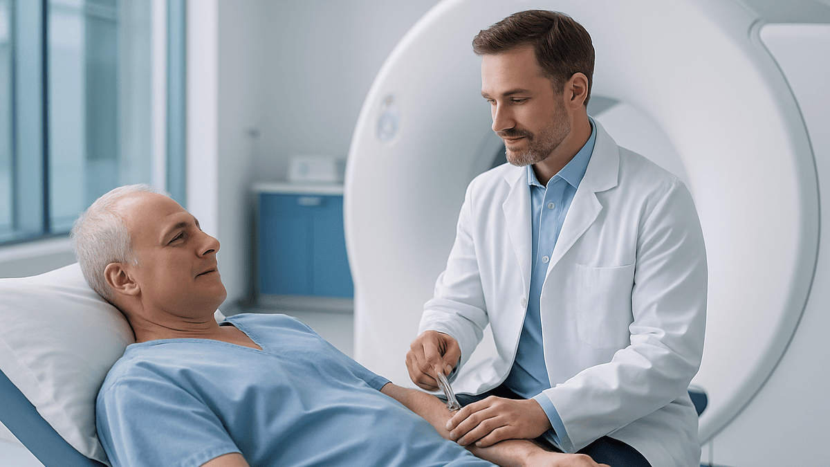
During coronary CT angiography, you lie on a narrow table while a small cannula in an arm vein delivers iodine-based contrast that briefly feels warm. Chest electrodes track your heartbeat, you may be given medicines to slow the pulse and widen the arteries, and the scanner captures the heart in a few seconds while you hold your breath. Most visits take about an hour.
The waiting room is quiet at 7:40 in the morning, and the man in the third chair is thinking about coffee. He was told to skip it, along with breakfast, and now he is holding a gown in a plastic bag and reading the same poster about hand hygiene for the fourth time. His cardiologist said the test was quick. Quick is relative when someone is about to inject something into your arm and photograph your heart.
He is one of thousands of people every week who want a straight answer to a simple question: what happens during coronary CT angiography, minute by minute, and which parts are worth worrying about? Most of the anxiety comes from the unknowns: the dye, the medicine that slows the heart, the machine, the breath hold.
This explainer walks through the appointment in order, from the first blood pressure check to the moment the cannula comes out, with the evidence for each step. The decisions belong to your care team; the understanding can belong to you.
What happens during coronary CT angiography, step by step
Coronary CT angiography is a scan that uses X-rays and an injected dye to build detailed pictures of the arteries that feed the heart muscle. You are awake throughout, nothing is threaded into your heart, and the part that produces the images lasts only seconds (Mayo Clinic).
The visit unfolds in a predictable order. A nurse or radiographer checks your identity, pregnancy status where relevant, kidney function results, allergies and the medicines you take. You change into a gown and remove anything metal above the waist. A small plastic tube called a cannula is placed in a vein, usually at the elbow, because the contrast needs to travel quickly to the heart.
Sticky electrodes go on your chest so the scanner can read your heartbeat and time each picture to the quiet moment between beats. If your pulse is brisk, you may be offered a medicine to slow it; if the team wants the arteries as wide as possible, a nitrate spray or tablet under the tongue is common (Cleveland Clinic). The scanner itself looks like a large doughnut. You lie on a table that slides through the ring; you are never enclosed in a tunnel.
A short practice run checks that you can hold your breath on command. Then the contrast is injected by a pump, you hold your breath for a few seconds, and the pictures are taken (Mayo Clinic). After a brief observation period the cannula is removed and, for most people, the day carries on as normal.
That is the whole arc. The sections that follow explain why each step exists and what it genuinely feels like.
How does a coronary CT angiogram actually work?
The heart is a difficult subject to photograph. It moves constantly, and the vessels of interest are only a few millimeters wide. Two pieces of engineering solve this.
The first is speed. A computed tomography (CT) scanner spins an X-ray source around you and reconstructs hundreds of thin cross-sectional slices into a three-dimensional model. Modern cardiac scanners rotate fast enough to freeze the heart in a fraction of a beat, which is why the imaging portion lasts seconds rather than minutes (Mayo Clinic).
The second is timing. Those chest electrodes feed your electrocardiogram, the electrical trace of each heartbeat, into the scanner. The software aims the X-ray burst at diastole, the resting phase when the ventricles fill and the coronary arteries lie still. A slow, regular rhythm makes that window longer and the pictures crisper, which is the reason so much of the appointment is about your pulse (Cleveland Clinic).
Contrast makes the arteries visible. Iodine absorbs X-rays strongly, so when iodine-based dye flows through the coronary vessels they appear bright white against the darker heart muscle. Where plaque narrows a vessel, the white column thins or is interrupted. Calcified plaque shows up even brighter; softer, fatty plaque appears darker within the wall (American Heart Association).
Many services also run a quick non-contrast pass first, called a coronary calcium score, which counts the calcified deposits in the artery walls. That number helps the reporting doctor interpret the angiogram and, in some people, contributes to the overall risk picture (Cleveland Clinic).
The result is a map: where the arteries run, whether they are narrowed, and what the narrowing is made of. Interpreting that map, and deciding what it means for you, is the treating team’s job.
How to prepare for a coronary CT angiogram: the hours before
Preparation is mostly about your heart rate and your kidneys, and the instructions are simpler than they sound.
Caffeine is the big one. Coffee, tea, energy drinks, cola and, in some protocols, chocolate are usually off the menu for about 12 hours beforehand because caffeine speeds the heart and can blunt the medicines used to slow it (Mayo Clinic). Nicotine has a similar effect, so many services ask you not to smoke or vape on the morning of the test.
Eating is usually restricted for roughly four hours before the scan, though water is generally encouraged because being well hydrated helps the kidneys handle contrast (Mayo Clinic). Your team will confirm their own timings; follow those rather than a general rule.
Medicines deserve a specific conversation. Most regular prescriptions are taken as usual. A few, including some diabetes medicines and any medicine for erectile dysfunction, need special mention because they interact with contrast or with nitrates. Never stop or skip a prescribed medicine on your own; ask the team who booked the scan what they want you to do and when (Cleveland Clinic).
Practicalities matter too. Wear a top that is easy to remove and avoid jewelry above the waist. Bring a list of your medicines and any previous reaction to contrast. If you know you are, or might be, pregnant, say so at booking rather than on the day (Mayo Clinic).
Finally, plan the practice of breathing. Some radiographers ask you to rehearse a calm, gentle breath in and hold at home. A steady hold of a few seconds, without straining, is all the scanner needs.
What happens during coronary CT angiography when your heart beats too fast?
A racing pulse is the most common reason a scan is delayed or repeated, and it is also the most fixable. When your heart is quick, the resting phase between beats shrinks, and the scanner has less time to catch the arteries standing still. The pictures blur, much as a photograph of a sprinter blurs in low light.
Many imaging services aim for a resting rate near or below 60 beats a minute, though the exact target varies and your team will tell you theirs. Two approaches bring the rate down.
The first is simply time and calm. Anxiety in an unfamiliar room raises the pulse, and a few minutes of lying still often brings it down by itself.
The second is a beta-blocker, a class of medicine that slows the heart by blunting the effect of adrenaline on the heart’s pacemaker cells. It may be given as a tablet in advance or through the cannula on the day; the injected form acts within minutes, the oral form over about an hour (Mayo Clinic; Cleveland Clinic). This is a one-off use for the scan, not a new prescription, and the team checks that you have no reason to avoid it, such as certain asthma patterns or a very low blood pressure.
Separately, a nitrate such as glyceryl trinitrate is often given under the tongue just before the contrast run. Nitrates relax the muscle in artery walls so the coronary vessels widen, making narrowings easier to see and measure. A brief headache or a flushed feeling is a common, short-lived effect (Cleveland Clinic).
If the rate will not settle, the team may adjust the scan protocol or reschedule. That is a judgment call for the radiologist or cardiologist, and it is not a failure on your part.
The contrast injection: what the dye is and what it feels like
The word ‘dye’ misleads people. Nothing is colored. The contrast is a clear, iodine-containing liquid that shows up white on X-ray images and is cleared from the body by the kidneys, mostly within a day (Johns Hopkins Medicine).
It is delivered by a mechanical pump through the cannula in your arm, followed by a flush of saline to push it along. Three sensations are so common that radiographers usually warn you about them in advance. A wave of warmth spreads across the chest and often down toward the groin. A metallic taste appears at the back of the mouth. Many people feel briefly as though they have passed urine, though they have not (Mayo Clinic). All three fade within a minute or so.
Less often, people notice mild nausea, an itch or a few hives. These are minor reactions and the team is trained to manage them (NHS). Serious allergic reactions to modern contrast are rare, but because they can happen, staff, emergency medicines and monitoring are in the room, and you will be watched for a short period afterwards (Mayo Clinic).
Two groups are screened more carefully. People with reduced kidney function are at higher risk of a temporary dip in kidney performance after contrast, so a recent blood test for kidney function is standard and the volume of contrast is kept as low as the scan allows (Cleveland Clinic). People with a previous reaction to iodine contrast are assessed individually; a past reaction does not automatically rule the scan out, but it changes how the team prepares.
One myth is worth retiring here: a shellfish allergy is not a contrast allergy. The two are unrelated, and mainstream guidance does not treat shellfish allergy as a reason to avoid iodine contrast.
The scan itself: breath holds, the gantry, and the seconds that count
By the time the table slides into the ring, most of the work is already done. What remains is a sequence of short instructions and a few seconds of stillness.
The radiographer leaves the room and speaks to you through an intercom. You can see and hear them the whole time. First comes a quick planning scan to locate the heart. Then, often, the non-contrast calcium score pass. Each requires a breath hold of a few seconds; the recorded voice or the radiographer will tell you exactly when to breathe in and when to relax (Mayo Clinic).
The main run follows. Contrast starts pumping, the scanner waits for the dye to reach the coronary arteries, and you hear the instruction to breathe in and hold. The gantry, the rotating part of the machine, whirs. The table may move slightly. The X-ray burst is timed to your heartbeat, and the acquisition lasts a matter of seconds on current scanners (Mayo Clinic).
Why the breath hold? The diaphragm sits directly under the heart. Every breath lifts and lowers the heart by a centimeter or more, which would smear the images. Holding after a gentle breath in, without gulping air, keeps the chest still and avoids the pulse jump that comes with a deep gasp.
People often ask whether the machine is loud or enclosed. Cardiac CT is quieter than MRI and the ring is open at both ends; your head is usually outside it during the heart scan. If claustrophobia worries you, tell the radiographer, who can talk you through each step or let you keep your eyes on the ceiling (Cleveland Clinic).
Once the images are checked for quality, you slide out. A repeat run is occasionally needed if the heart rate jumped or the breath hold slipped, which is why staying still and calm through the count matters more than anything else you do that day.
How long does a CT coronary angiogram take from door to door?
The honest answer separates the scan from the appointment, because people confuse the two and then feel misled.
The imaging itself takes seconds. Modern scanners capture the entire heart in a few heartbeats, sometimes within a single breath hold (Mayo Clinic). If you count the planning scan, the calcium score and the contrast run together, you may spend around ten minutes on the table (Cleveland Clinic).
The appointment is longer because of everything wrapped around those minutes. Checking in, changing, placing the cannula, attaching electrodes and confirming kidney results take time. If a beta-blocker is given by mouth, the team waits for it to work before scanning, and that wait can add an hour. After the scan you are observed briefly for any contrast reaction before the cannula is removed. Most services describe the whole visit as roughly an hour, with a wider range when heart-rate medicine is needed (Mayo Clinic).
A few situations stretch the day. A heart rate that will not settle may prompt a second dose or a rescheduled slot. An irregular rhythm may need a different scanning mode. A cannula that is hard to place adds minutes rather than hours. None of these is unusual and none changes the safety of the test.
Results take a separate track. The radiologist or imaging cardiologist reviews the three-dimensional reconstructions, often measures narrowings and describes plaque type, and issues a written report to the doctor who requested the scan. Timing varies by service; ask on the day when to expect the report and who will discuss it with you.
Practically, block out a morning or an afternoon, bring something to read for the pre-scan wait, and treat the drive home as ordinary unless the team tells you otherwise.
Who is coronary CT angiography usually for, and who is usually asked to wait
The test has a well-defined sweet spot. Current guidelines from cardiology societies in the United States and Europe, and NICE guidance in the United Kingdom, position coronary CT angiography as a first-line investigation for people with stable chest pain whose likelihood of coronary artery disease is low to intermediate (American Heart Association). Its particular strength is ruling disease out: a clean scan with normal arteries is reassuring and often ends the diagnostic pathway.
Typical reasons a doctor requests it include new or unexplained chest discomfort on exertion, an inconclusive exercise test, breathlessness where a heart cause is possible, checking the arteries before certain non-cardiac surgery or valve procedures, and clarifying anatomy when arteries may follow an unusual course (Mayo Clinic; Cleveland Clinic).
Some people are usually steered elsewhere or asked to wait. A suspected heart attack in progress is an emergency handled by other pathways, not a scheduled scan. Pregnancy prompts a careful discussion because of fetal radiation exposure; the test is generally deferred or an alternative used (Mayo Clinic). Severely reduced kidney function raises the risk from contrast and may tip the balance toward a different test. A previous severe contrast reaction, a persistently irregular rhythm such as atrial fibrillation that cannot be controlled, an inability to lie flat or hold the breath, and a very high heart rate that does not respond to medicine all make good images harder to obtain.
Heavy calcification and existing stents are a subtler issue. Both scatter X-rays and can make the inside of a vessel hard to judge, so the report may be less definitive and an invasive angiogram may be suggested instead (Cleveland Clinic).
None of these is an absolute rule. The requesting doctor weighs your history, your kidney results and the question being asked, and chooses the test most likely to answer it safely.
CT angiogram vs cardiac catheterization: how the tests compare
People are often offered one and wonder why not the other. The two tests look at the same arteries from very different angles, and the table below sets them side by side with a common third option, the exercise stress test.
| Feature | Coronary CT angiography | Invasive coronary angiography (cardiac catheterization) | Exercise stress test |
|---|---|---|---|
| What it shows | Artery anatomy, narrowings and plaque type in 3D | Artery anatomy from inside the vessel, in real time | Whether the heart shows strain under effort |
| How it is done | Contrast through an arm vein; scanner outside the body | Thin tube passed through a wrist or groin artery to the heart | Walking on a treadmill or cycling with ECG monitoring |
| Radiation | Yes, generally low with modern scanners | Yes, during X-ray imaging | None |
| Contrast | Yes | Yes | No |
| Can treat at the same time | No | Yes, a stent can be placed if needed | No |
| Typical recovery | Same day, normal activity | Several hours of observation; puncture-site care | Immediate |
Cardiac catheterization, described by MedlinePlus and Mayo Clinic as the reference standard for seeing the arteries, involves passing a catheter into an artery and injecting contrast directly. It carries the small risks of any invasive procedure, such as bleeding or vessel injury at the puncture site, but it allows the operator to treat a blockage on the spot.
The CT approach avoids the artery puncture and the recovery time, and it sees the wall of the vessel as well as the channel, which is why it can detect soft plaque that an invasive study would miss. Its limits are image quality in heavily calcified or stented arteries and the fact that it cannot treat what it finds.
Which test comes first depends on how likely disease is, how urgent the question is and what your kidneys and heart rhythm allow. That sequencing is a clinical decision for your team.
Radiation, contrast and other risks, in plain numbers where they exist
Risk conversations go best when the risks are named, sized honestly and set beside the risk of not knowing.
Radiation is the one most people ask about. Coronary CT angiography does use X-rays, and any X-ray exposure adds a very small theoretical increase in lifetime cancer risk. Doses have fallen substantially as scanners have become faster and as software has learned to reconstruct sharp images from less X-ray energy; mainstream sources now describe the exposure from a modern coronary CT scan as low, and the team tailors the protocol to your size and heart rate to keep it as small as possible (Mayo Clinic; American Heart Association). Everyone receives natural background radiation from soil, air and food every year, and a single cardiac CT is commonly compared with a period of that background exposure rather than with anything dramatic.
Contrast carries two kinds of risk. Allergic-type reactions range from a few hives to, rarely, a severe reaction affecting breathing or blood pressure; monitoring and emergency medicines are on hand for this reason (Mayo Clinic). Kidney stress is the other. Contrast can temporarily reduce kidney performance, particularly in people whose kidneys already work below normal, which is why kidney function is checked beforehand and fluids are encouraged afterwards (Cleveland Clinic).
The medicines used on the day have their own short-lived effects. Beta-blockers can lower blood pressure and cause light-headedness; nitrates commonly cause a brief headache or flushing (Cleveland Clinic). Both wear off within hours.
Finally, there is the risk of an unclear or incidental finding. A scan of the chest sees lungs, bones and large vessels as well as the heart, and it may reveal something unrelated that needs follow-up. Some people welcome that; others find it unsettling. It is worth knowing before you lie down.
What the hours and days after a coronary CT angiogram usually look like
Recovery is brief because nothing was cut, threaded or sedated. Still, a few things are worth planning.
Immediately afterwards, you sit or lie for a short observation period so staff can watch for any delayed contrast reaction. The cannula is removed and a small dressing applied. A bruise at the site is common and harmless; it fades over several days.
Drink water through the rest of the day. The kidneys clear iodine contrast into the urine, and good hydration supports that process (Mayo Clinic). Unless your team says otherwise, you can eat normally, return to work and drive; most people resume ordinary activity the same day (Mayo Clinic). If you received a beta-blocker, you may feel a little slower or lighter-headed for a few hours, so some services advise arranging a lift rather than driving yourself. Ask on the day.
The nitrate headache, if you had one, usually settles within the hour. Flushing and the metallic taste are gone by the time you dress.
Over the following days, the main event is the report. A radiologist or cardiologist reviews the images, measures any narrowings, comments on plaque type and calcium, and notes incidental findings. The report goes to the doctor who requested the scan, and that person, not the imaging department, discusses what it means for you. Outcomes range from reassurance and discharge from the chest pain pathway, to lifestyle and medication discussions, to a recommendation for further testing such as a functional stress test or an invasive angiogram (Cleveland Clinic).
If you are breastfeeding, ask your team about contrast; mainstream guidance generally allows feeding to continue, but the conversation should happen with them rather than online.
What people often get wrong about coronary CT angiography
Myths cluster around this test because its name sounds like the invasive version and because ‘dye’ and ‘radiation’ are loaded words. Here are the corrections the evidence supports.
‘They go into my heart.’ No. Nothing enters the heart or its arteries. The only puncture is a vein in your arm for the cannula; the scanner works entirely from outside the body (Mayo Clinic).
‘The dye is radioactive.’ It is not. Iodine contrast simply absorbs X-rays so that blood-filled arteries appear white. The radiation comes from the scanner, not the liquid (Johns Hopkins Medicine).
‘I am allergic to shellfish, so I cannot have contrast.’ Shellfish allergy is a reaction to a muscle protein, not to iodine, and mainstream guidance does not treat it as a contrast allergy. Tell the team about every allergy anyway; a previous contrast reaction is the one that changes the plan.
‘A normal scan means my heart is fine forever.’ A normal scan means the arteries showed no significant narrowing at the time of the test. It does not measure blood pressure, rhythm, valve function or how arteries will behave over the coming decades, and it does not replace attention to cholesterol, smoking, activity and blood sugar.
‘An abnormal scan means I need a stent.’ Plaque on a scan is common, particularly with age, and its presence alone does not mean a procedure is needed. Whether a narrowing limits blood flow, and whether treatment should be medicines, further testing or intervention, is a clinical judgment for your team (Cleveland Clinic).
‘The scan will be claustrophobic like an MRI.’ The CT ring is short and open, and for the heart scan your head is typically outside it. It is also far quieter (Cleveland Clinic).
Questions to ask your care team before and after the scan
A good appointment is a two-way conversation. These questions help you get the most from the people running the test and the doctor who requested it.
Before the scan, focus on preparation and safety:
- Why has this test been chosen for me rather than a stress test or an invasive angiogram?
- Which of my regular medicines should I take as usual on the day, and are there any you want me to time differently?
- Do my recent kidney results allow contrast, and what precautions are you taking?
- Will I be given a beta-blocker or a nitrate, and should someone drive me home?
- What exactly should I avoid eating or drinking, and for how long before the appointment?
- Does the scan include a calcium score, and what will that number add?
On the day, ask the radiographer how the breath-hold commands will sound and roughly how many runs to expect. If you are anxious about the enclosed feeling or the contrast sensations, say so; the team can adjust pacing and talk you through it.
After the scan, the questions shift to interpretation:
- Who will explain the report to me, and when?
- If plaque is found, how will you decide whether it needs treatment, more testing or monitoring?
- If the scan is normal, does anything else on my risk profile still need attention?
- Were there incidental findings outside the heart, and do they need follow-up?
- How, and to whom, should I report any late reaction to the contrast?
Write the answers down or bring someone who can. The details that feel obvious in the room are the ones that blur by the evening.
When to call your doctor after a coronary CT angiogram
Serious problems after this scan are uncommon, but a short list of red flags deserves prompt action rather than watchful waiting.
Call emergency services immediately if you develop swelling of the face, lips or tongue, difficulty breathing, wheeze, a widespread rash with faintness, or a feeling that your throat is closing. These can indicate a severe allergic reaction to contrast, which is rare but treatable when acted on quickly (Mayo Clinic). Do the same for crushing or spreading chest pain, sudden severe breathlessness, or collapse; the scan does not cause heart attacks, but the symptoms that led to the scan still need emergency care if they escalate.
Contact your care team the same day for hives or itching that appear hours after you left, persistent nausea or vomiting, marked light-headedness or a very slow pulse that does not settle after a beta-blocker, or a headache from the nitrate that is severe or lasts beyond a few hours.
Watch the cannula site over the next few days. Spreading redness, increasing pain, warmth or pus suggests infection or inflammation of the vein and should be checked. A soft bruise that fades is normal.
Kidney warning signs matter most if your kidney function was already reduced. Passing much less urine than usual, new swelling of the ankles, or unusual fatigue in the days after contrast should prompt a call, since a temporary dip in kidney performance is possible and your team may want a repeat blood test (Cleveland Clinic).
If you are unsure whether something is significant, call. Imaging departments and cardiology teams expect these questions and would far rather hear from you than have you sit at home wondering.
Frequently asked questions
What does 'during' cover here: the whole appointment or just the scan?
In this article, ‘during’ means the entire appointment, from check-in to cannula removal, not just the seconds of X-ray exposure. People searching what happens during coronary CT angiography usually want the full sequence: preparation, heart-rate medicine, contrast injection, breath holds and observation afterwards. The imaging portion is the shortest part of the visit by a wide margin (Mayo Clinic).
How long does a CT coronary angiogram take from arrival to leaving?
Most services describe the visit as roughly an hour, though the scan itself takes only seconds (Mayo Clinic). The time is spent on checks, placing the cannula, attaching electrodes and, if needed, waiting for a beta-blocker to slow the heart. A brief observation period follows the contrast injection. Ask your team for their typical timing on the day.
What are the common CT coronary angiogram contrast dye side effects?
The usual effects are a warm flush across the chest, a metallic taste and a brief sensation of having passed urine, all fading within about a minute (Mayo Clinic). Mild nausea, itching or a few hives occur less often. Serious allergic reactions are rare, and staff monitor you afterwards for that reason. People with reduced kidney function are checked beforehand because contrast can temporarily affect kidney performance.
Can I drive home after a coronary CT angiogram?
Usually yes, since no sedation is used and most people resume normal activity the same day (Mayo Clinic). The exception is if you were given a beta-blocker or nitrate and feel light-headed, when some services suggest arranging a lift. Ask the radiographer before you leave; their answer will reflect the medicines you actually received and how you feel.
What is the difference between a CT angiogram and cardiac catheterization?
A CT angiogram images the arteries from outside the body using contrast injected into an arm vein, while cardiac catheterization passes a thin tube through a wrist or groin artery into the heart. The CT version avoids an arterial puncture and shows plaque in the vessel wall; catheterization allows a blockage to be treated on the spot. Which comes first is a clinical decision (Mayo Clinic; MedlinePlus).
Do I need to stop my medicines before the scan?
Do not stop or change any prescribed medicine on your own. Most regular medicines are taken as usual, but a few, including some diabetes medicines and medicines for erectile dysfunction, need specific instructions because they interact with contrast or nitrates (Cleveland Clinic). Bring a full list to your booking conversation and follow exactly what your prescribing team tells you.
What if I am claustrophobic?
The CT scanner is a short, open ring rather than a tunnel, and during the heart scan your head is typically outside it. It is also much quieter than MRI (Cleveland Clinic). Tell the radiographer in advance; they can explain each step through the intercom, pace the breath holds and keep talking to you. Most people who dread it find the reality far easier than expected.
What happens if my heart rate will not come down?
The team may allow more time, give a further dose of the heart-slowing medicine, switch to a scanning mode that copes better with faster rhythms, or reschedule the appointment (Cleveland Clinic). A persistently fast or irregular pulse blurs the images, so the decision is about picture quality, not about you doing anything wrong. Avoiding caffeine and nicotine beforehand helps prevent this problem.
Is the radiation from a coronary CT angiogram dangerous?
The exposure from a modern coronary CT scan is generally described as low and has fallen substantially with faster scanners and better software (Mayo Clinic; American Heart Association). Any X-ray adds a very small theoretical lifetime risk, which your doctor weighs against the value of the information. Pregnancy changes that calculation, so tell your team if you are or might be pregnant.
How do I prepare for a coronary CT angiogram if I have diabetes or kidney problems?
Tell the booking team about both conditions in advance. A recent kidney function blood test is standard because contrast can temporarily stress the kidneys, and the team may adjust contrast volume or hydration accordingly (Cleveland Clinic). Some diabetes medicines need specific timing instructions around contrast; follow your prescriber’s advice rather than changing anything yourself. Fasting and caffeine rules still apply.
This article is for general information only and is not a substitute for professional medical advice. Please consult a qualified doctor about your individual situation.

More from the Blog

Living With Cardiorenal Syndrome: Daily Weight, Fluid Limits and the Kidney-Friendly Plate
Living well with cardiorenal syndrome usually rests on three daily habits: weighing yourself each morning to catch fluid gain early, keeping to the fluid…

High Ferritin and Iron Overload: When Stored Iron Becomes a Problem
A high ferritin result means the body is storing more iron than usual or, just as often, that inflammation, liver strain, alcohol, obesity or…

Wrist or Groin Access for Coronary Angiography: What Changes for Your Recovery
For a coronary angiogram, wrist (radial) access usually means less bleeding, no hours of lying flat, and an earlier walk to the car, while…

Is It Safe to Fly After a Pulmonary Embolism? Timing and Preparation for the Journey Home
Most people can fly again after a pulmonary embolism once they are stable on anticoagulant treatment, breathing comfortably at rest and cleared by their…

How Often Can You Donate Blood? Rules, Recovery and Benefits
In the United States, most healthy adults can donate whole blood every 56 days, up to six times a year. Platelet donations can be…

What Is Inflammation? Friend, Foe and the Chronic Middle
Inflammation is the immune system's built-in response to injury and infection, marked by redness, heat, swelling, pain, and loss of function. Short-term (acute) inflammation…