

What If ESWL Does Not Break the Stone? Repeat Sessions, Ureteroscopy and What Comes Next
Key Takeaways
- ESWL breaks stones into fragments but removes nothing, so a session only counts as successful once follow-up imaging shows the kidney and ureter are clear.
- Stones made of cystine, brushite or dense calcium oxalate frequently resist shock waves, which is why CT density and prior stone analysis guide the choice of a second treatment.
- Fragments under about 4 mm usually pass on their own within a few weeks, while larger pieces or a column of fragments in the ureter (steinstrasse) typically need ureteroscopy.
- Repeat ESWL makes most sense when the first session visibly cracked the stone; a stone that shows no fracture after a full session is unlikely to behave differently next time.
- Ureteroscopy with laser lithotripsy reaches the lower pole of the kidney and the pelvic ureter, the two places where shock waves most often fall short, and a prior ESWL session does not reduce its chance of clearing the stone.
- Fever with flank pain after any stone procedure can signal infection behind a blocked kidney and needs same-day medical assessment.
If ESWL did not break a kidney stone, or left fragments that will not pass, the usual next steps are a repeat shock wave session when the stone is still a good candidate, ureteroscopy with laser lithotripsy for most stones that resisted shock waves, or percutaneous surgery for large or very hard stones. Follow-up imaging guides the choice, and your urologist decides based on stone size, hardness and location.
The follow-up X-ray is up on the screen, and the radiologist’s cursor is hovering over the same pale smudge that was there six weeks ago. You did everything asked of you: lay still through the thudding of the shock waves, drank until you were tired of water, strained your urine through the little paper funnel. And the stone, apparently, did not get the memo.
If you are sitting with the news that ESWL did not work, you are not an unusual case and you have not run out of options. Shock wave lithotripsy is the gentlest tool urologists have for stones, which is exactly why it is often tried first and why it sometimes falls short against stones that are large, dense or awkwardly placed.
This explainer walks through what a failed session really means, why it happens, and the realistic paths that follow: another round of shock waves, a ureteroscope with a laser, or keyhole kidney surgery. The decision belongs to you and your treating team; the goal here is to make that conversation easier.
What is ESWL and how does it actually break a stone?
ESWL stands for extracorporeal shock wave lithotripsy: “extracorporeal” means from outside the body, and “lithotripsy” means stone crushing. A machine called a lithotripter generates pressure waves, focuses them through a water cushion pressed against your flank, and aims them at the stone using X-ray or ultrasound guidance. Nothing enters the body. The waves pass through skin and muscle largely unnoticed and release their energy where the tissue changes abruptly, which is at the surface of the stone.
Two things happen at that surface. The leading edge of each wave compresses the stone and the trailing edge pulls on it, creating tiny cracks that widen with every pulse. At the same time, microscopic bubbles form in the urine around the stone and collapse violently, chipping away at the surface like a sandblaster. Over the course of a session, which Mayo Clinic describes as typically lasting about 45 to 60 minutes, those cracks join up and the stone falls apart into gravel.
That is the crucial point most people miss. ESWL does not remove anything. It converts one stone into many smaller pieces and then relies on you to pass them in your urine over the following days or weeks. The procedure is judged a success only when the kidney and ureter are clear, or when what remains is small enough to be harmless.
Doctors usually offer ESWL for stones that are not too big, not too hard and sit where the waves can reach them cleanly. Cleveland Clinic and the NIH both note that it works best on stones up to about 2 cm. Outside that sweet spot, the odds shift, and that is where the rest of this article begins.
How do you know ESWL did not work?
There is no alarm bell when shock waves fail. The stone does not feel any different the next morning, and pain alone is an unreliable guide, because a stone that shattered beautifully can still hurt as the fragments move, while one that did not budge may sit quietly.
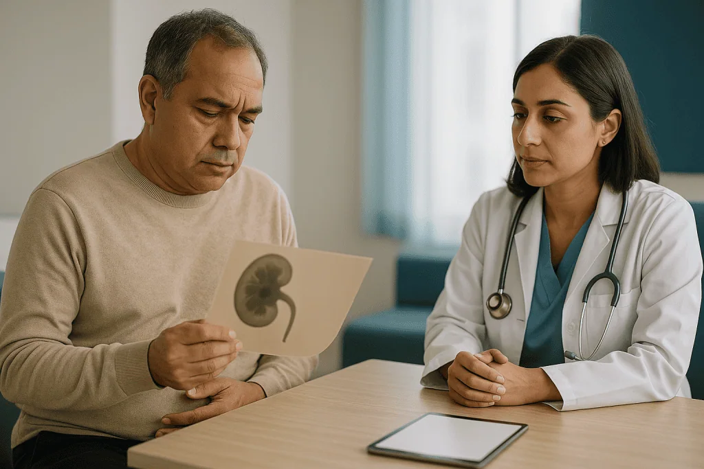
The verdict comes from imaging. Most urology services arrange a plain X-ray, an ultrasound or a low-dose CT scan some weeks after treatment, once the fragments that are going to pass have had time to do so. The MedlinePlus lithotripsy page notes that fragments can take anywhere from a few days to a few weeks to clear, which is why the check is not scheduled immediately. Three results are possible:
- The stone is unchanged. Same size, same place. The waves did not fracture it, or barely marked it.
- The stone broke but the pieces are stuck. Fragments line up in the ureter, the narrow tube between kidney and bladder, or settle in the lowest cup of the kidney where gravity works against them.
- Small residual fragments remain. Often described as clinically insignificant, these are tiny specks that may pass unnoticed or may sit harmlessly for years, though they can also seed a future stone.
Only the first two usually count as “ESWL did not work” in the sense that something further is needed. The third is a judgment call your urologist will talk through with you, weighing fragment size, your stone history and how much the stone has troubled you.
Many people are also asked whether they saw gravel in the strainer. It is useful information, but absence of visible grit proves nothing: fragments can be fine as sand or can still be waiting in the kidney. Trust the scan, not the sieve.
Why did ESWL not work? The usual reasons
Shock waves are a physics treatment, and physics is unforgiving about a handful of variables. When a session falls short, the explanation almost always lies in one of these.
The stone was too hard. Stone chemistry matters more than most people realize. Calcium oxalate monohydrate, brushite and cystine stones are notoriously resistant; they absorb energy without cracking. Mayo Clinic and the NIH both single out cystine stones, which form in an inherited condition, as poor candidates for shock waves. Dense stones also show up brighter on CT, and radiologists can estimate hardness from that density before anyone fires a single pulse.
The stone was too big. Beyond roughly 2 cm, the threshold Cleveland Clinic and the NIH cite, a stone may fragment but produce so much gravel that the ureter cannot cope, or it may need multiple sessions with diminishing returns.
The stone was in the wrong place. The lower pole of the kidney, its bottom-most chamber, drains uphill. Fragments created there must climb out against gravity, and many simply settle back where they started. Stones low in the ureter can also be shielded by the pelvic bones.
The waves had too far to travel. Energy fades with distance. In people with a greater distance between skin and stone, the focal point may arrive weaker than intended, and the imaging used to aim can be less precise.
Anatomy got in the way. A narrowed drainage channel, a kidney that sits in an unusual position or a previous operation can all mean fragments have nowhere to go.
None of these is a failure on your part. They are reasons to choose a different tool, and your urologist will usually be able to point to which one applied in your case.
Who is usually offered ESWL first, and who is usually asked to wait or skip it
Guideline-level practice, reflected in the NHS and NIH overviews of stone treatment, favors ESWL as a first option for people whose stones sit in the kidney or upper ureter, measure under about 2 cm, are not known to be of a hard type, and who have no reason to avoid the procedure. The appeal is obvious: no incision, often no general anesthetic, and a same-day discharge for most.

Some people are steered elsewhere from the start, or asked to wait, because the odds or the risks do not favor shock waves:
- Pregnancy. ESWL is not performed during pregnancy; the energy and the X-ray guidance are both concerns.
- Bleeding problems or blood-thinning medicines. Shock waves bruise the kidney. Anyone with a clotting disorder, or taking an anticoagulant that cannot safely be paused, is usually offered ureteroscopy instead. Whether to pause a medicine is strictly a decision for the prescribing clinician.
- An active urinary infection. Breaking up an infected stone can release bacteria into the bloodstream, so infection is treated first.
- A blockage below the stone. If urine cannot drain, fragments cannot either.
- Certain body shapes or anatomy. Where the stone is too deep to target reliably, or the kidney is unusually positioned, aiming becomes guesswork.
- An aneurysm near the kidney or a pacemaker may require extra precautions or a different approach; the team will advise individually.
Then there is the person who has already had one unsuccessful session. Here the question is not eligibility but likelihood. If the first attempt produced no visible fracture at all, the stone is telling you something about its hardness, and most urologists will lean toward a direct approach rather than hoping the second round behaves differently.
Does shockwave therapy break up kidney stones every time?
No, and anyone who tells you otherwise is selling simplicity. Shock waves are effective for the right stone, but “the right stone” is a narrower category than the brochure version suggests.
It helps to separate two different measures. The first is fragmentation: did the stone crack? The second is stone-free status: is the kidney actually clear on follow-up imaging? A session can score well on the first and poorly on the second, because fragments that form in a low-draining part of the kidney may never leave. When urology guidelines compare treatments, they tend to focus on that second, stricter measure, and by that standard ESWL generally clears fewer stones in a single session than ureteroscopy does, especially for lower-pole and larger stones. The trade-off is that ESWL is less invasive and carries a lower chance of needing a temporary internal tube afterward.
Honest expectation-setting also means acknowledging that a proportion of people need more than one shock wave session even when the stone is a good candidate. The NHS describes ESWL as sometimes requiring repeat treatment to clear a stone fully. That is not a sign the procedure went wrong; it is built into how the treatment works.
What shock waves cannot do is overcome stone chemistry. A cystine or brushite stone may emerge from a full session essentially intact, and repeating the experience adds bruising to the kidney without changing the outcome. Modern practice tries to predict this beforehand using CT density and, where a previous stone has been analyzed, its composition.
So the realistic answer to the question is: often, for smaller and softer stones in favorable positions; less reliably, for everything else. Knowing which group your stone belongs to is the single most useful piece of information when planning what comes next.
Repeat ESWL: when a second session is reasonable and when it is not
A second round of shock waves is a legitimate option, not a consolation prize. The case for it is strongest when the first session clearly did some work. If follow-up imaging shows the stone has cracked into a few large pieces, or shrunk noticeably, the stone has proven it will fragment and a further session may finish the job. Stones still in the kidney, rather than lodged in the ureter, also tend to favor another attempt, because fragments created there have room to settle before passing.
Urologists generally leave an interval between sessions to let the kidney recover from the bruising that shock waves inevitably cause. Your team will set that timing based on your imaging and how you tolerated the first treatment; it is not something to rush.
The case against repeating is just as clear:
- The stone shows no fracture lines at all after a full-energy session.
- CT density or a previous stone analysis points to a hard composition.
- Fragments have already piled up in the ureter and are not moving.
- You have a solitary kidney, where any prolonged obstruction is more dangerous.
- Pain, infection or bleeding after the first session was significant.
There is also a ceiling. Repeated sessions on the same kidney carry cumulative risk of tissue injury, and most guideline frameworks encourage switching strategy rather than persisting indefinitely. If your urologist suggests stopping after two attempts, that reflects the evidence on diminishing returns rather than impatience.
One practical note: if you are offered repeat ESWL, ask what changed. Different energy settings, a different patient position, or treating after a temporary stent has been placed can all shift the odds compared with the first attempt. If nothing is different, it is fair to ask why the result would be.
Kidney stone not passing after ESWL: fragments, steinstrasse and what to do
The most common way ESWL “fails” is not that the stone survives but that the gravel it leaves behind refuses to move. Understanding why points to the fix.
Fragments leave the kidney through the ureter, a muscular tube only a few millimeters wide. The NHS notes that stones under about 4 mm usually pass on their own, often within a few weeks, while larger pieces struggle. A stone that fragments into a dozen 3 mm pieces may clear uneventfully. The same stone fragmenting into three 7 mm chunks has created three new problems.
Sometimes many fragments reach the ureter at once and stack up in a column. Urologists call this steinstrasse, German for “stone street.” It can be silent, or it can cause the same colicky flank pain as the original stone, and if it blocks urine flow entirely it needs prompt attention. The NIH and Cleveland Clinic both list blockage by fragments among the recognized complications of ESWL.
What people often ask is whether they can hurry things along. The evidence supports a few sensible measures and discourages others:
- Fluids help keep urine flowing, though drinking extreme volumes adds nothing.
- Activity, within comfort, is fine; there is no need to stay in bed.
- Medicines that relax the ureter, a class called alpha-blockers, are sometimes prescribed to ease fragment passage. Whether one is appropriate for you is a decision for your prescribing clinician.
- Inversion therapy, lying head-down to tip lower-pole fragments out, has been studied with mixed results and is not a routine recommendation.
If the fragments have not moved by the follow-up scan, or if they block the kidney, the usual next step is ureteroscopy: going up the ureter to retrieve or laser the pieces directly. Waiting longer rarely helps once the stone street has formed.
Ureteroscopy after ESWL: what actually happens
Ureteroscopy is the workhorse of modern stone surgery and the most common destination after shock waves disappoint. A ureteroscope is a very thin telescope, rigid or flexible, passed through the urethra and bladder and up the ureter to wherever the stone sits. Because it follows the body’s own plumbing, there is no cut at all.
The procedure is done under general or spinal anesthesia. Once the scope reaches the stone, the surgeon has two choices: lift it out whole in a tiny wire basket if it is small enough, or break it with a laser fiber threaded through the scope and then basket the pieces or leave dust to wash out. Flexible scopes can bend into the lower pole of the kidney, which is exactly the region where ESWL fragments tend to strand, so a stone that shock waves could not shift from that pocket can usually be reached directly.
Most people go home the same day, as Cleveland Clinic and Mayo Clinic describe. Many leave with a ureteral stent, a soft plastic tube running from kidney to bladder that keeps the passage open while swelling settles. Stents are the part patients remember: they can cause urgency, a dull ache and blood-tinged urine until removed, usually within a few days to a few weeks, depending on your team’s plan.
Risks are real but uncommon: urinary infection, a scraped or, rarely, perforated ureter, and later narrowing of the ureter from scarring. Your surgeon will put these in context for your anatomy.
One reassurance worth having in writing: having had ESWL first does not make ureteroscopy harder or less likely to clear the stone. The two approaches are routinely used in sequence, and a prior shock wave session does not close the door on anything.
What is laser lithotripsy used for in the treatment of kidney stones?
Laser lithotripsy is the step that turns ureteroscopy from a retrieval tool into a stone-destroying one. A hair-thin optical fiber is passed down the working channel of the scope until its tip rests against the stone. Pulses of laser light, most commonly from a holmium laser, are absorbed by the stone’s surface and by a microscopic layer of water around it, generating heat and tiny shock waves at the point of contact. The stone either shatters into fragments that can be basketed, or is slowly ground into fine dust that flushes out with urine.
Because the energy is delivered from millimeters away under direct vision, laser lithotripsy sidesteps the two main weaknesses of ESWL. Distance is irrelevant; the fiber is already there. Hardness matters far less; the laser breaks cystine, brushite and dense calcium oxalate stones that shrug off external shock waves. That is why, in guideline frameworks from the NIH and others, laser ureteroscopy is the usual recommendation for stones that resisted ESWL or were never suitable for it.
Its range of use is broad:
- Stones anywhere in the ureter, including the lower segment shielded by the pelvis.
- Kidney stones up to moderate size, including lower-pole stones.
- Fragments and steinstrasse left after shock waves.
- Stones in people who cannot stop blood-thinning medicines, because bleeding risk is lower than with ESWL or percutaneous surgery.
- Stones in people with anatomy that makes shock wave targeting unreliable.
Its limits are mainly about volume. Dusting a very large stone takes a long time under anesthesia and may need staging across two procedures, which is where percutaneous surgery, discussed next, comes in. The surgeon in the room, seeing the stone directly, decides how far to push and when to stop.
When is percutaneous surgery the next step, and how do the options compare?
Percutaneous nephrolithotomy, usually shortened to PCNL, means removing a stone through a small incision in the back directly into the kidney. A tract about the width of a pencil is created under imaging guidance, a telescope is passed through it, and the stone is broken up with ultrasonic or laser energy and suctioned out. It is the most invasive of the three paths, generally needs a hospital stay of a night or more as Mayo Clinic and the NIH describe, and carries a higher bleeding risk, but it clears large stone burdens in one sitting better than anything else.
After failed ESWL, PCNL tends to be chosen when the stone is large, often above 2 cm, when it fills much of the kidney’s internal branches, when it sits in a lower pole with unfavorable drainage, or when previous ureteroscopy has been unable to reach or clear it.
| Option | How it reaches the stone | Usually chosen when | Main trade-offs |
|---|---|---|---|
| Repeat ESWL | Shock waves from outside the body | First session partly fragmented the stone; stone under about 2 cm; kidney rather than ureter | Least invasive; may still need more sessions; relies on fragments passing |
| Ureteroscopy with laser | Thin scope up the ureter | Stone resisted shock waves; fragments stuck in ureter; hard stone types; blood thinners | Day case; often a temporary stent; direct vision and high clearance for moderate stones |
| PCNL | Small tract through the back into the kidney | Large or complex stones; large residual burden after other treatments | Hospital stay; higher bleeding risk; best single-session clearance for big stones |
These are not rungs on a ladder to be climbed in order. A urologist may move straight from one failed ESWL session to PCNL if the stone is big enough, or combine ureteroscopy and PCNL in one anesthetic. The right sequence is the one that matches your stone, and only your team can see that picture in full.
What the following days and weeks usually look like
Whatever the next procedure, the rhythm of recovery is broadly similar and worth knowing in advance so that ordinary symptoms do not feel alarming.
In the first day or two, expect some blood in the urine. After ESWL this comes from bruising of the kidney; after ureteroscopy it comes from the scope and the stent; after PCNL, from the tract. Pink or rusty urine is normal; urine that looks like red wine or contains clots is not. Flank discomfort is common and usually eases steadily. If a stent is in place, bladder irritability, a sense of needing to go constantly and a tugging ache when you empty are the typical complaints, and they generally settle once the stent comes out.
Across the first couple of weeks, fragments and dust continue to pass. MedlinePlus notes this can take from a few days to a few weeks after shock waves, and the same holds after laser dusting. Most people return to desk work within a few days of ESWL or ureteroscopy; PCNL takes longer, with Mayo Clinic describing a hospital stay and a recovery of a week or more before normal activity.
The stent removal, if you have one, is usually quick and done in clinic, sometimes by a string left attached that you or a nurse can pull. Timing varies with the reason the stent was placed.
Then comes the piece people skip: follow-up imaging and prevention. A scan confirms the kidney is clear. Any stone or fragment retrieved should be sent for analysis, because knowing whether it was calcium oxalate, uric acid or cystine changes the prevention advice entirely. Blood and urine tests may follow. The NIH and NHS both emphasize that fluid intake and diet tailored to stone type reduce the chance of making another, and that conversation is as important as the procedure itself.
What people often get wrong about ESWL that did not work
“A failed session means the machine or the operator was bad.” Occasionally targeting is imperfect, but far more often the stone was always a marginal candidate. Hard composition, lower-pole position and distance from the skin are predictable headwinds, and a stone that ignores shock waves is giving useful information, not evidence of malpractice.
“More sessions will eventually work.” Not for hard stones. Repeated ESWL on a stone that showed no fracture adds kidney bruising without changing the physics. Guidelines steer toward a different approach rather than persistence.
“If I felt nothing pass, the stone must still be whole.” Fragments can be as fine as sand and pass unnoticed. Equally, feeling a lot of pain does not prove the stone shattered. Only imaging answers the question.
“Ureteroscopy is a step down, a sign things went badly.” For many stones ureteroscopy with laser would have been a reasonable first choice; it clears lower-pole and hard stones more reliably in a single procedure. Moving to it after ESWL is a change of tool, not an escalation of danger.
“Having ESWL first makes the next procedure harder.” The evidence does not support this. Prior shock waves do not reduce the chance that ureteroscopy clears the stone.
“Drinking gallons of water will flush out stuck fragments.” Good hydration helps; heroic volumes do not, and can leave you feeling unwell. A fragment physically wedged in the ureter needs a scope, not a flood.
“Once the stone is gone, the problem is over.” Roughly half of people who form one stone form another within a decade, according to the NIH. Stone analysis, metabolic testing and tailored prevention are where the long-term win lies, and skipping them is the most common mistake of all.
Questions to ask your care team when ESWL has not cleared the stone
A follow-up appointment after a disappointing result is short and easy to leave without the information you need. These questions are designed to pull out the specifics that shape the decision.
- Did the stone fragment at all? A partly broken stone and an untouched one lead to different recommendations.
- Do you know, or can you estimate, what the stone is made of? CT density and any previous stone analysis predict whether repeat ESWL has a realistic chance.
- Where exactly is the stone or the fragments now? Kidney versus ureter, and upper versus lower pole, matter enormously.
- Is my kidney draining normally at the moment? A blocked kidney changes the urgency of everything.
- If you suggest repeat ESWL, what will be different this time? Settings, positioning, a stent beforehand, or nothing.
- If you suggest ureteroscopy, will I likely need a stent, and for how long? Stent symptoms are the main thing people wish they had been warned about.
- Is percutaneous surgery on the table, and why or why not?
- How soon does this need to happen? Some situations can wait weeks; an obstructed or infected kidney cannot.
- Will the stone be sent for analysis, and will I have metabolic testing afterward?
- What should I do about my regular medicines before the next procedure? Especially blood thinners; never adjust these yourself.
- What symptoms should make me call before my next appointment?
Write the answers down or bring someone who will. It is also entirely reasonable to ask how many of each procedure the service performs and what its usual pathway is for stones like yours; this is routine information, not a challenge to anyone’s competence. The best decisions tend to come from patients who understand why a particular tool is being chosen, rather than simply which one.
When to call your doctor
Most of the days after ESWL, or after the ureteroscopy or surgery that follows it, are uncomfortable rather than dangerous. A small number of symptoms signal a blocked or infected kidney, significant bleeding or a serious complication, and these need same-day medical attention rather than waiting for the scheduled follow-up.
Seek urgent care, or call emergency services if you cannot reach your team quickly, if you notice any of the following:
- Fever or shaking chills. Infection behind a blocked kidney can progress to sepsis within hours. This is the single most important warning sign on this list.
- Severe pain that is not controlled by the medicines you were given, or pain that suddenly worsens.
- Inability to pass urine, or passing only tiny amounts over several hours.
- Heavy bleeding: urine that is dark red, contains clots, or does not lighten over a day or two.
- Persistent vomiting that prevents you keeping fluids down.
- Feeling faint, confused or unusually breathless.
- If you have a stent: it appears to have moved, you can see it, or the string has come away before planned removal.
- If you have a single kidney and develop any new flank pain or drop in urine output, regardless of severity.
Less urgent but still worth a call within a day or two: pain that is improving more slowly than you were told to expect, cloudy or foul-smelling urine without fever, or burning on urination that is getting worse rather than better.
The NHS and Mayo Clinic both flag fever with flank pain as a reason for immediate assessment in anyone with a known stone, and that advice applies doubly after a procedure. When in doubt, make the call; your team would far rather hear about a symptom that turns out to be nothing than miss one that mattered.
Frequently asked questions
What is ESWL?
ESWL, or extracorporeal shock wave lithotripsy, is a non-surgical treatment that uses focused pressure waves generated outside the body to crack a kidney stone into small fragments, which then pass in the urine. The session typically lasts about 45 to 60 minutes according to Mayo Clinic, most people go home the same day, and it is generally reserved for stones under about 2 cm.
Does shockwave therapy break up kidney stones?
Yes, for suitable stones: smaller, softer stones in the kidney or upper ureter often fragment well. It is less reliable for stones that are large, very dense, made of cystine, sitting in the lower pole of the kidney, or far from the skin surface. Follow-up imaging, not symptoms, confirms whether the stone broke and whether the fragments have cleared.
What do I do if a kidney stone doesn't pass after ESWL?
Keep your follow-up imaging appointment, maintain a normal fluid intake, and report fever, severe pain or reduced urine output immediately. If fragments remain stuck in the ureter or kidney on the scan, the usual next step is ureteroscopy, where a thin scope retrieves or lasers the pieces directly. Waiting longer rarely helps once fragments have lodged.
How many times can ESWL be repeated on the same stone?
There is no fixed number, but most urologists switch strategy after one or two unsuccessful sessions rather than persisting, because repeated shock waves bruise the kidney and hard stones do not become softer with time. A repeat is most reasonable when the first session clearly fragmented the stone; your urologist decides based on your imaging.
Is ureteroscopy after ESWL more difficult or less likely to work?
No. Published evidence indicates that a previous shock wave session does not reduce the chance that ureteroscopy clears the stone, and the two treatments are routinely used in sequence. Ureteroscopy reaches the stone under direct vision, so the hardness and distance problems that limit ESWL do not apply.
What is laser lithotripsy used for in the treatment of kidney stones?
Laser lithotripsy breaks stones from the inside using a thin fiber passed through a ureteroscope. It is used for stones anywhere in the ureter, for kidney stones of small to moderate size, for hard stone types that resist shock waves, for fragments left after ESWL, and for people who cannot pause blood-thinning medicines, because bleeding risk is lower than with other methods.
What is steinstrasse and is it dangerous?
Steinstrasse is a column of stone fragments that stacks up in the ureter after lithotripsy, named from the German for stone street. It can be painless, but if it blocks urine flow it can cause colicky pain and raise the risk of infection. Many clear on their own; those that obstruct the kidney or persist usually need ureteroscopy.
How long after a failed ESWL will I need another procedure?
It depends on whether the kidney is draining. An obstructed or infected kidney needs treatment urgently, often within days. If the kidney is draining and you are comfortable, your team may schedule repeat ESWL or ureteroscopy over the following weeks, after follow-up imaging. Your urologist sets the timing based on your scan and symptoms.
Will I need a stent after ureteroscopy following a failed ESWL?
Often, though not always. A ureteral stent is a soft tube that keeps the ureter open while swelling settles, and it is commonly placed when the ureter was irritated, when fragments remain, or when a second procedure is planned. Stents typically stay in from a few days to a few weeks and can cause urgency and mild ache until removed.
Can a kidney stone that did not break with ESWL dissolve with medicine instead?
Only in a limited situation. Pure uric acid stones can sometimes be dissolved by medicines that make the urine less acidic, a decision for your prescribing clinician after stone analysis and urine testing. Calcium-based stones, which are the majority, and cystine stones cannot be dissolved this way and need physical removal if they do not pass.
References
- Kidney stones: Treatment (NHS)
- Lithotripsy (MedlinePlus Medical Encyclopedia)
- Treatment for Kidney Stones (NIH, NIDDK)
This article is for general information only and is not a substitute for professional medical advice. Please consult a qualified doctor about your individual situation.

More from the Blog
Why Is My Pee Cloudy? Common Causes of Cloudy Urine and When It Needs a Test
Cloudy pee is most often caused by concentrated urine from drinking too little, harmless phosphate crystals after a meal, or a urinary tract infection,…

When Pelvic Pain Points to Prostatitis: Why Seeing a Urologist Early Shapes Treatment
See a urologist for prostatitis when pelvic, perineal or urinary symptoms last beyond a few weeks, keep returning after a course of antibiotics, or…

How Do You Prepare for a Day of Urology Tests? Full Bladder, Fasting and Medicines to Pause
Preparing for urology tests usually means three things: arriving with a comfortably full bladder if an ultrasound or urodynamic study is planned, fasting for…

Inside a Hypospadias Repair: How Surgeons Rebuild the Urethra and Straighten Curvature
Hypospadias surgery is usually a single outpatient operation under general anesthesia, most often done when a child is between about 6 and 18 months…
Stress Incontinence After Pregnancy and Childbirth: When to Wait, Train or Consider Surgery
Stress incontinence after pregnancy is leaking urine when you cough, sneeze, laugh, lift or run, because pregnancy and birth stretch the pelvic floor and…
Ureteroscopy Myths Corrected: Stents, Scarring and Being Put to Sleep
Ureteroscopy is a scope procedure that reaches a stone or blockage through the urethra and bladder, with no skin incision. It is usually done…