

Why Follow-Up Imaging Is Scheduled After Endovascular Surgery to Confirm Blood Flow

Key Takeaways
- Endovascular repair leaves the aneurysm sac in place and excludes it from blood flow, so imaging is the only way to confirm the sac is still protected.
- An endoleak is blood entering the sac outside the graft; type I and III leaks pressurize the sac directly and are treated urgently, while many type II leaks are watched and may close on their own.
- Society for Vascular Surgery guidance recommends imaging at about one month and twelve months after repair, then usually yearly, with an extra scan around six months if an early endoleak is seen.
- CT angiography is the baseline and problem-solving scan; duplex ultrasound, with no radiation or contrast, is the usual tool for routine long-term checks once the repair looks stable.
- A reassuring first CT with long seal zones can justify longer intervals between scans under European guidance, but no major guideline ends surveillance altogether.
- Complications such as graft migration, limb narrowing and sac growth are usually painless until advanced, which is why a quiet scheduled scan can prevent an emergency.
Follow-up imaging after endovascular surgery is scheduled because a stent graft can develop problems that cause no symptoms: blood leaking back into the aneurysm sac (an endoleak), the graft shifting or kinking, or a limb of the graft narrowing. Scans such as CT angiography or duplex ultrasound confirm blood is flowing only through the graft and the sac is stable or shrinking, so problems are caught early.
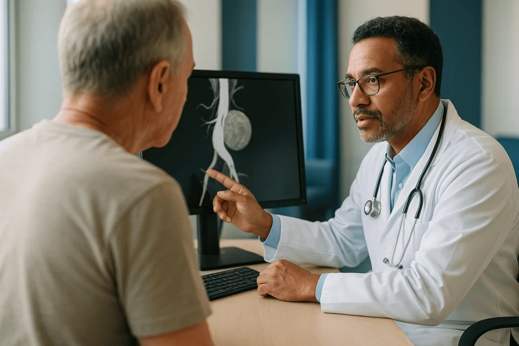
Ray is 74, walks the dog twice a day, and has not thought about his aorta in months. Then a letter arrives: another scan, a year after the repair that went so smoothly he was home in two days. He turns the envelope over. Nothing hurts. The incisions in his groin faded long ago. So why does the hospital keep asking him to lie inside a scanner?
His question is one of the most common ones vascular teams hear, and it deserves a real answer rather than a brochure line. Follow up imaging after endovascular surgery is not a formality or a box-ticking exercise. It exists because the repair he received works in a fundamentally different way from the open operation his father might have had, and that difference is exactly what makes a quiet, scheduled scan so valuable.
This article explains what those scans look for, how often they usually happen, which type of scan suits which situation, and what to do if something changes between appointments.
What follow-up imaging after endovascular surgery is actually checking
Picture the aneurysm as a balloon blown into the wall of a garden hose. An endovascular repair does not cut the balloon away. It threads a fabric-covered metal tube, the stent graft, through the hose so water bypasses the weak spot. The balloon is still there; it simply has no pressure inside it anymore.
Follow up imaging after endovascular surgery answers three questions about that balloon and that tube. First, is blood flowing only through the graft, with none slipping around or behind it? Second, is the graft still sitting where it was placed, with both ends sealed firmly against healthy artery? Third, is the aneurysm sac, the bulge the graft is meant to protect, staying the same size or shrinking?
Those questions matter because the answers can change silently. A sac that keeps growing after repair is a sac still under pressure, and pressure is what causes aneurysms to rupture. Clinical sources from Mayo Clinic and Johns Hopkins both describe lifelong imaging as part of the procedure itself, not an optional add-on.
Scans also check the smaller branches of the graft, the limbs that extend into each leg artery, because a narrowed or kinked limb can reduce blood flow to a leg long before it causes pain. And they look at the arteries above and below the graft for new bulging.
Think of each appointment less as a test you might fail and more as a reading on a gauge. A stable or shrinking sac with no leak is the reading everyone hopes to see, and most of the time that is what the report says. When it is not, the point of the schedule is that the team finds out while there is time to act calmly.
How an endovascular repair works, and why the seal is everything
Endovascular aneurysm repair, usually shortened to EVAR, is a procedure in which a stent graft is delivered to the aorta through small punctures or cuts in the groin arteries rather than through an open incision in the abdomen. The surgeon or interventional radiologist guides the folded device up the femoral artery using live X-ray pictures, then releases it so it springs open inside the aorta, as described by MedlinePlus.

The graft stays in place through friction and radial force. Its upper end presses against the healthy aorta just below the kidney arteries; its lower limbs press against the iliac arteries that carry blood toward each leg. These contact areas are called the landing zones or seal zones. If they hold, blood flows through the fabric tube and the aneurysm sac around it is excluded from the circulation.
Nothing is stitched. That is the trade-off at the heart of this approach. Open repair sews a graft directly to the artery wall, so once it heals the join is permanent. EVAR relies on a mechanical seal in a living, aging artery that may keep widening over the years. The aorta above the graft can dilate, the iliac arteries can lengthen and twist, and the graft itself can migrate a few millimeters downstream under the constant push of each heartbeat.
Those mechanical realities are why imaging exists. A seal can loosen without any sensation at all. Blood pressure inside the sac can rise again while the person feels perfectly well. The only way to know the repair is still doing its job is to look.
It helps to keep this balance in mind: the less invasive route brings a shorter hospital stay and a gentler recovery, and in exchange asks for a lifetime of periodic checks. Most people, once they understand the mechanism, find the bargain reasonable.
What is an endoleak after EVAR?
An endoleak is blood flowing into the aneurysm sac outside the stent graft. It is the single most common reason follow-up scans lead to further treatment, and the word covers several distinct situations that vascular teams label by type.
A type I endoleak happens at a seal zone: blood squeezes past the top or bottom of the graft where it meets the artery wall. Because this directly pressurizes the sac at full arterial pressure, it is treated as urgent.
A type II endoleak is different in character. Small branch arteries that used to come off the aneurysm, such as lumbar arteries in the back or the inferior mesenteric artery to the bowel, can flow backward into the sac. This is the most frequent type, and many type II leaks seal on their own or persist without the sac growing. Guideline bodies including the Society for Vascular Surgery, in guidance indexed on PubMed, advise watching these with imaging and intervening mainly when the sac enlarges.
A type III endoleak is a defect in the graft itself: a tear in the fabric or a separation between overlapping components. Like type I, it means direct pressure and is treated promptly.
Type IV refers to blood seeping through the pores of graft fabric, usually only seen at the time of implantation with blood-thinning medicine on board, and it typically resolves. Type V, often called endotension, describes a sac that keeps growing with no visible leak on any scan.
Why does the type matter to you as a patient? Because the response differs enormously. One type may mean a repeat procedure within weeks; another may mean a repeat scan in six months and nothing more. Imaging is what tells them apart, and the distinction is almost impossible to make from symptoms alone.
What is the best imaging for aortic aneurysm follow-up?
There is no single best scan; there is a best scan for a given person at a given point in their surveillance. Vascular teams generally choose among four tools, and the comparison below shows why they rarely rely on just one.
| Imaging method | What it shows well | Main limitations | Typical role |
|---|---|---|---|
| CT angiography (CTA) | Graft position, all endoleak types, sac diameter, limb narrowing, surrounding arteries | Ionizing radiation; iodine contrast can strain kidneys | Baseline scan after repair; problem-solving when other tests are unclear |
| Duplex ultrasound | Sac diameter, flow direction, many endoleaks | Operator-dependent; limited by bowel gas or body habitus; may miss graft migration | Routine yearly surveillance in stable cases |
| Contrast-enhanced ultrasound | Endoleak detection comparable to CTA in many studies | Same acoustic limits as duplex; not universally available | Alternative when kidney function makes iodine contrast undesirable |
| MR angiography | Endoleaks and sac size without radiation | Metal in some grafts causes artifact; longer scan; not suitable for every implant | Selected patients, especially younger ones facing decades of imaging |
| Plain abdominal X-ray | Metal frame integrity, migration, component separation | Shows nothing about blood flow or sac size | Sometimes paired with ultrasound |
CT angiography is a CT scan timed to capture arteries filled with an injected iodine dye, and it remains the reference standard because it answers every question at once. Duplex ultrasound combines ordinary ultrasound pictures with Doppler, which measures the speed and direction of blood flow, and it has become the workhorse of long-term follow-up precisely because it involves no radiation and no contrast.
Mayo Clinic lists CT, ultrasound and MRI as the standard tools for assessing the aorta. Which one you receive depends on your kidney function, your body shape, the type of graft you have, and what earlier scans have shown.
CT angiography after EVAR: what the scan involves
Most people have at least one CT angiogram after their repair, and the first one is usually the most informative. It sets the baseline against which every later image is compared.
You will be asked about kidney function beforehand, often with a blood test, because the iodine-based contrast is cleared by the kidneys and can temporarily reduce their performance in people whose kidneys are already under strain. If your function is borderline, the team may give fluids before and after, use less contrast, or switch to ultrasound-based surveillance. People who take metformin, a diabetes medicine, are sometimes asked to pause it around the scan; that instruction comes from the prescribing clinician, never from a leaflet.
The scan itself is quick. A small cannula goes into an arm vein, you lie on a table that slides through a ring-shaped machine, and the contrast is injected while images are captured over the course of a few breath-holds. A warm flush spreading through the body as the dye goes in is normal and passes within a minute or so. The whole appointment often takes well under an hour, most of it preparation.
Radiologists typically acquire images at more than one moment: before contrast, during the arterial phase when the dye is bright in the aorta, and sometimes a delayed phase a minute or two later. That delayed set is what catches slow type II endoleaks that fill the sac gradually and would be invisible on an arterial-only picture.
Radiation is the honest downside. Each CTA delivers a measurable dose, and someone repaired in their sixties may face two or three decades of scans. This is the main reason guideline writers and vascular teams have moved toward ultrasound for routine checks once a stable pattern is established, reserving CT angiography after EVAR for the baseline, for unclear findings, and for planning any repeat procedure.
A typical EVAR follow-up schedule, and why it changes
Schedules vary between countries, hospitals and individuals, so treat what follows as the shape of a typical plan rather than a promise about yours.
The Society for Vascular Surgery guideline, indexed on PubMed, recommends imaging at about one month after repair and again at twelve months, with a further scan at around six months if the first one shows a type II endoleak or an area of concern. After the first year, surveillance usually continues yearly, commonly with duplex ultrasound once the team is satisfied the graft is behaving.
The European Society for Vascular Surgery takes a risk-stratified view in its guidance, also indexed on PubMed. Its authors argue that a person whose early CT angiogram shows no endoleak and generous, well-sealed landing zones is at low risk of later trouble and may safely have longer gaps between scans, while someone with a short seal zone, a leak, or an aorta that is already widening above the graft needs closer attention.
So an EVAR follow up schedule is not fixed on day one. It is re-drawn after each result. Three stable yearly ultrasounds may earn a longer interval; a sac that has grown a few millimeters may trigger an earlier CT. This is the opposite of a rigid protocol, and it is why your letters may arrive at different intervals than a friend’s.
The one constant across every guideline is that surveillance does not end. The repair is designed to last a lifetime, but the artery it sits in keeps changing, and the only reliable way to know the two are still working together is to keep looking at regular intervals, however widely spaced those intervals eventually become.
Who needs closer surveillance, and who may be asked to space scans out
Not every person who leaves hospital with a stent graft carries the same risk, and teams try to match the intensity of follow-up to the anatomy they are watching.
Closer surveillance is usually recommended when the first scan shows any endoleak, when the aorta just below the kidney arteries was short, angled or cone-shaped at the time of repair, when the graft was placed in an emergency with less time for precise sizing, or when the iliac arteries were very tortuous. People whose aneurysm was unusually large before repair also tend to be watched more often, because a big sac has more room to behave unpredictably. Someone with a known type II endoleak will typically be imaged more frequently until the sac stops growing or the leak disappears.
Spacing out, by contrast, tends to be offered when the one-month CT angiogram is reassuring on every count: no leak of any kind, long seal zones at both ends, a sac that is already smaller, and no sign of kinking in the limbs. Guideline writers in both the American and European societies describe this group as candidates for less frequent imaging, though they stop short of saying it can be abandoned.
Kidney function shapes the plan as much as anatomy does. A person with reduced kidney performance may be steered firmly toward ultrasound to avoid repeated contrast, while someone with healthy kidneys and a difficult-to-scan body shape may need periodic CT because ultrasound simply cannot see enough.
Age and overall health matter too. For a very frail person, the team may weigh whether a finding on a scan would realistically lead to further treatment; if not, surveillance may be gentler. These are shared decisions, and the right place to have them is in clinic with the team that knows your pictures.
What recovery and the first weeks after EVAR usually look like
Most people are surprised by how quickly the first phase passes. MedlinePlus describes a hospital stay measured in days rather than weeks for an uncomplicated endovascular repair, with many people walking the day after the procedure. The groin punctures are small, so the early discomfort is usually bruising and stiffness rather than the deep incisional pain of an open operation.
The first week is about the access sites. A firm lump or spreading bruise around the groin is common and generally settles; a rapidly growing, pulsating or very tender swelling is not expected and needs a call. Short walks several times a day help circulation and reduce the risk of blood clots in the legs, which is why nursing staff are so insistent about getting people up.
Between roughly two and six weeks, energy returns in steps. Tiredness after an anesthetic is normal and fades. Driving usually resumes once you can brake sharply without groin pain and are no longer taking sedating medicines; the exact timing comes from your team and, in some places, from licensing rules.
On restrictions, the common advice from sources such as MedlinePlus is to avoid heavy lifting and strenuous straining for several weeks while the artery punctures heal fully, then gradually rebuild activity. Walking, light housework and gentle cycling on flat ground are generally encouraged early. Contact sports and heavy weights wait for the team’s clearance.
Somewhere in this window sits the first follow-up scan, typically at about a month according to the Society for Vascular Surgery guidance indexed on PubMed. It is worth seeing that appointment as the real end of the procedure. Until the baseline images have been reviewed, the team has not yet confirmed the repair is sealed and the sac is quiet.
What are the common complications that can occur after EVAR surgery?
Any honest explainer has to describe what can go wrong, and the useful framing is this: most of the problems that follow EVAR are mechanical, slow to develop, and visible on imaging long before they are felt.
Endoleaks lead the list, as described earlier. Next comes graft migration, where the whole device slides a short distance downstream over months or years as the aorta changes shape; a small shift may be harmless, a larger one can break the top seal. Limb occlusion is the blockage of one of the graft’s leg branches by a kink or clot, and it can present as a cold, pale or aching leg, or it can be picked up on a routine ultrasound before any symptom. Sac enlargement, with or without a visible leak, is the finding that most often prompts a repeat procedure because a growing sac is a sac at renewed risk of rupture.
Early after the procedure, access-site problems are the most frequent: bruising, a false aneurysm at the puncture, or an infection. Kidney function can dip after the contrast used during the repair. Rarely, graft infection develops months or years later and can cause fever, back pain or weight loss without an obvious source.
NHS guidance notes that endovascular repair carries a lower risk of early complications than open surgery but a higher chance of needing a further procedure later, and that regular scans are how those later needs are identified. Johns Hopkins makes the same point in describing the trade-offs of the endovascular stent graft.
No source can tell you your personal odds, and this article will not try. What the evidence does support is that a surveillance program turns most of these complications from emergencies into scheduled conversations.
What happens if a scan shows a problem
A phone call after a scan is unsettling, so it helps to know in advance that an abnormal report rarely means an ambulance. It usually means a decision.
If the finding is a type II endoleak with a sac that has not grown, the most common plan is simply to repeat imaging sooner, often in six months rather than a year, and see whether the leak closes on its own. Many do.
If the sac has enlarged, or the leak is at a seal zone or within the graft itself, the team will usually request a CT angiogram if the finding came from ultrasound, and then discuss a repeat endovascular procedure. These are typically smaller than the original repair: a short extension cuff to lengthen a seal zone, a stent placed inside a kinked limb, or a catheter guided into the sac to block the feeding branch of a type II leak with coils or glue. Each is done through the groin under local or regional anesthesia with a short stay.
Open conversion, converting the repair to a traditional open operation, is reserved for situations an endovascular fix cannot address, such as infection or a graft that cannot be re-sealed. It is uncommon and the decision is weighed carefully against a person’s overall health.
Sometimes the plan is to do nothing beyond watching. A sac growing by a millimeter a year in a person with significant other illness may be judged lower risk than another procedure. The vascular team weighs the size, the rate of change, the type of problem, and your own priorities.
Whatever the outcome, the point is that you reach it with information rather than with pain. That is the whole argument for the schedule, and it is strongest precisely at the moment a result is not what anyone hoped.
Aortic stent graft surveillance for life: why people stop going, and why it matters
Here is the uncomfortable finding that runs through the research on this topic: a large share of people who have had EVAR drift out of surveillance within a few years. Studies of health system records, including several indexed on PubMed, consistently describe incomplete follow-up as the norm rather than the exception, even though guidelines are unanimous that it should continue.
The reasons are human and understandable. Feeling well is the biggest one. When nothing hurts, a scan appointment feels like a chore rather than a safeguard. Distance, transport, cost of time off work, other medical appointments competing for attention, a move to a new area, and simple loss of contact with the hospital all play a part. Some people assume that a reassuring result means they are finished.
The problem is that the complications aortic stent graft surveillance exists to catch do not announce themselves. A person can have a type I endoleak and a sac growing back toward its original size while feeling entirely normal. The first symptom of a failed seal can be rupture.
What helps, practically? Ask your team to write down the next scan interval before you leave each appointment. Keep a copy of your reports and the make and model of your graft, which is on the implant card you should have been given; this information travels with you if you move or are seen elsewhere. If an appointment letter does not arrive when expected, call rather than wait. Tell your primary care clinician you have a stent graft so it appears in your record and can be flagged.
If you have already missed a year or two, the right response is to re-engage, not to feel embarrassed. Vascular units see this constantly and would far rather re-scan you late than not at all.
What people often get wrong about follow up imaging after endovascular surgery
Myths gather around any procedure that is common but rarely explained in full. These are the ones vascular teams correct most often.
The first is that the aneurysm has been removed. It has not. EVAR leaves the sac in place and takes the pressure out of it. Understanding this single fact makes the rest of surveillance make sense.
The second is that a good first scan means you are done. A reassuring one-month CT angiogram lowers risk and may earn longer intervals, as the European guidance indexed on PubMed describes, but the aorta keeps aging and every guideline keeps surveillance going.
A third misunderstanding is that pain is the warning sign to wait for. Endoleaks, migration and sac growth are typically painless until they are advanced. By the time an aneurysm hurts, the window for a calm, planned fix may have closed.
Many people also believe ultrasound is a second-rate substitute for CT. In stable surveillance it is often the preferred tool, because it has no radiation and no contrast and is good at measuring sac size and detecting flow. CT is reserved for questions ultrasound cannot answer.
Another assumption is that a stent graft rules out MRI. Most modern aortic grafts are MRI-conditional, meaning they can be scanned under specified conditions; your implant card and the radiology team settle this, not folklore.
Finally, some people think a repeat procedure means the first one failed. More accurately, it means the surveillance program worked. Endovascular repair was always expected to need occasional maintenance in a proportion of people; finding the need early and fixing it with a short procedure is the system functioning as designed, not a verdict on you or your original team.
Questions to ask your care team
A follow-up appointment goes better when you arrive with a short list. These questions are the ones that tend to produce the most useful answers, and none of them is a challenge to your team; good clinicians welcome them.
- What did my most recent scan show about the size of the aneurysm sac, and how does that compare with the measurement before my repair?
- Is there any endoleak, and if so, which type is it and what does that mean for my plan?
- How long are my seal zones, and does my anatomy put me in a lower-risk or higher-risk group for later problems?
- Which imaging method will you use for my routine checks, and why that one for me?
- How is my kidney function, and does it change the type of scan I should have?
- When is my next scan, and who is responsible for sending the appointment?
- What symptoms should prompt me to call before the next scheduled scan?
- Can I have copies of my reports and the details of my graft to keep at home?
- What activities, if any, do you want me to avoid, and for how long?
- If I move or travel for an extended period, how should my surveillance continue?
Write the answers down or ask whether you can record them. Many people remember only a fraction of what is said in a clinic room, especially when the news is reassuring and the mind relaxes.
One more worth asking, particularly if you take medicines for blood pressure or cholesterol: how do these fit with protecting the rest of my arteries? The graft addresses one segment of aorta. The habits and treatments that slow arterial disease elsewhere, which your prescribing clinician manages, protect the artery above and below it. Decisions about those medicines rest with that clinician, but understanding their purpose makes it easier to keep taking them.
When to call your doctor
Scheduled imaging is designed to catch slow changes. Some changes are not slow, and for those you should not wait for the next appointment.
Call emergency services immediately if you develop sudden, severe pain in the abdomen, back or side, especially if it is accompanied by lightheadedness, fainting, a racing heartbeat, cold clammy skin or a feeling of impending collapse. These can signal rupture or bleeding and are time-critical. NHS guidance describes this combination as a medical emergency.
Seek urgent same-day care if one leg becomes suddenly cold, pale, numb, weak or intensely painful, because this may indicate a blocked graft limb or a clot cutting off blood flow to the leg. Do the same for a groin puncture site that swells rapidly, pulses, bleeds through a dressing, or becomes hot, red and increasingly painful.
Contact your vascular team within a day or two for a fever without an obvious cause that lasts more than a couple of days, unexplained weight loss or night sweats in the months after repair, new persistent dull back or abdominal pain even if mild, a marked drop in how much urine you pass, or a pulsing sensation in the abdomen that you did not notice before.
Also call, without alarm, if your expected scan letter has not arrived, if you have been told a result and did not understand it, or if you are considering stopping a medicine because of side effects. Adjustments to prescriptions belong with the prescribing clinician.
A simple rule serves well: anything sudden and severe is an emergency call; anything new and persistent is a prompt call to the team; anything uncertain is a reason to ask rather than to wonder.
Frequently asked questions
How long does it take to recover from endovascular aneurysm repair?
Most people leave hospital within a few days of an uncomplicated endovascular repair and return to everyday activities over the following weeks, according to MedlinePlus. Groin bruising and tiredness are the main early complaints. Heavy lifting and strenuous straining are usually avoided for several weeks while the artery punctures heal. Your team sets the exact limits, and the first follow-up scan at about a month marks the real completion of the procedure.
What is the best imaging for aortic aneurysm follow-up?
There is no single best scan; CT angiography gives the most complete picture and is used as the baseline, while duplex ultrasound is preferred for routine yearly checks because it has no radiation or contrast. Contrast-enhanced ultrasound and MR angiography are alternatives for people with kidney concerns or those facing decades of scans. The choice depends on kidney function, body shape, graft type and earlier results, which is why it varies between people.
What restrictions should I follow after aortic aneurysm surgery?
For the first several weeks, teams generally advise avoiding heavy lifting, intense straining and contact sports while the groin punctures heal, and resuming driving only when you can brake sharply without pain and are off sedating medicines. Walking is encouraged from the first days to reduce clot risk and rebuild stamina. After the early weeks most people return to full activity, but your own surgeon’s instructions take priority over any general guidance.
What is a typical EVAR follow up schedule?
A common pattern is imaging at about one month after repair, again at about twelve months, and then yearly, with an extra scan around six months if the first shows an endoleak, as set out in Society for Vascular Surgery guidance indexed on PubMed. European guidance allows longer gaps for people whose early CT is reassuring with generous seal zones. The schedule is redrawn after each result rather than fixed at the start.
Is CT angiography after EVAR safe for my kidneys?
For most people with normal kidney function the iodine contrast used in CT angiography is well tolerated, but it can temporarily reduce kidney performance in people whose kidneys are already under strain. Teams usually check a blood test beforehand and may give fluids, reduce the contrast volume, or switch to ultrasound-based surveillance if function is borderline. Any instruction to pause a medicine around the scan comes from your prescribing clinician.
Do I still need scans if I feel completely well?
Yes, because the complications that surveillance looks for, including endoleaks, graft migration and sac growth, are typically painless until they are advanced. Feeling well is the most common reason people stop attending, and research indexed on PubMed shows incomplete follow-up is widespread. Every major guideline keeps surveillance going for life, although the interval between scans may lengthen if your repair has looked stable on several occasions.
What does endoleak after EVAR mean, and is it always treated?
An endoleak is blood flowing into the aneurysm sac outside the stent graft, and whether it is treated depends on its type. Leaks at a seal zone (type I) or through the graft (type III) pressurize the sac directly and are usually fixed promptly. Type II leaks from small branch arteries are the most common, often watched with repeat imaging, and treated mainly if the sac enlarges. Many type II leaks seal on their own.
What if I have missed a follow-up scan or two?
Re-engage with your vascular team as soon as you can; late surveillance is far better than none, and vascular units see this situation regularly. Call the clinic, explain that you have a stent graft and have not been imaged for a while, and ask for a new appointment. Bring your implant card and any old reports. Your team will usually arrange an updated scan and reset the schedule from that result.
Can I have an MRI with an aortic stent graft?
Most modern aortic stent grafts are classed as MRI-conditional, meaning they can be scanned safely under specified conditions, but this depends on the exact device. Your implant card lists the make and model, and the radiology team checks it against the manufacturer’s conditions before any MRI. Some grafts produce artifact that limits how well MR angiography shows the aorta, which is one reason CT and ultrasound remain more common for surveillance.
Is it safe to fly or travel after endovascular aneurysm repair?
Air travel is generally possible once you have recovered from the procedure, your groin sites have healed and your first follow-up scan has been reviewed; your team will advise on timing for your situation. On any long journey, walk the aisle regularly, keep well hydrated and move your ankles to reduce clot risk. If you will be away for an extended period, arrange how your surveillance scans will continue before you leave.
References
This article is for general information only and is not a substitute for professional medical advice. Please consult a qualified doctor about your individual situation.

More from the Blog

Statin Alternatives: Red Yeast Rice, Plant Sterols, Ezetimibe and PCSK9: What Works
Statin alternatives range from well-tested medicines to modest supplements. Ezetimibe, PCSK9 inhibitors and bempedoic acid have randomized trials showing they lower LDL cholesterol and…

Heart Disease in Women: The Different Symptoms, Risk Factors and Tests
Heart disease is the leading cause of death in women, yet it is often recognized later than in men. Chest pain remains the most…

Heart Bypass Recovery Week by Week: From Intensive Care to Your First Walks Outside
Recovery after heart bypass surgery typically unfolds over about 12 weeks. Most people spend a day or two in intensive care, around a week…

Living With a Cardiometabolic Disorder: What Regular Monitoring Visits Actually Track
Regular monitoring visits for a cardiometabolic disorder track a small set of linked measures: blood pressure, blood sugar (usually A1c), a cholesterol panel, weight…

Permanent Pacemaker Recovery: Why Your Arm Stays Below Shoulder Level for Several Weeks
After a permanent pacemaker is implanted, most people are asked to keep the arm on the device side below shoulder height for roughly four…

The First 48 Hours After Carotid Angiography: Rest, Fluids and the Lifting Limit
After a carotid angiogram, most people lie flat for a few hours while the puncture site seals, drink extra fluids to help the kidneys…