Aponeurosis: Early Signs, Risk Factors, and How It Is Treated

An aponeurosis is a flat tendon-like tissue that supports muscles and movement. Commonly affected areas include the foot, palm, scalp, and abdominal wall.
Key Takeaways
- An aponeurosis is a flat tendon-like tissue that supports muscles and movement.
- Commonly affected areas include the foot, palm, scalp, and abdominal wall.
- Symptoms often include localized pain, tenderness, stiffness, or weakness with activity.
- Diagnosis usually relies on a physical exam and, when needed, ultrasound or MRI.
- Treatment may include rest, physical therapy, orthotics, medication, injections, or surgery.
An aponeurosis is a broad, strong sheet of connective tissue that helps attach muscles to other structures and supports movement. Problems involving an aponeurosis can cause pain, stiffness, swelling, or reduced function, and treatment depends on which part of the body is affected and what is causing the injury or disease.
Overview: What the aponeurosis is and why it matters
The aponeurosis is a tough, fibrous sheet of connective tissue. It is similar to a tendon, but instead of forming a rope-like cord, it spreads out more broadly across an area. Its role is to help attach muscle to bone, fascia, or other tissues and to distribute force during movement.
Several parts of the body contain an aponeurosis. Well-known examples include the plantar aponeurosis on the sole of the foot, the palmar aponeurosis in the hand, the galea aponeurotica of the scalp, and the abdominal aponeuroses that help form the front and sides of the abdominal wall. Because these tissues are involved in support and motion, irritation or injury can interfere with daily activities such as walking, gripping, bending, or exercise.
When people search for “aponeurosis,” they are often trying to understand pain or stiffness in a specific area rather than a single disease. In practice, aponeurosis-related problems range from overuse injuries and small tears to chronic degeneration or thickening of the tissue. The outlook is often good with a clear diagnosis and appropriate treatment.
Early signs and symptoms of aponeurosis problems
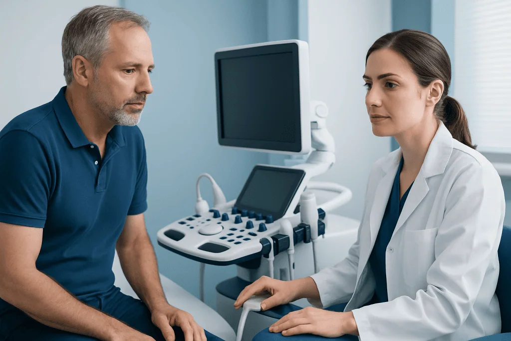
Early signs depend on which aponeurosis is involved, but several patterns are common. Many people notice a gradual onset of localized pain, tenderness when pressing the area, and discomfort that worsens with activity. Stiffness after rest, especially first thing in the morning or after prolonged sitting, can also occur.
In the foot, irritation of the plantar aponeurosis may cause heel or arch pain, especially with the first steps of the day. In the hand, thickening of the palmar aponeurosis can begin as a small lump or cord in the palm and may slowly affect finger extension over time. In the abdominal wall, strain may cause pain when coughing, twisting, or exercising.
Other symptoms can include swelling, a sense of tightness, reduced flexibility, and weakness during movement. A sudden injury may produce sharp pain, bruising, or an audible pop, although this is less common than gradual overuse symptoms. Persistent symptoms deserve medical assessment to rule out related conditions such as plantar fasciitis or more complex tendon and soft-tissue disorders.
- Pain with movement or weight-bearing
- Tenderness over a defined area
- Morning stiffness or stiffness after rest
- Reduced grip, push-off strength, or flexibility
- Visible thickening, pulling, or deformity in some cases
Where aponeurosis disorders happen most often
Although aponeurotic tissue is found in many parts of the body, some regions are affected more often in clinical practice. The plantar aponeurosis in the foot is commonly involved in overuse-related heel and arch pain. Repeated loading from running, prolonged standing, poor footwear, or changes in activity can contribute to irritation in this area.
The palmar aponeurosis in the hand can become abnormally thickened, leading to nodules and cords that may gradually pull one or more fingers toward the palm. This pattern is classically seen in Dupuytren’s contracture. Not every palm nodule progresses, but monitoring changes in hand function is important.
The abdominal wall aponeuroses may be strained during sports, heavy lifting, pregnancy, surgery recovery, or activities that sharply increase pressure in the core. Less commonly, the scalp aponeurosis can be involved in lacerations, trauma, or certain surgical procedures. The exact symptoms, seriousness, and treatment approach vary by location, so evaluation should always be individualized.
Causes and risk factors
Aponeurosis problems may arise from overuse, sudden strain, age-related tissue changes, or underlying medical conditions. Repetitive stress is a major factor, especially in the foot and abdominal wall. Activities that involve running, jumping, rapid directional changes, prolonged standing, or repetitive gripping can place repeated tension on these tissues.
Biomechanics also matter. Flat feet, high arches, tight calf muscles, reduced joint mobility, muscle imbalance, poor technique in sports, and inadequate footwear may increase strain on the plantar aponeurosis. In the hand, age, family history, male sex, diabetes, smoking, alcohol use, and certain connective-tissue tendencies have been associated with palmar aponeurosis disorders.
Additional risk factors include obesity, sudden increases in training intensity, physically demanding work, prior injury, and reduced recovery time. In some cases, an aponeurosis is not the primary problem but becomes painful because nearby muscles, tendons, or joints are overloaded. For that reason, clinicians usually assess the whole movement pattern rather than only the painful spot.
How aponeurosis conditions are diagnosed
Diagnosis starts with a medical history and physical examination. A doctor asks where the pain or tightness is located, how long symptoms have been present, what movements worsen them, and whether the symptoms began gradually or after a specific injury. Functional questions are also important, such as difficulty walking, gripping, exercising, or straightening a finger.
During the examination, the clinician checks tenderness, tissue thickening, range of motion, strength, gait, posture, and any visible pulling or deformity. This is often enough to identify common problems affecting the plantar or palmar aponeurosis. When the diagnosis is uncertain, imaging may help confirm inflammation, degeneration, tearing, or involvement of nearby structures.
Ultrasound can be useful for evaluating soft tissue in motion, while MRI provides detailed images when a deeper injury or alternative diagnosis is suspected. X-rays do not show the aponeurosis itself but may help rule out bone problems. If symptoms involve the foot, a patient may be assessed by specialists in orthopedics or rehabilitation to guide a structured treatment plan.
Treatment options for aponeurosis injuries and disorders
Treatment depends on the location, severity, and cause. Many overuse-related aponeurosis problems improve with conservative care. This often includes modifying activity, reducing repetitive strain, using supportive footwear or splints when appropriate, applying ice after aggravating activity, and taking clinician-guided pain relief measures. Physical therapy can be especially helpful for stretching tight structures, improving strength, and correcting movement patterns that place excess stress on the tissue.
For foot-related problems, orthotic support, calf and foot stretching, and gradual return to activity are commonly recommended. In hand conditions such as palmar aponeurosis thickening, treatment may range from observation to injections, needling techniques, hand therapy, or surgery when finger contracture affects function. Patients with persistent pain may also benefit from evaluation by physical therapy and rehabilitation specialists.
When a significant tear, chronic disabling pain, or advanced contracture is present, procedural treatment may be considered. This can include image-guided injections in selected cases or surgical release/repair when less invasive options have not provided enough improvement. For selected hand problems, referral for hand surgery may be appropriate. Treatment plans are individualized, and recovery timelines vary based on the tissue involved and the person’s activity level.
Near the end of the care pathway, some patients need multidisciplinary review to coordinate diagnosis, imaging, rehabilitation, and surgery if required. Acibadem International’s multidisciplinary specialists and JCI-accredited hospitals diagnose and treat aponeurosis-related conditions for international patients when advanced evaluation or treatment is needed.
Self-care, recovery, and prevention
Recovery often improves when stress on the affected tissue is reduced without stopping all movement. Relative rest is usually more helpful than complete inactivity. This means avoiding activities that sharply worsen symptoms while maintaining gentle motion and following a clinician’s exercise plan. Sudden return to full activity can delay healing.
Supportive self-care may include stretching, strengthening, good footwear, warm-up before exercise, and gradual progression of training loads. People who stand for long periods may benefit from cushioned shoes, rest breaks, and attention to posture and lower-limb mechanics. Those with hand symptoms should avoid forceful repetitive gripping if it increases discomfort.
Prevention focuses on managing the factors that overload the tissue. This may include maintaining a healthy weight, improving flexibility, treating muscle imbalance, using proper sports technique, and addressing early symptoms before they become longstanding. If symptoms persist despite home care, specialist assessment can help prevent chronic pain or loss of function.
When to seek medical care
Medical care is advisable if pain lasts more than a few weeks, keeps returning, or begins to affect work, walking, sleep, sports, or hand function. A person should also seek assessment if they notice a lump, thickened cord, increasing stiffness, or difficulty straightening a finger, since these changes may signal progression of a palmar aponeurosis disorder.
More urgent evaluation is appropriate after a sudden injury with marked swelling, bruising, severe pain, inability to bear weight, obvious deformity, or loss of movement. Medical review is also important if symptoms occur together with numbness, fever, redness, or a wound, because these features may suggest a different or more serious problem.
In general, early assessment can make treatment simpler and more effective. A qualified doctor can confirm whether the aponeurosis is the source of symptoms, identify contributing factors, and recommend the safest plan for recovery.
Frequently asked questions
What is an aponeurosis in simple terms?
An aponeurosis is a broad, strong sheet of connective tissue that helps attach muscles to other structures. It supports movement by spreading and transmitting force across an area rather than through a narrow cord like a typical tendon.
Is an aponeurosis the same as fascia or a tendon?
Not exactly. An aponeurosis is closely related to both fascia and tendon, but it is usually flatter and more sheet-like than a tendon. It can also blend with surrounding fascial tissue, which is why the terms may sometimes seem similar in everyday discussion.
Can aponeurosis pain go away on its own?
Mild overuse-related symptoms may improve with rest, activity modification, and simple self-care. However, ongoing pain, repeated flare-ups, or symptoms that limit function should be assessed by a doctor to confirm the diagnosis and guide treatment.
What is the most common aponeurosis problem?
In general practice, the plantar aponeurosis in the foot is one of the most commonly discussed sites because it is linked to heel and arch pain. The palmar aponeurosis in the hand is also important because thickening there can gradually affect finger movement.
Does every palmar aponeurosis lump mean surgery is needed?
No. Some people have small nodules or cords that change very little over time and may only need monitoring. Surgery or another procedure is usually considered when contracture significantly affects hand function or daily activities.
How long does recovery from an aponeurosis injury take?
Recovery time depends on the location, severity, and cause of the problem. Mild irritation may improve over weeks, while chronic degeneration, major tears, or contractures can take longer and may require structured rehabilitation or procedures.
References
- American Academy of Orthopaedic Surgeons
- National Institute of Arthritis and Musculoskeletal and Skin Diseases
- American Society for Surgery of the Hand
- Mayo Clinic
- MedlinePlus
This article is for general information only and is not a substitute for professional medical advice. Please consult a qualified doctor about your individual situation.
Explore treatments in Turkey — costs, top hospitals & a free quote
JCI-accredited · board-certified surgeons · reply within 24h