Bone Scan Procedure: Preparation, Procedure and Results
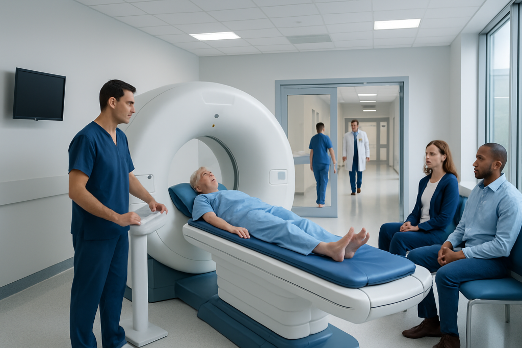
A bone scan detects changes in bone metabolism and may help evaluate fractures, infection, arthritis, cancer spread, and other bone conditions. Most people can eat and take usual medicines before a bone scan, but individual instructions from the imaging team should always be followed.
Key Takeaways
- A bone scan detects changes in bone metabolism and may help evaluate fractures, infection, arthritis, cancer spread, and other bone conditions.
- Most people can eat and take usual medicines before a bone scan, but individual instructions from the imaging team should always be followed.
- The camera usually takes about 30 to 60 minutes to obtain images, while the full appointment may be longer because the tracer needs time to circulate.
- Drinking water and emptying the bladder frequently after the tracer injection helps remove remaining tracer from the body.
- Results are usually interpreted by a nuclear medicine specialist or radiologist and sent to the referring doctor rather than provided immediately.
A bone scan procedure is a nuclear medicine imaging test that helps clinicians identify areas of unusually high or low bone activity. It usually involves an injection of a small radioactive tracer, a waiting period, and a scan that is painless and noninvasive.
Overview: What Is a Bone Scan Procedure?
A bone scan procedure, also called bone scintigraphy or a nuclear bone scan, is an imaging test that shows how actively different areas of bone are changing. It uses a very small amount of radioactive material, called a tracer, together with a special camera. The tracer collects in bones and is more visible in areas with increased or reduced bone turnover.
Clinicians may request this test when an X-ray or other scan does not fully explain bone pain or when they need to investigate a possible fracture, infection, inflammation, or bone disease. It can also be part of staging or follow-up for some cancers, because certain cancers can spread to bone. A bone scan is sensitive for detecting abnormal bone activity, but it does not always identify the exact cause of a finding.
The test is generally safe, painless apart from the brief injection, and performed by a nuclear medicine team. Findings are interpreted alongside symptoms, medical history, physical examination, laboratory tests, and other imaging when needed.
How a Bone Scan Works and Who May Need One
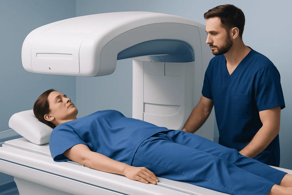
After the tracer is injected into a vein, it travels through the bloodstream and is taken up by bone tissue. Bones naturally renew themselves throughout life. When a bone is healing, inflamed, infected, under stress, or affected by certain tumors, its activity can change. These areas may take up more tracer and appear as “hot spots” on the images. Less commonly, reduced uptake may also be clinically important.
A doctor may recommend a bone scan to assess unexplained or persistent bone pain, a suspected stress fracture, possible bone infection, inflammatory joint disease, or complications of a known bone disorder. It may also help evaluate the extent of some cancers, including prostate cancer, when there is a clinical reason to check for bone involvement.
Bone scan preparation for prostate cancer is broadly similar to preparation for other indications. However, the oncology team may coordinate the timing with blood tests, CT, MRI, PET imaging, treatment plans, or prior scans. Patients should tell the team about any recent imaging studies, especially those involving barium or contrast agents, as these can occasionally affect image interpretation.
Bone Scan Procedure Preparation and Pre-Procedure Teaching

For most people, bone scan preparations are straightforward. Fasting is usually not required, and routine medicines can often be taken as prescribed. However, every imaging department may have specific instructions, particularly for patients having a specialized scan or multiple tests on the same day. The scheduling team should be asked for individualized guidance in advance.
During bone scan pre-procedure teaching, patients should tell the healthcare team if they are pregnant, might be pregnant, or are breastfeeding. The team will discuss whether the test should be delayed, modified, or performed with additional precautions. It is also important to mention allergies, kidney problems, recent surgery or injury, and all medicines, vitamins, and supplements.
Comfortable clothing without metal fasteners may be useful, although a gown may be provided. Jewelry, belts, watches, and metal objects may need to be removed before imaging. Bringing water, reading material, or a quiet activity can make the waiting period more comfortable.
What should you not do before a bone scan?
Patients should not make changes to prescribed medicines, stop eating, or restrict fluids unless their doctor or imaging center specifically tells them to do so. They should avoid arriving without disclosing a possible pregnancy, breastfeeding, recent nuclear medicine tests, or recent barium-based examinations. Following the center’s exact instructions is more important than using general advice found online.
How much water to drink before a nuclear bone scan?
There is no single amount of water that suits every person. Many centers encourage patients to drink several glasses of water during the waiting period after the injection, unless fluid intake is medically restricted for a condition such as heart failure or advanced kidney disease. The imaging team can give a personal recommendation and will usually ask the patient to empty their bladder before the pictures are taken.
Step by Step: What Happens During the Scan
The appointment commonly begins with a brief review of medical details and a small injection of tracer into a vein, usually in the arm or hand. The injection is similar to a blood test or intravenous line placement. Some scans include pictures immediately after injection, but many require a waiting period of roughly two to four hours so the tracer can reach the bones.
During the waiting period, patients can often leave the department or sit in a designated area, depending on local procedures. They are usually encouraged to drink fluids and urinate regularly. In selected cases, additional early images or blood-flow images may be taken. This is sometimes called a three-phase bone scan and can provide more information about infection, injury, or inflammation.
For the main imaging portion, the patient lies on a padded table while a gamma camera moves slowly around or close to the body. The camera does not use strong magnets and does not usually enclose the patient like some MRI scanners. Remaining still is important for clear images, but the technologist can help with positioning and comfort.
How long are you in the machine for a bone scan?
The camera portion usually lasts about 30 to 60 minutes, although a more detailed scan can take longer. The total visit may last several hours because of the time between tracer injection and delayed imaging. The technologist will explain the expected schedule and let the patient know if additional images are needed.
Benefits, Limitations and Possible Risks
A key benefit of a bone scan is that it can survey much of the skeleton in one examination and may identify bone changes before they are clearly visible on a standard X-ray. It can be particularly helpful when symptoms are difficult to localize or when clinicians need a whole-body view of bone activity.
The images are sensitive but not fully specific. For example, a hot spot can occur with healing after an injury, arthritis, infection, benign bone conditions, or cancer. For this reason, an abnormal result does not by itself diagnose cancer or another single condition. A doctor may recommend targeted X-rays, CT, MRI, PET imaging, blood tests, or follow-up imaging to clarify a result.
The radiation exposure is generally low and the tracer loses radioactivity naturally over time. Allergic reactions are uncommon, and serious reactions are rare. Mild bruising, discomfort, or temporary soreness can occur at the injection site. Patients who are pregnant or breastfeeding need individualized advice because the tracer may pose considerations for the baby.
Recovery Timeline and Aftercare
Most people return to usual activities immediately after a bone scan. There is generally no sedation, no incision, and no recovery period. Unless the care team gives different instructions, eating, drinking, driving, working, and taking regular medicines can continue as normal after the examination.
Drinking extra fluids for the rest of the day, when medically appropriate, and urinating regularly can help the body clear the remaining tracer. Careful handwashing after using the toilet is sensible. The nuclear medicine team may provide temporary guidance about close contact with infants, young children, or pregnant people, depending on the tracer used and local safety practices.
If redness, swelling, worsening pain, or other unexpected symptoms develop at the injection site, patients should contact the imaging department or their doctor. These concerns are uncommon, but it is appropriate to seek advice rather than ignore a change that persists.
Results and Next Steps
After the images are completed, a nuclear medicine physician or radiologist reviews them and prepares a report for the doctor who ordered the scan. The report describes whether tracer uptake appears typical or whether there are areas that may need correlation with other information. The referring clinician discusses what the findings mean in the context of the person’s health and reason for testing.
Do you get bone scan results immediately?
Usually, final bone scan results are not available immediately because image review and formal reporting take time. A technologist may confirm that the images have been completed, but they do not typically interpret the findings. The referring doctor may receive the report within a few days, although timing varies by hospital and clinical urgency.
If a scan identifies an area requiring further assessment, this does not necessarily mean a serious problem is present. Additional tests are often used to distinguish common causes, such as arthritis or a healing injury, from other conditions. Acibadem International’s multidisciplinary specialists and JCI-accredited hospitals can coordinate nuclear medicine imaging and follow-up assessment for international patients when appropriate.
When to Seek Medical Care
A bone scan itself usually does not require special medical care afterward. However, patients should contact their doctor if they develop unusual or persistent symptoms after the injection, such as significant swelling, rash, breathing difficulty, or feeling unwell. Severe symptoms, especially trouble breathing or facial swelling, require urgent medical attention.
More broadly, unexplained bone pain that is persistent, worsening, occurs at night, follows a significant injury, or is associated with fever, unexplained weight loss, or reduced ability to use a limb should be medically assessed. These symptoms have many possible causes, and early evaluation can help guide appropriate testing and treatment.
People with a known cancer diagnosis should also inform their oncology team about new, persistent bone pain, weakness, numbness, loss of bladder or bowel control, or sudden difficulty walking. These symptoms need timely assessment. Depending on the underlying concern, clinicians may coordinate imaging and care through services such as oncology treatment.
Frequently asked questions
Is a bone scan painful?
A bone scan is usually not painful. The tracer injection may cause a brief pinprick, similar to a blood draw, and lying still on the imaging table may feel uncomfortable for some people. The camera itself does not touch the body in most cases and does not cause pain.
Can you eat before a bone scan?
Most routine bone scans do not require fasting, so people can usually eat normally. Instructions may differ if another test is scheduled at the same time or if a specialized imaging protocol is being used. The imaging center’s instructions should always take priority.
Why do I need to wait after the tracer injection?
The waiting period gives the tracer time to circulate through the bloodstream and collect in the bones. This improves the clarity and usefulness of the delayed images. Patients are often asked to drink fluids and empty their bladder during this time.
What does a hot spot on a bone scan mean?
A hot spot is an area where more tracer has collected, suggesting increased bone activity. It can be caused by many conditions, including healing fractures, arthritis, infection, or cancer. Further clinical assessment or imaging may be needed to determine the cause.
Can a bone scan detect cancer?
A bone scan can show bone changes that may be associated with cancer spread to bone, but it cannot confirm cancer on its own. Noncancerous conditions can produce similar findings. Doctors interpret the scan with medical history, examination findings, laboratory results, and other imaging tests.
How long does radiation stay in the body after a bone scan?
The tracer gradually loses radioactivity and is also eliminated through urine, mostly over a short period after the test. Drinking fluids and urinating regularly, if appropriate for the person’s health, can support this process. The nuclear medicine team will provide any specific safety advice needed.
References
- RadiologyInfo.org
- Society of Nuclear Medicine and Molecular Imaging
- National Cancer Institute
- National Health Service
- Mayo Clinic
This article is for general information only and is not a substitute for professional medical advice. Please consult a qualified doctor about your individual situation.
Joint and spine care in Turkey — expert assessment & treatment
JCI-accredited · board-certified surgeons · reply within 24h
Add us as a Preferred Source to see more of our trusted health content across Google Search, AI Overviews and Discover.

More from the Health Library



Related Specialists

Dr. Eymen Aslan
Anesthesiology
Dr. Mustafa Mert Okumuş (m)
Aesthetic Plastic and Reconstructive Surgery
Prof. Dr. Meltem Vural
Physical Medicine & Rehabilitation
Prof. Dr. H. Armağan Arıcan
Radiation Oncology