Bone Tumour Surgery: Procedure, Recovery and Results
Bone tumours may be benign or malignant, and surgery is not needed for every tumour. Specialist imaging and biopsy findings guide the safest surgical plan.
Key Takeaways
- Bone tumours may be benign or malignant, and surgery is not needed for every tumour.
- Specialist imaging and biopsy findings guide the safest surgical plan.
- Limb-sparing surgery can often remove a tumour while retaining the limb, sometimes with reconstruction.
- Bone tumor surgery recovery time ranges from weeks to months and may be longer after major reconstruction.
- Follow-up imaging, rehabilitation and monitoring are important after surgery, especially for malignant tumours.
Bone tumour surgery is an operation to remove a tumour from bone, confirm its diagnosis and, when needed, rebuild the affected area. The approach, recovery and outlook depend on whether the tumour is benign or cancerous, its location and size, and whether reconstruction or additional cancer treatment is required.
Overview: What is bone tumour surgery?
Bone tumour surgery is performed to remove a growth arising in a bone or, less commonly, a tumour that has spread to bone from another part of the body. It may be used to treat a painful or weakening benign tumour, remove a suspected cancer, obtain tissue for diagnosis, prevent a fracture, or control symptoms caused by advanced cancer.
The aim is to remove the tumour as completely and safely as possible while protecting nearby muscles, joints, nerves and blood vessels. Many operations are planned by an orthopaedic oncologist together with radiologists, pathologists, medical oncologists, radiation oncologists and rehabilitation specialists. The final plan is individual because bone tumours vary greatly in behaviour and location.
Not every bone lesion requires bone tumor removal surgery. Some noncancerous lesions can be monitored with imaging, while others may be treated with medication or a minimally invasive procedure. A specialist assessment is important before deciding whether surgery offers the best balance of benefit and risk.
How serious is a bone tumor?
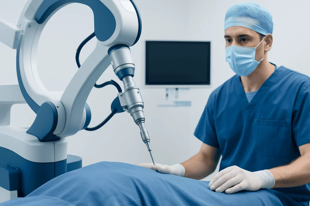
A bone tumour can range from harmless to life-threatening, so its seriousness cannot be judged from symptoms alone. Many bone tumours are benign, meaning they do not spread to distant organs. Even benign tumours can require treatment if they cause pain, affect growth in a child, damage a joint or make the bone more likely to break.
Malignant primary bone tumours, often called bone cancers, can invade nearby tissue and may spread to other parts of the body. Their outlook depends on the tumour type, grade, size, location, whether it has spread and how it responds to treatment. Common primary bone cancers include osteosarcoma, Ewing sarcoma and chondrosarcoma, but each has a different treatment pathway.
Sometimes imaging identifies a lesion that is not a tumour at all, such as a cyst, infection or stress-related bone change. For this reason, patients should avoid assuming a diagnosis based on an X-ray report alone. A review by a specialist team and, when appropriate, a carefully planned biopsy provide clearer answers.
Candidacy, diagnosis and planning before surgery
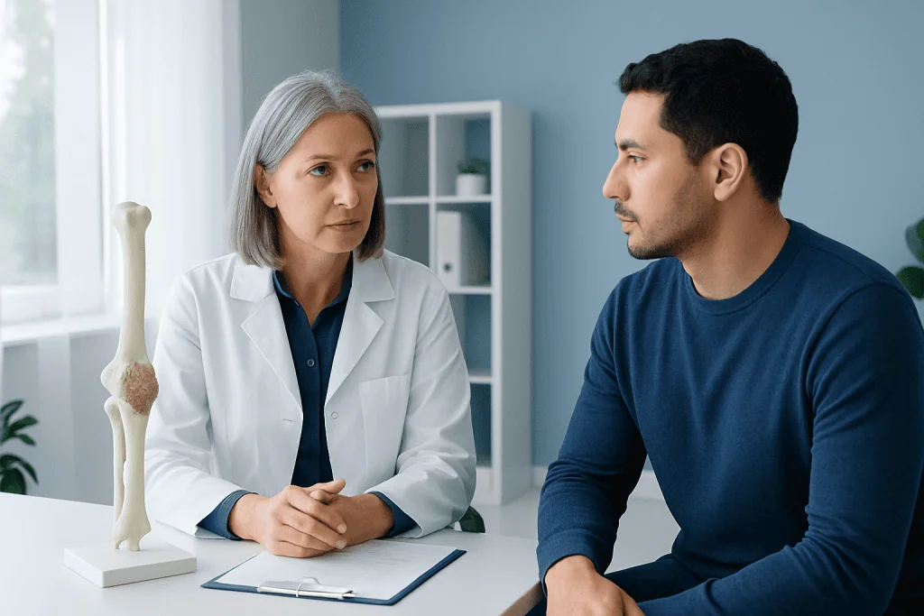
A person may be considered for bone tumor resection when a tumour is confirmed or strongly suspected, when it is growing or causing symptoms, or when it threatens the strength or function of a bone. Surgery may also be part of a combined treatment plan for cancer, before or after chemotherapy and sometimes radiotherapy. The timing depends on the exact diagnosis.
Assessment commonly includes an examination and imaging such as X-rays, magnetic resonance imaging (MRI) and computed tomography (CT). A chest scan, bone scan or PET scan may be used when doctors need to check for spread. Blood tests and an assessment of general health help prepare for anaesthesia and recovery.
If a biopsy is needed, it should ideally be planned by the team that may perform definitive surgery. The biopsy route matters because it may need to be removed during the operation to avoid leaving tumour cells along the needle or incision track. Pathology results identify the tumour type and support decisions about surgical margins and additional treatment.
- Wide excision: removal of the tumour with a surrounding margin of healthy-looking tissue, often used for malignant tumours.
- Curettage: scraping a tumour from inside the bone, sometimes followed by filling the space with bone graft, cement or another material.
- Stabilisation or reconstruction: use of plates, screws, rods, a prosthetic joint, bone graft or other reconstruction to restore support and movement.
How does bone tumour surgery work? Step by step
Before the operation, the surgical team reviews scans, pathology and the reconstruction plan. The patient receives anaesthesia, most often general anaesthesia, and the limb or affected area is positioned to allow safe access. In selected cases, surgeons may use image guidance, custom instruments or implants designed from detailed imaging.
During bone tumor removal surgery, the surgeon makes an incision and carefully reaches the affected bone while protecting surrounding structures. The tumour is removed by curettage or by resection of a section of bone, depending on its type and behaviour. For a cancerous tumour, the surgeon aims for an appropriate margin based on the diagnosis and treatment plan.
If removing the tumour leaves the bone unstable or creates a large defect, reconstruction may be carried out during the same procedure. Options include internal fixation, a metal implant, joint replacement component, bone graft or, in selected complex cases, reconstruction using the patient’s own tissue. The tissue removed is examined by a pathologist, including assessment of surgical margins when relevant.
Limb-sparing surgery is often possible, but it is not appropriate in every situation. Rarely, amputation may provide the safest way to fully treat a tumour that involves major nerves or blood vessels, severe infection, or extensive tissues. The team discusses these possibilities before surgery whenever feasible.
How long does it take to recover from bone tumor surgery?
Bone tumor surgery recovery time varies widely. A smaller curettage procedure may allow gradual return to daily activities over several weeks, while recovery after removal of a large tumour and limb reconstruction often takes several months. Recovery can take longer when chemotherapy, radiotherapy, wound healing concerns, infection or another health condition affects rehabilitation.
In the first days after surgery, care focuses on pain control, protecting the wound, preventing blood clots when indicated and safely starting movement. Some patients need crutches, a brace, walker or wheelchair for a period. The surgeon will explain how much weight can be placed through the limb, which depends on the operation and reconstruction.
Physical therapy is a central part of bone tumor surgery recovery. It helps restore range of motion, muscle strength, balance and confidence with walking or using the affected body part. Return to driving, work, sports and heavier activities should be guided by the treating team rather than a fixed timetable.
A bone tumor removal recovery time can also include emotional adjustment. Changes in mobility, appearance, uncertainty during follow-up and ongoing cancer treatment can be challenging. Support from rehabilitation professionals, mental health specialists, family and patient support services can be helpful throughout recovery.
What usually happens after a tumor is removed?
After surgery, the removed tissue is examined in detail by a pathologist. The final report confirms the tumour type and, for cancer, provides information such as grade and whether the edges of the removed tissue are free of tumour. These findings help the multidisciplinary team decide whether any further treatment is recommended.
For benign tumours, follow-up may involve wound checks, rehabilitation and periodic X-rays or scans to confirm healing and watch for recurrence. For malignant bone tumours, follow-up is usually more frequent and may include imaging of the surgical site and chest, since some bone cancers can spread to the lungs. The schedule is tailored to the diagnosis and treatment received.
Some patients require chemotherapy, radiotherapy or both as part of their care. These treatments may be given before surgery to shrink or control a tumour, or after surgery to reduce the risk of remaining microscopic disease in selected cancer types. They are not routinely needed for all benign tumours or all bone cancers.
Long-term care also monitors the reconstructed bone or implant, joint movement, nerve function and bone strength. Contact sports, high-impact activities or heavy lifting may need to be limited temporarily or permanently after certain reconstructions. The surgical and rehabilitation teams can provide practical guidance for each stage.
Can bone tumors be cured? Benefits, risks and outlook
Many benign bone tumours can be cured or effectively controlled with complete treatment, although a few can recur and need further monitoring. Some malignant primary bone tumours can also be cured, particularly when they are localised and treated with the appropriate combination of surgery and, when indicated, chemotherapy or radiotherapy. No individual outcome can be predicted without knowing the exact diagnosis and stage.
The potential benefits of surgery include removing the tumour, relieving pain, protecting the bone from fracture, preserving mobility and obtaining a definitive diagnosis. In cancer care, surgery may be the most important treatment for local control. The expected benefit should always be considered alongside the likely effect on function and quality of life.
Possible risks include bleeding, infection, blood clots, delayed wound or bone healing, stiffness, pain, nerve or blood-vessel injury, fracture, implant problems and the need for additional operations. There is also a risk of tumour recurrence, especially if a tumour cannot be removed with an adequate margin or has an aggressive biology. The surgeon discusses risks in relation to the planned operation.
For international patients, Acibadem International’s multidisciplinary specialists and JCI-accredited hospitals diagnose and treat bone tumours with coordinated surgical, oncology and rehabilitation care. A second opinion from a bone tumour specialist may be particularly useful before complex surgery or when treatment recommendations are uncertain.
When to seek medical care
Persistent bone pain should be assessed, especially if it becomes worse over time, occurs at night, is associated with swelling or affects normal activities. A new lump near a bone or joint, unexplained limping, reduced movement, or a fracture after a minor injury also deserves medical evaluation. These symptoms often have causes other than cancer, but prompt assessment can identify the right treatment.
After bone tumour surgery, patients should contact their surgical team urgently for fever, worsening redness or drainage from the wound, rapidly increasing pain or swelling, new numbness or weakness, shortness of breath, chest pain, or a sudden inability to use the operated limb. These symptoms need timely medical advice because they may indicate a complication.
Routine follow-up appointments should not be skipped, even when recovery appears to be going well. Imaging and clinical examinations help the team assess healing, rehabilitation progress and any evidence that a tumour has returned. Patients should ask their doctor which symptoms and activity limits apply to their specific procedure.
Frequently asked questions
How long does bone tumour surgery take?
The length of surgery depends on the tumour location, whether it can be removed by curettage or needs a wider resection, and whether reconstruction is required. A relatively limited procedure may take a few hours, while complex limb-sparing surgery with reconstruction can take much longer. The surgical team can give the most reliable estimate after reviewing imaging and the operative plan.
Is bone tumour surgery painful?
The operation is performed under anaesthesia, so the patient does not feel pain during surgery. Pain, swelling and stiffness are common afterward, but these are managed with a personalised pain-control plan and rehabilitation. Pain should gradually improve; worsening or unexpected pain should be reported to the care team.
Will I be able to walk after bone tumour removal surgery?
Many people can walk again after surgery on a leg or pelvis, but the pace and extent of recovery depend on the bone involved, the amount removed and the type of reconstruction. Temporary aids such as crutches, a walker or a brace are often needed. Physiotherapy helps patients regain safe movement and strength.
Does every bone tumour need to be removed?
No. Some benign bone tumours and incidental bone lesions can be safely monitored with periodic imaging if they are not causing symptoms or weakening the bone. Surgery may be recommended when a lesion is painful, enlarging, structurally risky, uncertain in diagnosis or cancerous. The decision should be made with a specialist familiar with bone tumours.
What are the chances that a bone tumour will come back after surgery?
Recurrence risk depends on the type of tumour, its grade, whether it was benign or malignant, and how completely it could be removed. Some benign tumours and some cancers have a higher tendency to return than others. Follow-up examinations and imaging are designed to detect recurrence early.
Can a biopsy spread a bone tumour?
A properly planned biopsy has a very low risk of causing tumour spread. For suspected bone cancer, it is important that the biopsy is performed or coordinated by the specialist team that would carry out definitive surgery. This allows the biopsy path to be placed where it can be removed during surgery if necessary.
References
- American Cancer Society
- National Cancer Institute
- American Academy of Orthopaedic Surgeons
- NHS
- European Society for Medical Oncology
This article is for general information only and is not a substitute for professional medical advice. Please consult a qualified doctor about your individual situation.
Joint and spine care in Turkey — expert assessment & treatment
JCI-accredited · board-certified surgeons · reply within 24h
Add us as a Preferred Source to see more of our trusted health content across Google Search, AI Overviews and Discover.