Double Vision: When Diplopia Needs a Neuro-Ophthalmology Evaluation

Double vision can be monocular, affecting one eye, or binocular, caused by the eyes not aligning properly. Binocular diplopia often needs a careful eye movement and neurologic assessment to identify nerve, muscle, or brain-related causes.
Key Takeaways
- Double vision can be monocular, affecting one eye, or binocular, caused by the eyes not aligning properly.
- Binocular diplopia often needs a careful eye movement and neurologic assessment to identify nerve, muscle, or brain-related causes.
- Sudden double vision with headache, weakness, drooping eyelid, speech difficulty, or imbalance should be assessed urgently.
- Diagnosis may include an eye examination, neurologic examination, blood tests, and sometimes MRI, CT, or vascular imaging.
- Treatment depends on the cause and may include observation, prisms, patching, medication, treatment of an underlying condition, or eye muscle surgery.
Double vision, medically called diplopia, may come from the eye itself or from problems in the nerves, muscles, or brain pathways that control eye movement. A neuro-ophthalmology evaluation is especially important when diplopia is sudden, persistent, associated with pain or neurologic symptoms, or improves when either eye is covered.
Overview
Double vision, or diplopia, means seeing two images of a single object. The images may appear side by side, one above the other, diagonally separated, or slightly overlapping. Some people notice it only when looking in a certain direction, when tired, or at distance; others may experience it constantly.
Doctors first determine whether the double vision is monocular or binocular. Monocular diplopia remains when the unaffected eye is covered and usually relates to an eye problem such as dry eye, cataract, corneal irregularity, or an uncorrected prescription. Binocular diplopia disappears when either eye is covered and usually means the two eyes are not pointing at the same target.
Binocular double vision may involve the eye muscles, the nerves that move the eyes, the neuromuscular junction, the eye socket, or brain pathways that coordinate eye movement. This is where neuro-ophthalmology becomes important. A neuro-ophthalmologist evaluates conditions that sit at the boundary between eye health and the nervous system, helping to identify causes that may not be obvious during a routine eye check.
Symptoms That May Accompany Double Vision
Diplopia itself can vary in pattern. Horizontal double vision is often described as two images side by side, while vertical diplopia is seen as one image above the other. Some patients tilt or turn the head to reduce the separation between images, and others may close one eye to read, walk, or drive more comfortably.
Associated symptoms provide important clues. Eye pain, pain with eye movement, headache, drooping eyelid, unequal pupils, bulging of one eye, nausea, dizziness, imbalance, facial numbness, or weakness can point toward nerve, muscle, orbital, or neurologic causes. Fluctuating double vision that is worse later in the day may suggest a disorder of the nerve-muscle connection, such as myasthenia gravis.
Double vision can also affect daily safety and comfort. It may make stairs, driving, reading, screen use, or sports difficult because depth perception is disrupted. Until a doctor identifies the cause and vision is stable, patients are generally advised to avoid driving or operating machinery if diplopia interferes with clear single vision.
Causes and Risk Factors

Many causes of diplopia are treatable, but they are different enough that accurate diagnosis matters. Eye-related causes of monocular diplopia include dry eye disease, irregular astigmatism, keratoconus, cataract, lens displacement, retinal disorders, or problems with glasses or contact lenses. These causes are usually assessed first by an ophthalmologist or optometrist.
Binocular diplopia is commonly related to eye misalignment. In adults, this may result from cranial nerve palsies affecting the third, fourth, or sixth cranial nerves; thyroid eye disease; myasthenia gravis; orbital inflammation; trauma; previous eye surgery; decompensated childhood strabismus; or, less commonly, tumors, aneurysms, stroke, or inflammatory neurologic disease. In children, sudden eye crossing or persistent double vision should also be assessed, as the causes and treatment priorities may differ by age.
Risk factors depend on the underlying condition. Diabetes, high blood pressure, high cholesterol, smoking, vascular disease, thyroid disease, autoimmune disease, migraine, head injury, and a history of cancer or neurologic illness can all be relevant. Older adults may develop small-vessel cranial nerve palsies, which often improve over time, but a doctor must still rule out other causes when the presentation is unusual or accompanied by warning signs.
- Sudden onset: may require urgent assessment, especially if new or severe.
- Pain or headache: can suggest inflammation, aneurysm, infection, or other serious causes.
- Pupil changes or drooping eyelid: can be important signs in third nerve palsy.
- Neurologic symptoms: weakness, speech trouble, confusion, or imbalance need immediate medical attention.
How Neuro-Ophthalmology Evaluation Diagnoses Diplopia
A neuro-ophthalmology evaluation begins with a detailed history. The doctor asks when the double vision started, whether it is constant or intermittent, whether covering one eye stops it, and whether it changes with gaze direction, fatigue, or head position. Information about recent infections, trauma, surgery, new medications, vascular risk factors, thyroid disease, autoimmune disease, and neurologic symptoms is also important.
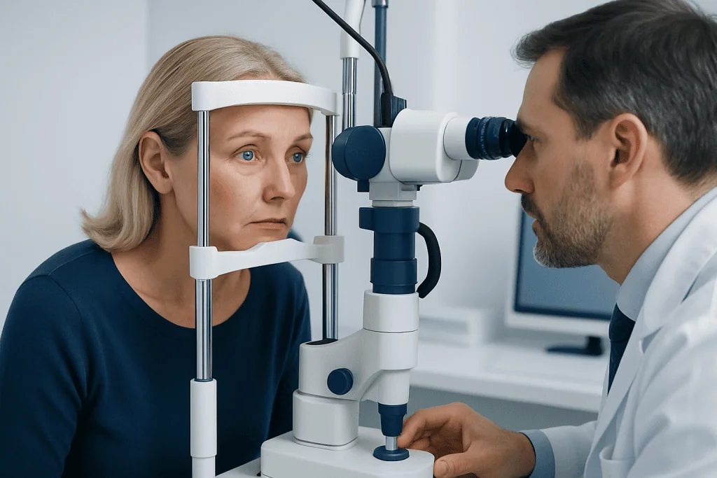
The examination typically includes visual acuity, refraction when needed, pupil testing, eyelid assessment, eye pressure, and a careful look at the front and back of the eyes. The specialist then evaluates eye alignment and eye movements in different directions of gaze. Tests such as cover testing, prism measurements, double vision mapping, color vision, visual fields, and optic nerve assessment may help localize the problem.
Depending on findings, additional testing may be recommended. Blood tests can check for thyroid disease, inflammation, infection, autoimmune markers, diabetes control, or myasthenia gravis antibodies. Imaging such as MRI or CT may be used to examine the brain, eye sockets, and cranial nerves. CT or MR angiography may be considered when a vascular cause, such as aneurysm, is a concern.
The goal is not only to confirm that the eyes are misaligned, but to understand why. A small change in eye movement may reveal a specific cranial nerve palsy, while variable weakness may suggest a neuromuscular junction disorder. In other cases, swelling or restriction of the eye muscles may point toward thyroid eye disease or orbital inflammation.
Treatment Options
Treatment for double vision depends on the cause, severity, and expected course. Some conditions improve with time and careful monitoring, while others require targeted treatment. For example, certain microvascular cranial nerve palsies may gradually recover, whereas thyroid eye disease, myasthenia gravis, inflammation, infection, aneurysm, or stroke-related causes require specific medical management.
Symptom relief is often possible while the underlying condition is being treated. Temporary patching of one eye can eliminate double vision, although it also reduces depth perception. Fresnel prisms, which are thin temporary prism films placed on glasses, may help align images in selected cases. Permanent prism glasses can be considered if the misalignment is stable.
Medical treatment may include managing vascular risk factors, treating thyroid disease, using medications for myasthenia gravis, addressing inflammation, or treating infection when present. If double vision is caused by an orbital or neurologic condition, care may involve ophthalmology, neurology, endocrinology, radiology, and sometimes neurosurgery or rheumatology.
When eye misalignment remains stable and troublesome despite medical care, eye muscle surgery may be considered. Surgery aims to improve alignment in the most useful gaze positions, but the decision depends on measurements over time, the underlying diagnosis, and the patient’s visual needs. The doctor will discuss realistic goals, alternatives, and follow-up before recommending any procedure.
Prevention and Self-Care
Not all causes of double vision can be prevented, but general health measures can reduce some risks. Good control of diabetes, blood pressure, cholesterol, and thyroid disease supports healthy blood vessels and nerves. Avoiding smoking, using protective eyewear during sports or high-risk work, and seeking care after head or eye injury are also practical steps.
Patients who already have diplopia can take simple precautions. If vision is double, driving should be avoided until a clinician confirms it is safe. Good lighting, handrails on stairs, removing trip hazards, and taking extra time when walking can help prevent falls. If a patch is used, alternating the patched eye as advised may reduce strain, but patients should follow their clinician’s instructions.
It is helpful to keep notes about the double vision before an appointment. Patients can write down when it occurs, whether it is horizontal or vertical, whether it improves when either eye is covered, and whether it changes with fatigue or gaze direction. Bringing current glasses, previous eye records, medication lists, and any recent imaging reports can make the evaluation more efficient.
When to See a Doctor
Any new, persistent, or unexplained double vision should be assessed by a qualified eye doctor. A routine ophthalmology examination may be enough when monocular diplopia is suspected, but binocular diplopia often benefits from neuro-ophthalmology evaluation, especially if it is new, worsening, or associated with neurologic features.
Urgent medical assessment is important if double vision begins suddenly or occurs with severe headache, eye pain, drooping eyelid, unequal pupils, weakness, numbness, trouble speaking, facial droop, confusion, difficulty walking, loss of vision, fever, or recent head trauma. These symptoms do not always mean a dangerous condition is present, but they require timely evaluation to protect vision and overall health.
Patients should also seek care if double vision interferes with reading, work, walking, or driving, even if symptoms are mild. Early assessment can provide reassurance, reduce safety risks, and identify treatable causes. At Acibadem International, multidisciplinary specialists in JCI-accredited hospitals evaluate and treat complex eye and neurologic causes of diplopia for international patients, using coordinated care when several specialties are needed.
Frequently asked questions
Is double vision always a sign of a serious problem?
No. Some causes, such as dry eye, cataract, or a glasses prescription issue, are common and treatable. However, new binocular double vision can sometimes reflect a nerve, muscle, or brain-related condition, so it should be medically evaluated.
How can a person tell if double vision is monocular or binocular?
A simple clue is to cover one eye, then the other. If the double vision remains when only one eye is open, it is likely monocular and often related to the eye itself. If it disappears when either eye is covered, it is likely binocular and usually means the eyes are not aligned.
When does diplopia need urgent care?
Urgent evaluation is needed when double vision starts suddenly or occurs with severe headache, eye pain, drooping eyelid, unequal pupils, weakness, numbness, speech problems, imbalance, confusion, or recent trauma. These symptoms require prompt assessment because some causes need time-sensitive treatment.
What does a neuro-ophthalmologist do for double vision?
A neuro-ophthalmologist examines both the visual system and the nervous system pathways that control eye movement. The evaluation may include eye alignment measurements, pupil and eyelid assessment, optic nerve testing, visual fields, blood tests, and imaging when needed.
Can double vision go away on its own?
Some types of double vision, such as certain small-vessel cranial nerve palsies, may improve over weeks to months. Even when improvement is likely, a doctor should confirm the cause and monitor recovery. Treatment may be needed if symptoms are severe, persistent, or due to another condition.
Can prism glasses help diplopia?
Prism glasses can help selected patients by shifting images so the brain can fuse them into one. Temporary prisms may be used while the condition is changing, and permanent prisms may be considered once measurements are stable. They are not suitable for every type of diplopia, so assessment is important.
Is it safe to drive with double vision?
Driving is generally unsafe if double vision affects clear single vision, depth perception, or reaction time. A patient should avoid driving until evaluated and advised by a qualified clinician. Temporary measures such as patching may reduce diplopia but can also affect depth perception.
References
- American Academy of Ophthalmology
- North American Neuro-Ophthalmology Society
- National Institute of Neurological Disorders and Stroke
- Mayo Clinic
- Merck Manual Professional Edition
This article is for general information only and is not a substitute for professional medical advice. Please consult a qualified doctor about your individual situation.
Explore treatments in Turkey — costs, top hospitals & a free quote
JCI-accredited · board-certified surgeons · reply within 24h
Check your numbers in seconds
BMI, calories, due date, blood pressure and 30+ more clinical calculators — free, instant, doctor-reviewed ranges.