Full Body MRI Scan: Preparation, Procedure and Results
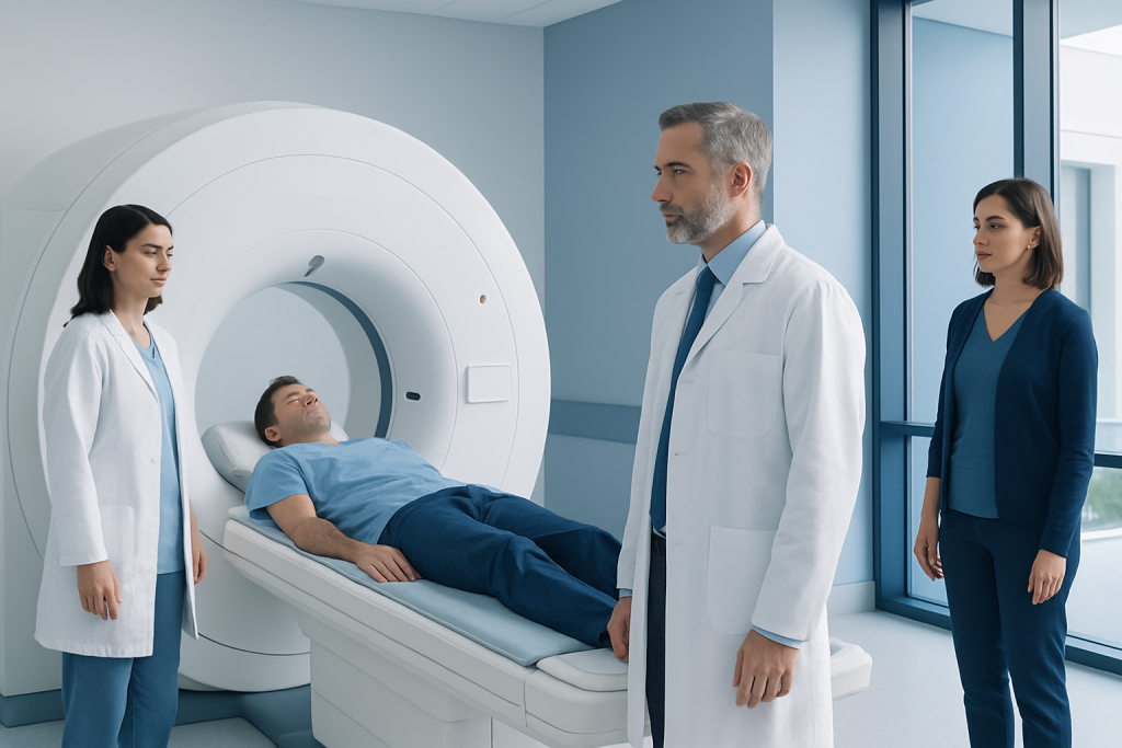
A full body MRI scan does not use X-rays or ionizing radiation. The examination commonly takes about 45 to 90 minutes, depending on the body areas and whether contrast is needed.
Key Takeaways
- A full body MRI scan does not use X-rays or ionizing radiation.
- The examination commonly takes about 45 to 90 minutes, depending on the body areas and whether contrast is needed.
- Metal implants, electronic devices and metal-containing clothing must be reviewed before entering the MRI room.
- MRI can identify some abnormalities, but incidental findings are common and may require further tests.
- Most people can return to normal activities immediately after a routine MRI.
A full body MRI scan is an imaging examination that uses a powerful magnet and radio waves to create detailed pictures of multiple areas of the body without ionizing radiation. It may be used for selected health concerns or surveillance plans, but it is not a substitute for routine preventive care or evaluation of new symptoms.
Overview: What Is a Full Body MRI Scan?
A full body MRI scan, also called whole-body magnetic resonance imaging, uses a strong magnetic field and radio waves to produce detailed images of several regions of the body. Unlike CT scans and standard X-rays, MRI does not use ionizing radiation. A radiologist reviews the images and prepares a report for the doctor who requested the examination.
The exact meaning of “full body” differs between imaging centers and clinical protocols. It may include the head and neck, chest, abdomen, pelvis and parts of the musculoskeletal system, but it does not necessarily provide the same detail as separate MRI examinations tailored to one organ or symptom.
Whole-body MRI can be appropriate in selected circumstances, such as monitoring certain inherited cancer syndromes, assessing some cancers, evaluating inflammatory conditions, or investigating symptoms that involve more than one body system. Its value is greatest when there is a clear medical question and the findings can be interpreted alongside a person’s history, examination and other tests.
How a Full Body MRI Scan Works and Who May Benefit
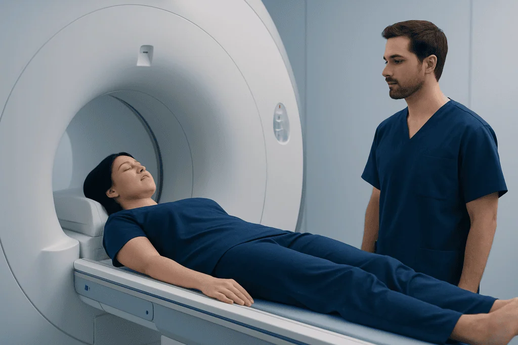
During MRI, hydrogen atoms in the body respond to magnetic fields and radiofrequency pulses. As these atoms return to their usual state, they release signals that a computer converts into detailed images. Different sequences highlight tissues such as fat, fluid, muscles, nerves, bone marrow and many internal organs.
A clinician may recommend a full body MRI scan for a person with a known condition requiring broad assessment, a carefully selected surveillance program, or symptoms that may involve several regions. For example, people undergoing assessment for suspected cancer may need imaging that is planned around the likely diagnosis and the question being asked. A targeted scan is often more useful than a general scan for localized symptoms.
MRI is not suitable for everyone without additional planning. People with pacemakers, cochlear implants, neurostimulators, aneurysm clips, infusion pumps, implanted metal fragments or certain joint replacements should tell the imaging team well in advance. Many modern implants are MRI-conditional, meaning scanning may be possible under specific safety conditions, but this must be confirmed by trained staff.
When a wider diagnostic evaluation is required, clinicians may also use targeted imaging and specialist assessment. For example, a patient may be referred for MRI scanning designed specifically for the brain, spine, joints or internal organs rather than relying on a broad screening examination.
Preparing for a Full Body MRI Scan
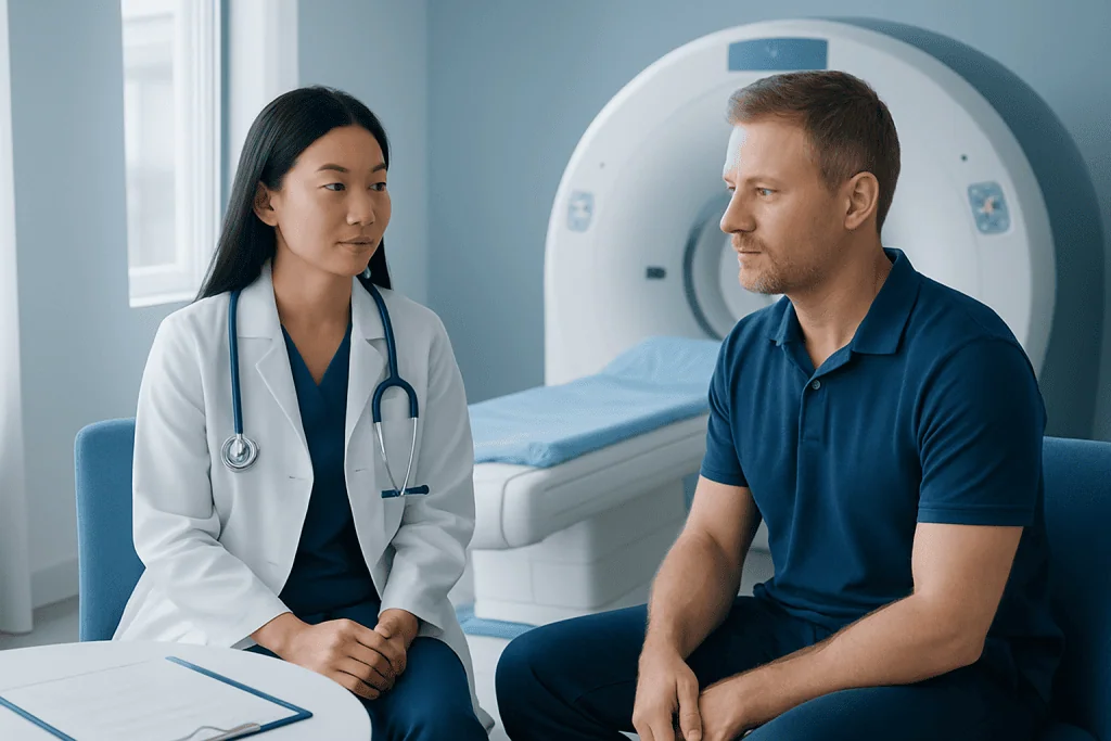
The imaging center will provide instructions based on the planned examination. Some full body MRI protocols require no special dietary preparation, while others may ask the patient to avoid food for several hours, particularly when the abdomen, liver, pancreas or pelvis will be evaluated. Drinking water may be encouraged or limited depending on the protocol.
Before the scan, patients complete a detailed safety questionnaire. It is important to report all implanted devices, previous surgery, possible metal fragments from work or injury, kidney disease, pregnancy, allergies, and any prior reaction to MRI contrast. People who have severe claustrophobia or difficulty lying still should discuss this early, as supportive measures or sedation may be considered in appropriate cases.
Patients should bring previous scan reports and images when available. These can help the radiologist compare new findings with earlier studies. If contrast material is planned, the team may review kidney function and medical history first. MRI contrast agents are not used for every full body MRI scan.
What cannot you do before an MRI?
Patients should not enter the MRI area with metal or electronic items unless the MRI team has confirmed they are safe. This includes jewelry, watches, hearing aids, credit cards, phones, keys, hairpins, removable dental appliances and some medication patches. They should also avoid applying products that may contain metallic particles, such as certain cosmetics, temporary tattoos or glitter-containing products, if instructed by the imaging center.
Step by Step: What Happens During the Procedure?
After check-in, the patient changes into an MRI-safe gown if needed and removes metal-containing items. A radiographer or MRI technologist confirms the safety information, explains the process and helps position the patient on a movable examination table. Coils, which are specialized receivers that improve image quality, may be placed around certain body areas.
The table moves gradually into the scanner. The MRI machine makes repetitive tapping, knocking or humming sounds while images are acquired, so earplugs or headphones are provided. The patient can communicate with the staff through an intercom and is usually given a call button. Remaining still is important because movement can blur the images and may prolong the examination.
If contrast is required, a small intravenous cannula may be placed in the arm or hand. The contrast is injected during part of the scan to help distinguish blood vessels, inflammation, tumors or other tissue changes. The team monitors the patient throughout the procedure and can pause if necessary.
How long does a full-body MRI typically take?
A full-body MRI typically takes about 45 to 90 minutes. The duration depends on the areas included, the scanner technology, whether contrast is used, the patient’s ability to remain still and whether additional sequences are needed after an initial image review. A targeted MRI may take less time, while complex protocols can take longer.
Benefits, Limits and Possible Risks
A major benefit of MRI is its ability to create detailed soft-tissue images without ionizing radiation. It can show information that may not be visible on an X-ray or, in some situations, a CT scan. Whole-body MRI may provide a broad overview when there is a medically appropriate reason to examine several body regions.
However, a full body MRI scan has important limits. It may detect incidental findings, meaning changes that are unrelated to symptoms and may never cause harm. These findings can lead to follow-up imaging, laboratory tests, specialist appointments or biopsy. MRI also cannot answer every clinical question, and a normal result does not rule out every disease.
Most people tolerate MRI well. The main concerns are safety issues related to metal or implanted devices, anxiety in enclosed spaces, discomfort from remaining still and rare reactions to contrast. MRI contrast agents are generally used carefully, particularly in people with significant kidney impairment. Any new rash, breathing difficulty or other concerning symptom after contrast should be reported promptly.
What two body parts do not appear in MRI?
This question can be misleading because MRI can image most body regions, including the brain, spine, chest, abdomen, pelvis and limbs. However, MRI is generally less effective for showing air-filled lungs and dense cortical bone in the same way as CT, because both provide relatively little MRI signal. MRI can still assess some lung and bone-related conditions, but the most appropriate imaging method depends on the clinical concern.
After the Scan and Understanding Results
After a routine MRI, most patients can eat, drink, drive and return to normal activities immediately. If sedation was used, the patient will need observation until fully alert and should arrange for a responsible adult to accompany them home. The imaging team will provide specific aftercare instructions when contrast or sedation has been used.
A radiologist interprets the images and sends a written report to the referring doctor. Results are usually discussed in the context of the person’s symptoms, physical examination, laboratory tests and previous imaging. A finding on MRI is not automatically a diagnosis, and further tests may be recommended to clarify uncertain or unexpected changes.
Can I keep my pants on during an MRI?
Whether pants can be worn depends on their material and the imaging center’s safety policy. Pants with zippers, buttons, rivets, drawstring tips, metallic fibers or hidden metal are not safe or may interfere with image quality. Even clothing without obvious metal can contain metallic threads, so many centers ask patients to change into an MRI gown for safety and consistency.
When to Seek Medical Care
A full body MRI scan should not delay medical assessment for concerning symptoms. A person should seek prompt medical care for symptoms such as sudden weakness or difficulty speaking, severe chest pain, new severe shortness of breath, coughing up blood, fainting, uncontrolled bleeding, or a severe headache that develops suddenly. Emergency services may be needed for acute or life-threatening symptoms.
A doctor should also evaluate persistent unexplained weight loss, a new or growing lump, ongoing fever, blood in urine or stool, persistent abdominal pain, new neurological symptoms, or pain that does not improve as expected. The clinician can decide whether MRI, another imaging test, laboratory testing or referral is most appropriate.
For international patients, Acibadem International’s multidisciplinary specialists and JCI-accredited hospitals can coordinate diagnostic imaging and follow-up care when clinically indicated. The most useful next step is a discussion with a qualified doctor who can match the imaging plan to the person’s symptoms, medical history and individual risk factors.
Frequently asked questions
Is a full body MRI scan safe?
MRI does not use ionizing radiation, and it is considered safe for many people when standard screening procedures are followed. The strong magnetic field can be hazardous for certain implants, devices or metal fragments, so a complete safety review is essential before the scan.
Does a full body MRI scan require contrast?
No. Some whole-body MRI protocols are performed without contrast, while others use contrast to answer a particular clinical question. The decision depends on the areas being assessed, the suspected condition and the patient’s medical history.
Can a full body MRI find cancer?
MRI may identify abnormalities that require further assessment, including some findings that could be related to cancer. It cannot diagnose every cancer or replace recommended cancer screening tests, and any suspicious result usually needs targeted follow-up.
Can I eat before a full body MRI scan?
Instructions vary by protocol. Some scans allow normal eating and drinking, while abdominal or pelvic imaging may require fasting for a period beforehand. Patients should follow the directions provided by their imaging center.
What should I wear for a full body MRI?
Wear comfortable clothing that is easy to change out of, and avoid garments with metal fasteners, zippers, underwires or decorative metallic elements. The imaging center may ask the patient to wear a gown to ensure safety and prevent image artifacts.
When will full body MRI results be available?
Timing varies by facility and the complexity of the examination. A radiologist must review many images carefully, and the referring doctor usually discusses the report after it is finalized. Urgent findings are typically communicated through established clinical pathways.
References
- American College of Radiology
- Radiological Society of North America
- U.S. Food and Drug Administration
- National Institute of Biomedical Imaging and Bioengineering
- National Health Service
This article is for general information only and is not a substitute for professional medical advice. Please consult a qualified doctor about your individual situation.
Explore treatments in Turkey — costs, top hospitals & a free quote
JCI-accredited · board-certified surgeons · reply within 24h