Ultrasound of Cleft Palate: Preparation, Procedure and Results
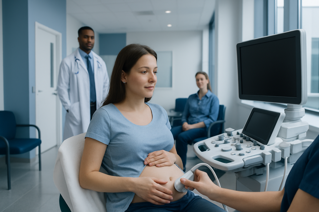
A routine pregnancy ultrasound can detect many cleft lips, but an isolated cleft palate may not be visible. A suspected facial cleft usually leads to a detailed fetal ultrasound and may lead to further testing or specialist counselling.
Key Takeaways
- A routine pregnancy ultrasound can detect many cleft lips, but an isolated cleft palate may not be visible.
- A suspected facial cleft usually leads to a detailed fetal ultrasound and may lead to further testing or specialist counselling.
- Ultrasound uses sound waves, not radiation, and is considered safe when performed for an appropriate medical reason.
- An ultrasound finding is not always a final diagnosis; image quality, fetal position and pregnancy stage can affect results.
- After birth, a careful examination of the mouth confirms whether a cleft palate is present and helps plan care.
Ultrasound of cleft palate is a prenatal imaging assessment that examines the developing baby’s lip, nose and mouth. It can identify many cleft lips, while cleft palate—especially when it occurs without a cleft lip—can be more challenging to see before birth.
Overview: What Is an Ultrasound of Cleft Palate?
An ultrasound of cleft palate is part of prenatal imaging used to examine a developing baby’s face and mouth. It is often performed during the detailed anatomy scan in the second trimester, when the clinician checks the facial profile, upper lip, nose and, where possible, the palate. The scan uses high-frequency sound waves to create images and does not use ionising radiation.
A cleft is an opening that occurs when structures of the face do not join fully during early fetal development. A cleft lip may affect one or both sides of the upper lip and may occur with or without a cleft palate. A cleft palate is an opening in the roof of the mouth. It can involve the hard palate at the front, the soft palate toward the back, or both.
Ultrasound is particularly useful for identifying a cleft of the lip. Seeing the palate is technically more difficult because it sits inside the mouth and can be obscured by bone, shadows, the baby’s position or surrounding tissues. Therefore, a normal prenatal scan cannot always rule out an isolated cleft palate.
How Prenatal Ultrasound Assesses the Face and Palate
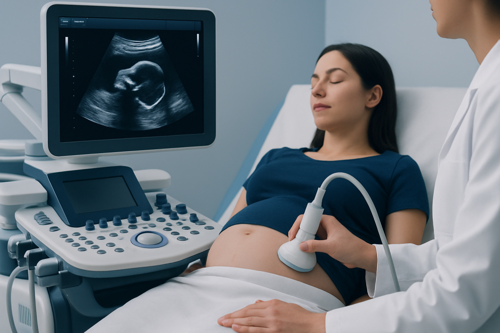
During a fetal anatomy scan, the sonographer obtains standard views of the baby’s face. These commonly include views of the profile, nose and lips. If there is a concern about a cleft, the examination may be extended to obtain targeted images of the upper jaw, mouth and palate from several angles.
Two-dimensional ultrasound is the standard method. In selected situations, three-dimensional ultrasound may provide additional surface images of the face and help parents and clinicians understand a suspected cleft lip. However, 3D imaging does not replace a careful diagnostic 2D examination and may still have limits in assessing the palate.
Image quality varies from one examination to another. It can be affected by fetal position, movement, gestational age, the amount and location of amniotic fluid, maternal body tissues and whether the baby’s hands or the umbilical cord are near the face. A repeat scan may be recommended simply to obtain clearer views.
Who May Need a Detailed Cleft Palate Ultrasound?
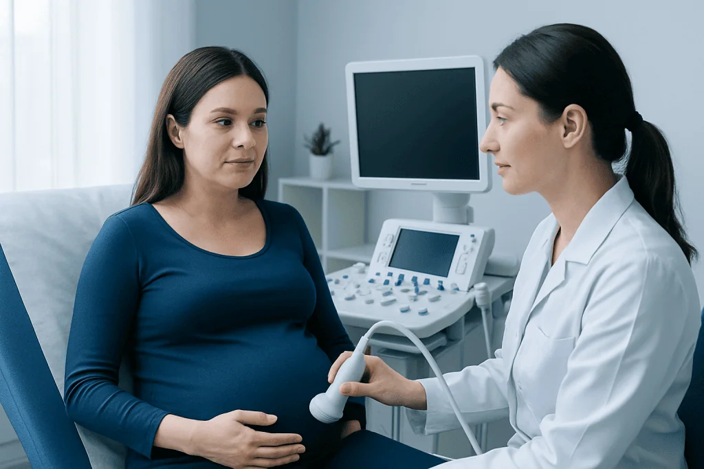
All pregnant people are generally offered a mid-pregnancy anatomy ultrasound, which includes an assessment of fetal facial structures where feasible. A more detailed facial examination may be advised if routine images suggest a possible cleft, if the face could not be adequately visualised, or if there are other ultrasound findings that need further assessment.
Additional imaging may also be considered when there is a personal or family history of cleft lip or palate, a previous pregnancy affected by a facial cleft, or a known medical or genetic factor associated with congenital differences. In many cases, however, a cleft occurs without a known family history or identifiable cause.
A suspected cleft lip or palate may prompt referral to a maternal-fetal medicine specialist, genetic counsellor, paediatric specialist or cleft care team. These discussions are intended to clarify what the scan shows, explore whether further testing is appropriate and prepare the family for care after birth.
Step by Step: What Happens During the Procedure?
No special preparation is usually needed for an ultrasound of cleft palate. The pregnant person may eat and drink normally unless their care team gives different instructions. Wearing comfortable clothing that allows access to the abdomen can make the appointment easier.
During the examination, the person lies on an examination couch. The sonographer applies a small amount of clear gel to the abdomen and moves a handheld transducer over the skin. The device sends and receives sound waves, and the resulting images are displayed on a monitor. The procedure is external, painless and generally takes place as part of a routine or targeted prenatal scan.
If the baby is facing away, has hands over the face or is in a position that limits the images, the sonographer may ask the person to change position, take a short walk or return for another appointment. This does not necessarily indicate a problem. A specialist may also use advanced imaging views when clinically appropriate.
There is no physical recovery period after an ultrasound. The person can return to usual daily activities immediately. Results may be discussed at the visit, although a formal review by the obstetrician or fetal medicine specialist may be needed when the images are complex or a possible difference is identified.
What Are the Ultrasound Findings for Cleft Lip and Palate?
On ultrasound, a cleft lip may appear as a break or discontinuity in the normally smooth line of the upper lip. It may be seen on one side, on both sides, or in the middle of the lip. The scan may also show changes involving the nostril or upper gum line, which can help clinicians assess how extensive the cleft may be.
When a cleft extends through the upper gum and into the palate, targeted views may suggest an opening in the front part of the palate. Findings can be clearer when a cleft lip is also present. In contrast, an isolated cleft of the soft palate, or a small opening within the mouth, may not be detectable on prenatal ultrasound.
Clinicians also look carefully for other fetal features. Most cleft lips and palates occur on their own, but some can occur alongside other structural or genetic conditions. Finding a cleft does not by itself establish a cause or predict a child’s overall health. Further assessment is individualized according to the ultrasound findings and family history.
What Is the Likelihood of a False Positive Cleft Palate Ultrasound Result?
A false positive result means that ultrasound suggests a cleft palate, but the condition is not confirmed after further assessment or birth. This can happen because fetal facial imaging is complex. Shadows from bone, fetal movement, the angle of the scan and limited views inside the mouth can sometimes resemble a gap or make normal structures difficult to interpret.
The likelihood of a false positive result differs according to the type of suspected cleft, the quality of the images and the experience of the imaging team. For this reason, a possible cleft palate finding is commonly reviewed with a detailed ultrasound by a fetal medicine specialist rather than being treated as a final diagnosis from one image alone.
Repeat imaging may clarify the anatomy. In some situations, fetal MRI may be discussed as an additional tool, particularly when ultrasound views remain limited and the result would meaningfully affect counselling or care planning. Confirmation after birth is still important, especially for isolated cleft palate.
How Accurate Is Ultrasound for a Cleft Lip?
Ultrasound is generally more accurate for a cleft lip than for a cleft palate. A well-positioned baby and clear imaging can allow clinicians to identify many cleft lips during the second-trimester anatomy scan. Detection is often easier for a larger or more extensive cleft and when it involves the lip and upper gum.
Accuracy is not absolute. A small cleft, a subtle notch in the lip, fetal position or less favourable imaging conditions may lead to a missed diagnosis. Conversely, an apparent irregularity may need specialist review before it can be confidently interpreted. Ultrasound can provide important prenatal information, but it does not replace the physical examination after delivery.
When a cleft lip is confirmed or strongly suspected, a specialist team can discuss expected feeding needs, possible involvement of the palate, timing of postnatal assessment and longer-term support. Children may benefit from coordinated care involving paediatrics, plastic and reconstructive surgery, dentistry, speech and language therapy, ear specialists and other professionals as needed.
Can You Pick Up a Cleft Palate on Ultrasound?
Yes, ultrasound can sometimes identify a cleft palate, particularly when it occurs together with a cleft lip or affects the front part of the palate. Detailed targeted scanning and carefully obtained facial views can improve assessment. However, the palate is not always fully visible on a standard prenatal examination.
An isolated cleft palate is one of the more difficult facial differences to detect before birth. The soft palate at the back of the mouth is especially challenging to visualise. Therefore, a normal appearance of the lip and face on ultrasound does not guarantee that an isolated cleft palate is absent.
If a cleft palate is diagnosed only after delivery, this does not mean that anyone did anything wrong during pregnancy. Some palatal clefts are simply not possible to identify reliably with available prenatal imaging. Early postnatal assessment helps ensure that feeding and specialist care are arranged promptly.
Benefits, Limitations and Next Steps After a Suspected Finding
The main benefit of prenatal ultrasound is that it can identify many facial clefts before birth and give families time to receive clear information, consider any recommended assessments and make a care plan. Prenatal recognition can also support planning for feeding guidance and specialist review after delivery.
Ultrasound is non-invasive and has no radiation exposure. Its main limitation is that it cannot detect every cleft, particularly an isolated palate cleft. The test also cannot always determine the full extent of a cleft or whether it is linked with another condition. These uncertainties can be addressed through specialist counselling and, when appropriate, additional tests.
After a suspected finding, the care team may recommend a detailed ultrasound, discussion of genetic screening or diagnostic testing options, and consultation with a paediatric cleft team. Treatment after birth is individualized and may include feeding support, hearing assessment, dental care, speech support and cleft lip and palate surgery when appropriate.
Acibadem International’s multidisciplinary specialists and JCI-accredited hospitals can support international patients with prenatal assessment, counselling and coordinated cleft care planning.
When to Seek Medical Care
Pregnant people should contact their obstetrician, midwife or fetal medicine team if an ultrasound report mentions a possible facial cleft, limited views of the face, or another fetal finding that needs review. A referral for specialist imaging can provide a more detailed assessment and an opportunity to ask questions in a supportive setting.
After birth, parents should seek prompt paediatric evaluation if a baby has difficulty latching or feeding, milk coming through the nose, frequent coughing or choking during feeds, poor weight gain, or a visible opening in the lip or mouth. These symptoms can have several causes, but early assessment is important.
Routine care remains important throughout pregnancy. A suspected cleft is not usually an emergency, but it should be followed up as advised so that the family receives accurate information and appropriate support.
Frequently asked questions
When is ultrasound used to check for cleft lip and palate?
The face is commonly assessed during the detailed anatomy ultrasound in the second trimester. If the images are incomplete or a possible cleft is seen, a targeted follow-up scan may be arranged. Timing can vary according to local practice and the baby’s position.
Does an ultrasound diagnosis of cleft palate always mean the baby has a cleft?
No. Ultrasound can raise a concern, but some findings need confirmation with repeat specialist imaging or examination after birth. The palate can be difficult to view, so clinicians interpret the result alongside the quality of the images and other findings.
Can a normal ultrasound rule out cleft palate?
A normal scan reduces concern for many visible facial clefts, particularly cleft lip, but it cannot fully rule out an isolated cleft palate. Small clefts and soft-palate clefts may not be visible before birth. Newborn examination remains important.
Is an ultrasound of cleft palate safe during pregnancy?
Diagnostic prenatal ultrasound uses sound waves rather than radiation and is widely used in obstetric care. It is considered safe when performed by trained professionals for an appropriate medical reason. The scan is external and does not enter the uterus.
Will more tests be needed if a cleft is suspected?
A detailed fetal ultrasound is commonly recommended to clarify the facial anatomy and check for other findings. Depending on the situation, the care team may discuss genetic counselling, screening, diagnostic testing or fetal MRI. Not every pregnancy requires all of these tests.
What happens after a baby is born with cleft lip or palate?
A paediatric team examines the baby and helps address immediate feeding needs. Ongoing care may involve a cleft team with surgical, dental, hearing, speech and developmental expertise. Treatment is planned according to the child’s specific type of cleft and needs.
References
- American College of Obstetricians and Gynecologists
- International Society of Ultrasound in Obstetrics and Gynecology
- Centers for Disease Control and Prevention
- American Cleft Palate-Craniofacial Association
This article is for general information only and is not a substitute for professional medical advice. Please consult a qualified doctor about your individual situation.
Ultrasound in Turkey — costs, top hospitals & a free quote
JCI-accredited · board-certified surgeons · reply within 24h

Check-up & Preventive Medicine
Comprehensive health screenings designed for early detection and prevention.
3 specialists in this unitMore from the Health Library


Related Specialists

Prof. Dr. Alpay Turan Sezgin
Cardiology
Assoc. Prof. Dr. Emre Özgü
Gynecology & Obstetrics
Dr. Cansu Hemşinlioğlu
Inpatient Clinic Physicians Clinical Service
Prof. Dr. Muzaffer Olcay Çizmeli
Interventional Neuroradiology