Knee Xray: What Patients Need to Know

A knee xray mainly shows bones, joint spacing, and alignment. It is often the first imaging test for knee pain, injury, or swelling.
Key Takeaways
- A knee xray mainly shows bones, joint spacing, and alignment.
- It is often the first imaging test for knee pain, injury, or swelling.
- A normal xray does not rule out soft tissue problems such as ligament or meniscus injuries.
- The test is quick, painless, and uses a small amount of radiation.
- Results are interpreted together with symptoms, physical examination, and sometimes other scans.
A knee xray is a common, fast imaging test used to look at the bones of the knee and the space within the joint. It can help doctors assess pain, injury, swelling, arthritis, and changes in alignment, while also showing when other tests may be needed.
What a Knee Xray Shows
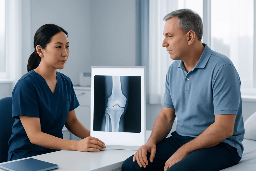
A knee xray is a quick, widely used imaging test that helps doctors look at the bones of the knee joint. It is often ordered for pain, swelling, injury, stiffness, or trouble bearing weight. In many cases, it is the first step in understanding whether symptoms are related to a fracture, arthritis, joint alignment, or other bony changes.
Unlike scans that focus on soft tissues, a knee xray is best at showing structures such as the femur, tibia, fibula, and kneecap. It can also show the space between bones, which may narrow with arthritis, as well as signs of dislocation, bone spurs, calcification, or changes after previous injury.
For patients, one of the most useful points to know is that a knee xray answers some questions very well, but not all of them. If pain appears to come from cartilage, ligaments, tendons, or the meniscus, the xray may be normal even when symptoms are real and significant. In that situation, a doctor may recommend additional assessment or MRI imaging to look more closely at soft tissues.
Why a Doctor May Order a Knee Xray
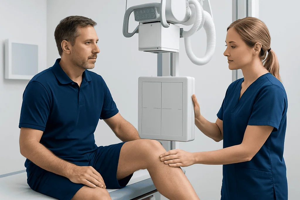
Doctors use knee xray findings together with a patient’s history and physical examination. The test may be recommended after a fall, sports injury, twisting motion, or direct blow to the knee. It is also commonly used when symptoms develop gradually, such as stiffness in the morning, long-term pain with walking, or visible changes in knee shape.
A knee xray may help evaluate:
- Possible fracture or crack in the bone
- Dislocation or poor joint alignment
- Wear-and-tear arthritis
- Inflammatory joint damage suggested by examination
- Swelling with concern for bone involvement
- Loose bodies or calcified fragments in or around the joint
- Follow-up after surgery or a known knee condition
In some cases, the xray helps confirm a likely diagnosis. In others, it helps rule out urgent bone problems and guides the next step. For example, if a person has persistent pain but the xray does not explain it, the clinician may then consider conditions such as meniscus tear or tendon-related problems that are better seen on other imaging tests.
How the Test Is Done and How to Prepare
A knee xray is usually performed in a radiology department, outpatient clinic, or emergency setting. The patient may be asked to stand or lie down while a technician positions the knee in one or more views. These images often include front, side, and angled views so the doctor can assess the joint from different directions.
The test itself is painless, although holding the knee in position may be uncomfortable if the joint is injured or swollen. The process is generally brief. Patients are usually asked to remove metal objects or clothing that could block the image. If there is any possibility of pregnancy, it is important to tell the healthcare team before the xray.
No special preparation is usually needed. Most people can eat, drink, and take regular medicines as usual unless told otherwise. If walking or standing is difficult, staff can often adjust positioning to make the test safer and more comfortable.
What the Results Can and Cannot Tell You
A knee xray can provide important information about bone health and the overall structure of the joint. It may show a fracture, signs of joint degeneration, reduced joint space, kneecap tracking problems, or changes related to prior trauma. In older adults, it is commonly used to assess osteoarthritis, where the xray may show narrowing of the joint, bone spurs, or hardening of bone beneath the cartilage.
At the same time, xray findings do not always match symptoms exactly. Some people have clear arthritis changes on imaging but only mild discomfort, while others have significant pain with only limited xray changes. This is one reason doctors do not rely on the image alone. A careful history, physical examination, and functional assessment remain essential.
It is also important to understand what a knee xray cannot fully show. Ligaments, cartilage, tendons, and the meniscus are not seen in detail on standard xray. If a doctor suspects a soft tissue injury, patellar tendon problem, or a condition such as ACL tear, other imaging may be more helpful. In selected cases, CT scanning may also be used when more bone detail is needed.
Common Conditions a Knee Xray May Help Assess
A knee xray is not a diagnosis by itself, but it plays a central role in assessing several common conditions. One of the most frequent reasons for ordering it is suspected osteoarthritis. The xray may show narrowing between the bones, bone spurs, or changes in alignment that help explain stiffness and pain during movement.
After an injury, the test can help identify fractures, dislocations, or avulsion injuries where a small piece of bone is pulled away by a tendon or ligament. It may also reveal chronic changes from previous trauma, including malalignment or signs that healing has occurred in an unusual position.
Doctors may also use a knee xray when evaluating persistent swelling, reduced range of motion, or deformity. Depending on the patient’s age and symptoms, it can contribute to the assessment of inflammatory arthritis, infection-related bone changes, or less common bone lesions. If severe joint damage is present, patients may later discuss options such as rehabilitation, medication, injections, or knee replacement with an orthopedic specialist.
Benefits, Limitations, and Radiation Safety
One of the main benefits of a knee xray is that it is fast, accessible, and useful in both urgent and routine care. It helps clinicians make decisions quickly, especially when they need to know whether a bone injury or major structural problem is present. Because the knee is a superficial joint, xray images can often provide clear basic information.
The main limitation is that xray is not designed to show every possible cause of knee pain. Soft tissue injuries, early cartilage wear, and some stress injuries may not appear clearly. When symptoms continue despite a normal or only mildly abnormal xray, further evaluation is often appropriate rather than dismissing the problem.
Knee xray uses a small amount of ionizing radiation. For most patients, the amount is low and considered acceptable when the test is medically indicated. Radiology teams aim to use the lowest dose needed to obtain useful images. Patients who have concerns about radiation exposure can discuss them with their doctor, especially if repeated imaging is being considered.
When to Seek Medical Care
Medical attention is appropriate if knee pain begins after a fall, twisting injury, sports impact, or any event that makes walking difficult. Prompt assessment is also sensible when there is marked swelling, inability to bear weight, visible deformity, locking, or a feeling that the knee is unstable. In these situations, a knee xray may be part of the early evaluation.
People should also consult a doctor if knee pain lasts more than a few days, keeps returning, limits daily activity, or is associated with stiffness, reduced movement, or gradual worsening over time. Even when symptoms seem mild at first, persistent problems can benefit from a clear diagnosis and treatment plan.
Urgent evaluation is especially important if the knee is red, very warm, accompanied by fever, or if severe pain develops suddenly without a clear explanation. Care is also important when symptoms occur in children, older adults, or anyone with known bone disease. Near the end of the care pathway, patients may be referred for specialist imaging, rehabilitation, or orthopedic treatment. Acibadem International’s multidisciplinary specialists at JCI-accredited hospitals diagnose and treat knee conditions for international patients when advanced evaluation is needed.
Frequently asked questions
Is a knee xray painful?
The xray itself is painless. Some people feel discomfort when the knee is positioned for the images, especially if the joint is swollen or injured. The test is usually very brief.
Can a knee xray show ligament or meniscus injuries?
A standard knee xray does not show ligaments or the meniscus in detail. It is mainly used to look at bones, joint space, and alignment. If a soft tissue injury is suspected, a doctor may recommend MRI or another imaging test.
How long does a knee xray take?
The imaging itself usually takes only a few minutes, although the full visit may be a little longer because of positioning and registration. In emergency settings, timing can vary depending on the patient’s condition and the department’s workload.
What does it mean if my knee xray is normal but I still have pain?
A normal xray means no clear bone abnormality was seen, but it does not rule out all causes of knee pain. Problems involving cartilage, tendons, ligaments, the meniscus, or early stress injuries may not appear on xray. Persistent symptoms should be discussed with a qualified doctor.
Is the radiation from a knee xray dangerous?
A knee xray uses a small amount of radiation, and for most patients the risk is very low when the test is medically needed. Radiology teams take steps to limit exposure. Patients who are pregnant or may be pregnant should inform the healthcare team beforehand.
Do I need a knee xray for every kind of knee pain?
Not always. Doctors decide based on symptoms, examination findings, age, injury history, and whether there are warning signs such as swelling, inability to bear weight, or deformity. In some cases, other tests or a period of observation may be more appropriate.
References
- American College of Radiology
- Radiological Society of North America
- National Institute of Arthritis and Musculoskeletal and Skin Diseases
- American Academy of Orthopaedic Surgeons
- MedlinePlus
This article is for general information only and is not a substitute for professional medical advice. Please consult a qualified doctor about your individual situation.
Joint and spine care in Turkey — expert assessment & treatment
JCI-accredited · board-certified surgeons · reply within 24h