MRI Spectroscopy: Preparation, Procedure and Results
MRI spectroscopy is usually performed during or after a standard MRI scan and does not use ionizing radiation. It measures chemical signals, or metabolites, rather than showing anatomy alone.
Key Takeaways
- MRI spectroscopy is usually performed during or after a standard MRI scan and does not use ionizing radiation.
- It measures chemical signals, or metabolites, rather than showing anatomy alone.
- MRS can help assess brain tumors, infection, stroke-related changes, epilepsy-related abnormalities and some metabolic or degenerative disorders.
- The examination is painless, but patients need to lie still in the MRI scanner and must complete metal-safety screening.
- Results are interpreted with MRI images, symptoms, examination findings and, when needed, laboratory tests or biopsy.
MRI spectroscopy, also called magnetic resonance spectroscopy or MRS, is a specialized addition to an MRI scan that measures certain chemicals within body tissue. It is most often used in the brain to provide extra information about abnormalities seen on imaging, supporting diagnosis and treatment planning alongside clinical assessment and other tests.
MRI Spectroscopy Overview
MRI spectroscopy is a noninvasive imaging technique that evaluates the chemical composition of selected tissue. It is commonly performed as an additional sequence during a magnetic resonance imaging scan, especially when doctors need more detail about a change seen in the brain. Unlike an X-ray or CT scan, MRI and MRI spectroscopy use a powerful magnetic field and radiofrequency energy rather than ionizing radiation.
Standard MRI produces detailed pictures of anatomy, including the structure of the brain, spinal cord, organs and soft tissues. MRI spectroscopy adds a different type of information: it identifies patterns of naturally occurring chemicals, called metabolites, in a small region or across a larger area. These patterns may help a radiologist and treating specialist understand whether tissue is more likely to reflect tumor activity, inflammation, injury, infection or another process.
MRS is a supportive diagnostic tool, not a stand-alone diagnosis. An abnormal chemical pattern does not always identify one specific disease, and a normal result does not exclude every condition. Doctors interpret it in context with a person’s symptoms, medical history, neurological examination and conventional MRI findings.
How MRI Spectroscopy Works
Hydrogen atoms are abundant in water and fat throughout the body. During MRI, the magnetic field aligns these atoms, and radiofrequency pulses temporarily alter their alignment. As the atoms return to their usual state, they release signals that can be measured and converted into images.
MRI spectroscopy analyzes small differences in these signals to estimate the relative amounts of certain metabolites. In brain MRS, commonly assessed compounds may include N-acetylaspartate, choline, creatine, lactate and lipids. For example, changes in choline or N-acetylaspartate may provide clues about cell membrane turnover or neuronal health, but they must never be interpreted alone.
The scan may assess one carefully chosen small area, known as single-voxel spectroscopy, or create a metabolic map of a broader region, called multivoxel spectroscopy. The best method depends on the location and size of the area being evaluated, the clinical question and image quality.
What Is the Difference Between MRI and MRI Spectroscopy?

Standard MRI is primarily an anatomical test. It shows the location, shape and internal features of structures and can reveal swelling, bleeding, scars, masses or changes in tissue appearance. Different MRI sequences highlight different tissue characteristics, helping doctors identify where an abnormality is located.
MRI spectroscopy is a functional and biochemical complement to MRI. Rather than focusing mainly on appearance, it evaluates chemical signals from a selected area. This can be useful when two conditions look similar on routine images or when clinicians need more information about the biological behavior of a lesion.
In practice, MRI spectroscopy is usually performed alongside conventional MRI rather than replacing it. Depending on the clinical situation, specialists may also use other advanced imaging methods, such as contrast-enhanced MRI, diffusion imaging or perfusion imaging, to build a clearer overall picture.
Who May Need MRI Spectroscopy and What Conditions Can It Detect?
MRI spectroscopy is most frequently used to assess an unusual area in the brain when standard MRI alone cannot fully clarify its cause. It may be considered for adults or children, provided they can safely undergo MRI and remain sufficiently still for imaging. The referring clinician decides whether it is likely to add useful information.
It can support assessment of brain tumors and help distinguish active tumor tissue from treatment-related changes in selected cases. It may also contribute to the evaluation of brain infections or abscesses, inflammatory conditions, tissue injury after reduced blood flow, seizure-related abnormalities and certain inherited or metabolic brain disorders. Its value differs between conditions, and additional testing may still be needed.
MRS does not reliably detect every disease and cannot by itself confirm cancer. If a brain mass is suspicious or treatment decisions depend on knowing the exact tissue type, a specialist may recommend monitoring, further imaging or tissue sampling. Readers looking for information about diagnosed brain tumors may also find brain tumor care helpful.
Although brain imaging is the most established use, spectroscopy may sometimes be used in other body areas in specialist settings. Its role outside the brain is more selective and depends on local expertise, equipment and the specific clinical question.
Preparation and Step-by-Step MRI Spectroscopy Procedure
Before the appointment, the imaging team asks about implants, previous surgery, metal fragments, medical devices, pregnancy and kidney problems. Some devices are MRI-safe or MRI-conditional, while others may make MRI unsuitable. Patients should also mention severe claustrophobia, allergies, hearing difficulties and any difficulty lying flat.
Most MRI spectroscopy examinations do not require special dietary preparation. Instructions may differ if the scan includes contrast material, sedation or another examination on the same day. Patients should follow the center’s individual guidance and bring information about implants, prior scans and relevant medical records when requested.
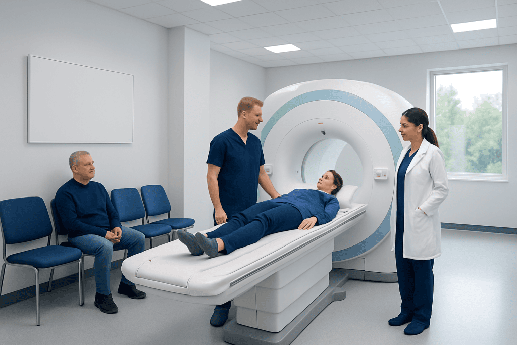
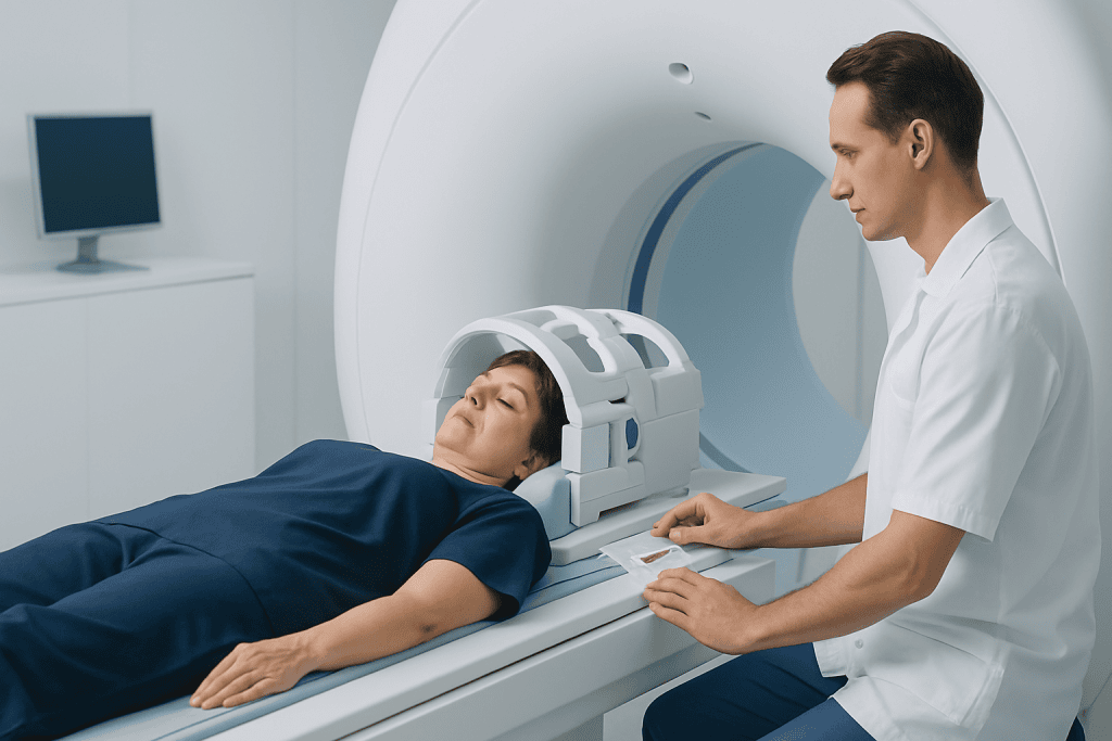
During the scan, the patient changes into suitable clothing and removes metal items such as jewelry, watches, hair accessories, hearing aids, removable dental appliances and bank cards. They lie on a padded table that moves into the scanner. A head coil may be placed around the head for brain imaging; it does not cause pain, but it helps obtain accurate signals.
The machine makes loud tapping and knocking sounds while images and spectroscopy data are collected, so earplugs or headphones are provided. The patient can communicate with the radiography team through an intercom and is usually given an alert device. Remaining still is important because movement can reduce image quality. If contrast is needed for the MRI portion, it is given through a small intravenous line; spectroscopy itself does not require contrast.
How Long Does an MRI Spectroscopy Take?
The spectroscopy portion of an examination often takes about 10 to 30 minutes, depending on the number of areas being assessed and the type of technique used. When it is added to a conventional MRI, the complete appointment may take longer, commonly around 30 to 90 minutes in total.
Timing can vary because careful positioning, safety checks, intravenous contrast, repeat sequences for movement and the complexity of the clinical question all affect the schedule. The imaging team can provide the most accurate estimate before the appointment.
Patients should plan to arrive early enough for screening and registration. If sedation is arranged because of severe anxiety or an inability to stay still, additional preparation, observation and transport home may be needed.
Results, Benefits, Risks and Recovery
After the scan, a radiologist reviews the MRI images and spectroscopy data. The report may describe metabolite levels or ratios and explain whether the pattern supports particular possibilities. Results are sent to the referring doctor, who discusses what they mean in relation to the individual’s health situation. Reporting times vary by center and by the complexity of the findings.
The main benefit of MRI spectroscopy is that it can add biochemical information without surgery or radiation exposure. This may help clinicians narrow possible explanations for an abnormality, select the most appropriate next test, target an area for biopsy or monitor changes over time. However, technical limitations, small lesion size, nearby bone or air, and movement can affect data quality.
MRI spectroscopy is generally safe for people who are appropriately screened. The principal risks relate to the MRI environment: some implanted devices or metal objects can be unsafe, and the confined space and noise can be uncomfortable. If contrast is used, there is a small possibility of an allergic-like reaction, and the team considers kidney health when relevant. Sedation, when required, has its own risks and is managed by trained professionals.
No recovery time is usually needed after MRI spectroscopy. Patients can generally return to normal activities immediately unless they received sedation, in which case they should follow discharge instructions and avoid driving or making important decisions until the effects have fully worn off. Acibadem International’s multidisciplinary specialists and JCI-accredited hospitals assess and treat neurological conditions for international patients, including through appropriate diagnostic imaging and follow-up care.
When to Seek Medical Care
A person should arrange medical assessment for persistent or worsening neurological symptoms, such as new seizures, unexplained weakness or numbness, ongoing severe headaches, changes in vision or speech, balance problems, new confusion or a noticeable change in memory or behavior. These symptoms have many possible causes, and only a clinician can determine whether imaging, including MRI spectroscopy, is appropriate.
Urgent medical care is important for sudden symptoms that may indicate a neurological emergency, including a sudden severe headache, weakness on one side of the body, facial drooping, trouble speaking, loss of consciousness or a seizure that is prolonged, repeated or a first seizure. Emergency services should be contacted according to local guidance.
People who have already had an MRI or spectroscopy scan should contact their clinician if they have questions about the findings or if symptoms change while awaiting follow-up. The referring team can explain whether further evaluation, specialist consultation or repeat imaging is needed.
Frequently asked questions
Is MRI spectroscopy painful?
MRI spectroscopy is not painful. The person must lie still in the scanner and may notice loud noises, but there are no injections or sensations specifically caused by the spectroscopy measurements. An intravenous injection may be needed only if contrast is used for the accompanying MRI scan.
Do patients need contrast for MRI spectroscopy?
MRI spectroscopy itself does not require contrast material. However, contrast may be recommended as part of the standard MRI examination if it can help evaluate blood vessels, inflammation, a tumor or another abnormality. The clinician and imaging team decide whether contrast is appropriate.
Can MRI spectroscopy diagnose a brain tumor?
MRI spectroscopy can provide information that supports the assessment of a suspected brain tumor, including clues about tissue metabolism. It cannot confirm the exact tumor type on its own. Doctors combine it with MRI findings, symptoms and sometimes biopsy results.
Are there radiation risks from MRI spectroscopy?
No. MRI spectroscopy does not use ionizing radiation, unlike X-rays and CT scans. It uses magnetic fields and radiofrequency energy, so safety screening for implants and metal is still essential.
Can someone with claustrophobia have MRI spectroscopy?
Many people with claustrophobia can complete the scan with planning and support. The imaging team may offer communication throughout the examination, comfort measures, breaks when possible or, in selected cases, medication or sedation arranged in advance. It is important to mention claustrophobia before the appointment.
What can affect the accuracy of MRI spectroscopy results?
Movement during scanning can reduce data quality, which is why remaining still is important. Very small abnormalities, lesions near bone or air spaces, technical factors and overlapping changes from different conditions can also limit interpretation. Results are therefore considered alongside the complete MRI study and clinical information.
References
- Radiological Society of North America
- American College of Radiology
- National Institute of Biomedical Imaging and Bioengineering
- National Cancer Institute
- World Health Organization
This article is for general information only and is not a substitute for professional medical advice. Please consult a qualified doctor about your individual situation.
Explore treatments in Turkey — costs, top hospitals & a free quote
JCI-accredited · board-certified surgeons · reply within 24h