Optic Neuritis MRI: Preparation, Procedure and Results
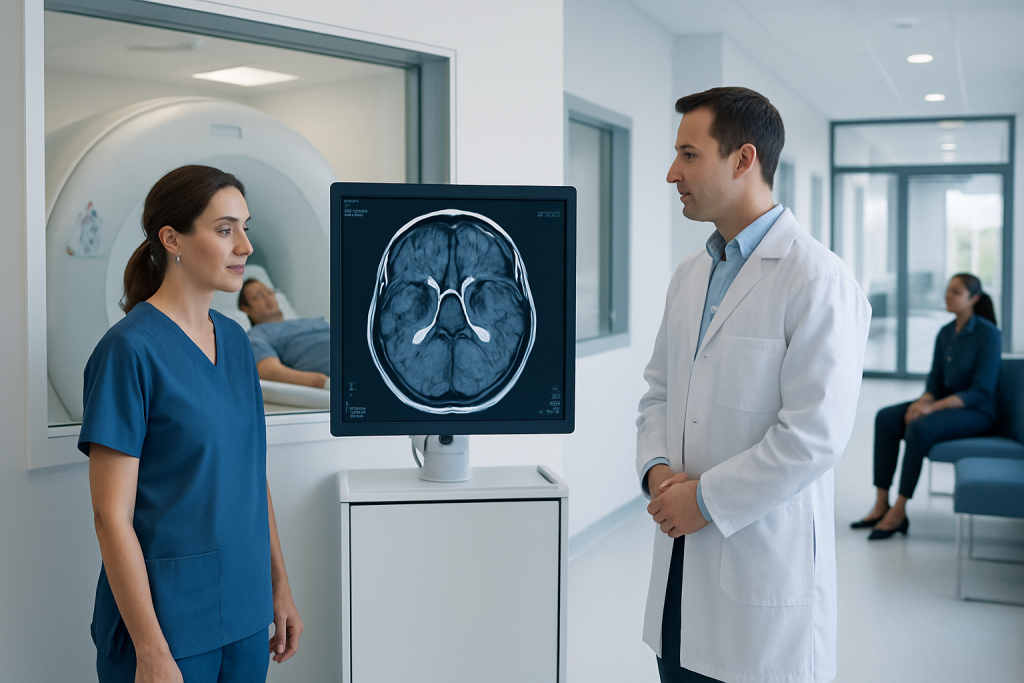
MRI can identify optic nerve inflammation and assess the brain for changes associated with multiple sclerosis or other inflammatory conditions. A normal MRI does not always exclude optic neuritis, especially if imaging is performed later in the illness or the scan protocol is not focused on the optic nerves.
Key Takeaways
- MRI can identify optic nerve inflammation and assess the brain for changes associated with multiple sclerosis or other inflammatory conditions.
- A normal MRI does not always exclude optic neuritis, especially if imaging is performed later in the illness or the scan protocol is not focused on the optic nerves.
- The examination usually takes about 30 to 60 minutes, with additional time needed for registration, screening and preparation.
- Contrast-enhanced MRI can help show active inflammation, but it is not appropriate for every person and requires safety screening.
- Optic neuritis needs prompt assessment because sudden visual changes can also have other causes that require different treatment.
An optic neuritis MRI is a scan of the optic nerves, brain and sometimes spinal cord that can show inflammation and help clinicians evaluate possible underlying causes. It is painless and does not use radiation, although contrast dye may be recommended to improve the visibility of active inflammation.
Overview: What an Optic Neuritis MRI Shows
Optic neuritis is inflammation of the optic nerve, the pathway that carries visual information from the eye to the brain. It often causes reduced vision in one eye, pain that is worse with eye movement, colours appearing less vivid, or a patch of missing vision. An optic neuritis MRI is an important imaging test that helps clinicians look for inflammation of this nerve and evaluate structures elsewhere in the nervous system.
The scan commonly includes MRI images of the brain and eye sockets, also called the orbits. In some situations, the spinal cord may be imaged as well. MRI does not use X-rays or radiation. Instead, it uses a strong magnetic field and radio waves to create detailed pictures of soft tissues, including the optic nerves, brain and spinal cord.
MRI is not used in isolation. Eye examination findings, the pattern and timing of symptoms, and targeted laboratory or nerve-function tests all help a neuro-ophthalmology or neurology team determine whether optic neuritis is likely and whether another condition needs to be considered.
How MRI Works and Who May Need It
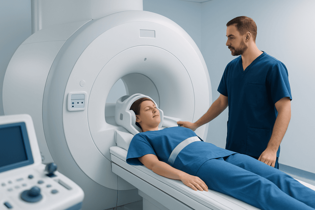
During MRI, the person lies on a moving table that slides into a tunnel-shaped scanner. Special coils receive signals from the body and help create high-resolution images. For suspected optic neuritis, the imaging team may use sequences designed to show swelling, changes within the optic nerve and abnormalities in the brain’s white matter.
A gadolinium-based contrast agent may be injected into a vein during the examination. Active inflammation can cause the optic nerve to enhance after contrast, helping distinguish recent inflammation from older changes. The decision to use contrast is individual and is made after reviewing kidney health, previous contrast reactions, pregnancy status and other relevant factors.
MRI is often recommended for people with symptoms that suggest optic neuritis, particularly a first episode of unexplained visual loss. It can be especially helpful when symptoms are atypical, severe, recurrent, affect both eyes, or occur alongside numbness, weakness, balance difficulties or other neurological symptoms. The scan may also support evaluation for multiple sclerosis and related inflammatory disorders.
Preparing for an Optic Neuritis MRI

Most people can eat, drink and take their usual medicines before an MRI unless their clinical team gives different instructions. It is helpful to bring details of previous scans, eye test results, implant cards and a current list of medicines. If contrast may be used, the team may ask about kidney disease, dialysis, previous contrast reactions or pregnancy.
Before the scan, staff will ask detailed safety questions. Metallic or electronic devices can be affected by the magnetic field, so it is important to report pacemakers, cochlear implants, neurostimulators, aneurysm clips, shrapnel injuries, insulin pumps and any past surgery. Many modern implants are MRI-compatible under specific conditions, but this must be confirmed in advance.
Jewellery, watches, hearing aids, removable dental items, bank cards and clothing with metallic parts are removed before entering the scan room. People who feel anxious in enclosed spaces should mention this when booking. The imaging team may discuss comfort measures, and in selected cases a clinician may consider medication for claustrophobia; this requires planning because a sedative can affect the ability to drive afterward.
Step-by-Step: What Happens During the Procedure
After check-in and safety screening, the person changes into suitable clothing if needed and lies on the MRI table. A lightweight head coil is placed around the head to improve image quality. Cushions, padding and straps may be used to help keep the head comfortably still, as movement can blur detailed images of the small optic nerves.
The table moves into the scanner. The machine makes repeated loud tapping, knocking or humming sounds while images are acquired, so earplugs or headphones are provided. Staff can see and speak with the person throughout the scan, and an alarm device is available if help is needed. Remaining still and keeping the eyes relaxed can improve the quality of the images.
If contrast is planned, a small intravenous cannula is inserted, usually in the arm or hand. The contrast is given partway through the scan, followed by additional images. The scan itself is painless. Some people notice a brief cool sensation at the injection site, but significant discomfort is uncommon.
Following the examination, a radiologist reviews the images and prepares a report for the referring clinician. Imaging findings are interpreted alongside the eye examination and other test results. If optic neuritis is confirmed or strongly suspected, treatment may include optic neuritis treatment tailored to the person’s symptoms, examination findings and likely cause.
What Are the Typical MRI Results for Optic Neuritis?
Typical MRI findings may include a segment of the optic nerve that appears swollen or has a higher signal on fluid-sensitive images. After gadolinium contrast, an actively inflamed optic nerve may show enhancement, meaning it takes up contrast more than expected. These changes may be visible in the part of the nerve behind the eye, within the optic canal or nearer the brain.
The brain MRI is also reviewed for white-matter lesions. In a person with a typical first episode of optic neuritis, certain lesion patterns can indicate a higher likelihood of a demyelinating condition such as multiple sclerosis. However, lesions are not diagnostic on their own, and some people with optic neuritis have no brain lesions at the time of scanning.
An MRI can be normal despite genuine optic neuritis. This may happen when inflammation is mild, imaging is done after active enhancement has faded, the protocol is not optimized for the optic nerves, or the affected area is difficult to visualize. Conversely, unusual patterns—such as inflammation affecting a long segment of nerve or nearby structures—may lead clinicians to investigate other inflammatory, infectious, vascular or compressive causes.
What Is the “10-20-40-60 Rule” for Optic Neuritis?
The “10-20-40-60 rule” is not a universally accepted medical guideline, formal MRI scoring system or diagnostic rule for optic neuritis. It may be used informally in some online discussions, but its meaning can vary and it should not be used to interpret symptoms, MRI findings or an individual outlook.
Clinicians assess optic neuritis using a broader clinical picture: the speed and pattern of visual loss, eye-movement pain, colour vision, pupil responses, visual-field testing, retinal imaging, MRI findings and any neurological symptoms. They also consider whether the episode follows the usual pattern or has features that need urgent assessment for another diagnosis.
When questions arise about numbers, timelines or prognostic rules, it is best to ask the treating ophthalmologist or neurologist what they mean in that person’s situation. Individual recovery and future risk depend on the cause of optic nerve inflammation and the results of the complete evaluation, not on one simplified rule.
How Long Does an Optic Nerve MRI Take?
An optic nerve MRI commonly takes around 30 to 60 minutes once the person is positioned in the scanner. The exact duration depends on whether images of the brain, orbits and spinal cord are needed, whether contrast is used, and whether additional sequences are required after the radiologist reviews preliminary images.
The full appointment may take longer because it includes registration, MRI safety screening, changing clothes, placing an intravenous line if contrast is planned and answering questions. People should allow enough time for these steps and for possible delays in a busy imaging department.
After the scan, most people can return to normal activities immediately. If sedation was used for anxiety or claustrophobia, an adult companion may be needed, and driving or making important decisions should be avoided for the period advised by the clinical team. The referring clinician will explain when and how results will be discussed.
What Tests Are Done for Optic Neuritis?
Assessment usually starts with a detailed history and eye examination. This may include checking visual acuity, colour vision, pupil responses, eye movements and the appearance of the optic disc at the back of the eye. Visual-field testing can identify areas of reduced peripheral or central vision, while optical coherence tomography (OCT) measures layers of the retina and optic nerve head.
MRI of the brain and optic nerves is often part of the assessment, but other tests may be selected according to the clinical situation. Blood tests can help investigate infection, inflammation, nutritional problems or autoimmune conditions. Specific antibody tests may be considered when the clinical pattern raises concern for disorders such as neuromyelitis optica spectrum disorder or myelin oligodendrocyte glycoprotein antibody-associated disease.
Visual evoked potentials may measure how quickly visual signals travel from the eyes to the brain. In selected cases, lumbar puncture testing of cerebrospinal fluid may provide additional information about inflammation or infection. Not everyone needs every test; clinicians choose investigations based on the person’s symptoms, examination and MRI findings.
Benefits, Limitations, Risks and When to Seek Medical Care
The main benefit of optic neuritis MRI is the detailed assessment it provides without radiation exposure. It can support diagnosis, identify active inflammation, help rule out some alternative explanations for visual symptoms and guide discussions about further neurological assessment. It may also provide a useful baseline for future comparison when follow-up imaging is needed.
MRI has limitations. Findings must be interpreted carefully, as a scan cannot always establish the cause of optic nerve inflammation or predict an individual outcome. Contrast is generally well tolerated, but allergic-type reactions are possible and are usually mild; serious reactions are uncommon. Gadolinium contrast is used cautiously in people with severe kidney impairment, and MRI itself may not be suitable with certain implants or retained metal fragments.
Urgent medical assessment is appropriate for sudden or worsening vision loss, pain with eye movement, new double vision, a new blind spot, significant headache with neurological symptoms, weakness, numbness, difficulty speaking or poor balance. Prompt evaluation is important because visual symptoms can have several causes, some requiring time-sensitive treatment.
Acibadem International’s multidisciplinary ophthalmology, neurology and radiology specialists can assess optic neuritis and related neuro-ophthalmic concerns for international patients in JCI-accredited hospitals. Care plans are based on the clinical assessment, imaging findings and the person’s individual needs.
Frequently asked questions
Can an MRI diagnose optic neuritis?
MRI can strongly support a diagnosis of optic neuritis by showing inflammation or contrast enhancement in the optic nerve. However, diagnosis also depends on symptoms, eye examination findings and, when appropriate, other tests. A normal MRI does not completely rule out optic neuritis.
Does optic neuritis always show on an MRI with contrast?
No. Active inflammation often enhances after contrast, but enhancement may not be seen in every case. Timing of the scan, scan technique and the severity or location of inflammation can all affect visibility.
Do people need contrast for an optic nerve MRI?
Contrast is often useful because it can show active optic nerve inflammation more clearly. It is not necessary or suitable for everyone, and the decision is made after medical and MRI safety screening.
Is an optic neuritis MRI painful?
The MRI scan itself is painless. The machine is loud and requires the person to remain still, and an intravenous injection may be needed if contrast is used. Staff provide hearing protection and communication throughout the examination.
Can optic neuritis occur without multiple sclerosis?
Yes. Optic neuritis can occur as an isolated episode and does not automatically mean a person has multiple sclerosis. MRI findings, antibody tests and follow-up evaluation can help clinicians assess the likelihood of underlying causes.
When will optic neuritis MRI results be available?
A radiologist must review the images and issue a formal report, so timing varies by imaging service and clinical urgency. The referring ophthalmologist or neurologist will explain the results in the context of the person’s symptoms and eye examination.
References
- American Academy of Ophthalmology
- National Eye Institute
- National Multiple Sclerosis Society
- Radiological Society of North America
- Mayo Clinic
This article is for general information only and is not a substitute for professional medical advice. Please consult a qualified doctor about your individual situation.
Joint and spine care in Turkey — expert assessment & treatment
JCI-accredited · board-certified surgeons · reply within 24h
Add us as a Preferred Source to see more of our trusted health content across Google Search, AI Overviews and Discover.