Subchondral Sclerosis: Symptoms, Causes, and Treatment Options

Subchondral sclerosis means the bone beneath joint cartilage has become denser or harder on imaging. It is commonly associated with osteoarthritis, aging, prior injury, and ongoing joint overload.
Key Takeaways
- Subchondral sclerosis means the bone beneath joint cartilage has become denser or harder on imaging.
- It is commonly associated with osteoarthritis, aging, prior injury, and ongoing joint overload.
- Some people have no symptoms from the bone change itself; symptoms usually come from the underlying joint condition.
- Diagnosis is based on symptoms, examination, and imaging such as X-rays or MRI when needed.
- Treatment focuses on the cause and may include exercise, weight management, medicines, physical therapy, injections, or surgery in selected cases.
Subchondral sclerosis is a bone change seen under joint cartilage, most often as part of osteoarthritis and long-term joint stress. It is not a diagnosis on its own, but an imaging finding that helps explain pain, stiffness, and reduced movement in some people.
Overview: what subchondral sclerosis means
Subchondral sclerosis is a term doctors use when the bone just beneath the cartilage of a joint looks denser than usual on imaging, especially on X-rays. In plain language, it refers to hardening or thickening of the supporting bone under the smooth cartilage layer that helps a joint move easily.
This finding is most often seen in osteoarthritis, where cartilage gradually wears down and the bone underneath responds to repeated stress. It can appear in weight-bearing joints such as the knees and hips, but it may also affect the spine, hands, feet, or shoulder depending on the underlying joint problem.
Subchondral sclerosis is not always the direct cause of pain. Instead, it is usually one part of a broader pattern of joint change that can include cartilage loss, narrowing of the joint space, bone spurs, and sometimes small fluid-filled cysts in the bone. That is why treatment is guided by symptoms and the overall joint condition rather than the imaging finding alone.
How it develops inside a joint
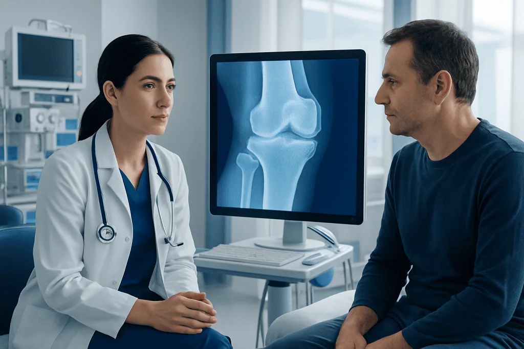
Healthy joints distribute force through cartilage and the underlying subchondral bone in a balanced way. When cartilage becomes thinner or damaged over time, the forces passing through the joint change. The supporting bone may adapt by becoming denser and less elastic, which is what appears as subchondral sclerosis on imaging.
This process is often gradual. Repeated loading, past injury, alignment problems, or long-term inflammation can all alter how a joint absorbs impact. The body responds by remodeling bone. While this may be an attempt to stabilize the area, the stiffer bone can also contribute to ongoing joint dysfunction.
Subchondral sclerosis is commonly discussed together with osteoarthritis, but it can also be seen with other conditions that affect joints and bone structure. Understanding this context helps explain why two people with similar X-ray findings may have very different symptoms and treatment needs.
Symptoms and where it commonly occurs
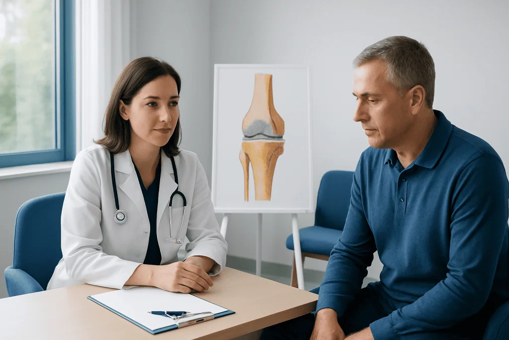
Subchondral sclerosis itself is an imaging finding, so symptoms usually reflect the underlying joint disease rather than the bone change alone. Many people notice aching pain with activity, stiffness after rest, tenderness around the joint, or a reduced range of motion. In osteoarthritis, discomfort often worsens gradually and may become more noticeable with walking, climbing stairs, standing for long periods, or repetitive hand use.
The knee and hip are especially common sites because they bear body weight and absorb repeated mechanical stress. In the knee, symptoms may include pain when bending, climbing, or rising from a chair. In the hip, groin pain, buttock discomfort, or stiffness when putting on shoes may occur. In the spine, related degenerative changes can contribute to back or neck pain and stiffness.
Some people hear grinding or clicking, notice swelling after activity, or feel less stable on the affected side. Others may have clear sclerosis on an X-ray but only mild symptoms. Because symptoms vary widely, the degree of pain does not always match how advanced the imaging changes appear.
- Common symptoms: pain with use, stiffness, reduced flexibility, tenderness
- Possible associated signs: swelling, grinding sensation, limited function
- Common locations: knees, hips, spine, hands, and feet
Causes and risk factors
The most common cause of subchondral sclerosis is joint degeneration from osteoarthritis. As cartilage wears down, the bone beneath it is exposed to greater stress and adapts by becoming denser. This is why the finding is frequently reported on knee, hip, or spinal imaging in middle-aged and older adults.
Other causes and contributors include previous joint injury, repetitive impact from work or sports, obesity, and joint misalignment. For example, a past meniscus tear, ligament injury, or poorly healed fracture can change joint mechanics for years. Family history and natural aging also play a role in how quickly cartilage and bone remodeling occur.
Less commonly, inflammatory or structural joint disorders may be involved, so doctors consider the full clinical picture. Risk factors that increase the chance of symptomatic joint degeneration include:
- Older age
- Excess body weight
- History of joint trauma or surgery
- Repetitive heavy loading at work or in sport
- Poor joint alignment or muscle weakness
- Underlying cartilage or inflammatory joint disease
Diagnosis and what imaging can show
Diagnosis starts with a medical history and physical examination. A doctor asks where the pain is felt, what activities make it worse, how long stiffness lasts, and whether there has been any injury, swelling, locking, or instability. The joint is then examined for tenderness, movement limits, alignment changes, and signs of inflammation.
X-rays are often the first imaging test because they can show classic joint changes, including subchondral sclerosis, narrowing of the joint space, and bone spurs. MRI may be used if symptoms are out of proportion to X-ray findings, if soft tissue injury is suspected, or if the diagnosis is uncertain. In selected cases, CT can provide a more detailed view of bone structure.
Doctors also look for conditions that may mimic osteoarthritis-related pain. Depending on the symptoms, they may consider tendon injuries, bursitis, inflammatory arthritis, or osteonecrosis. Blood tests are not usually needed for simple osteoarthritis, but they may help if an inflammatory or autoimmune condition is possible.
Treatment options and symptom management
Treatment for subchondral sclerosis focuses on the underlying joint disorder, the person’s symptoms, and the effect on daily life. For many people, a combination of exercise therapy, activity adjustment, weight management, and pain relief can improve comfort and function. Gentle strengthening around the joint can reduce load and support movement more efficiently.
Physical therapy is often helpful, especially for knee, hip, and spine involvement. A therapist may recommend mobility work, muscle strengthening, balance training, and strategies to reduce joint strain. When symptoms flare, doctors may suggest short-term pain medicines, topical treatments, or in some cases joint injections. In the right setting, options such as knee replacement or hip replacement may be discussed if joint damage is advanced and daily activities remain limited despite conservative care.
Supportive devices can also help. Braces, orthotics, a cane, or footwear changes may improve alignment and reduce stress on the affected joint. If arthritis affects the spine or mobility has declined, a tailored physical therapy and rehabilitation program may support pain control and safe movement.
Surgery is not based on the presence of sclerosis alone. It is generally considered when there is clear structural joint disease, persistent pain, and reduced quality of life despite well-planned non-surgical treatment. The decision depends on symptoms, imaging, age, activity goals, and overall health.
Prevention, self-care, and protecting joint health
Not every cause of joint degeneration can be prevented, but many people can lower stress on their joints and slow symptom progression. Regular low-impact exercise such as walking, cycling, swimming, or strength training helps support cartilage health, muscle balance, and everyday function. Rest is important during painful flare-ups, but long periods of inactivity can worsen stiffness and weakness.
Weight management is especially important for the knees, hips, and lower back because extra body weight increases mechanical load. Small, sustainable changes in diet and activity may reduce symptoms over time. Good footwear, proper lifting technique, and gradual progression in sport or exercise can also help protect joints.
Self-care measures often include heat for stiffness, ice after activity-related soreness, pacing daily tasks, and avoiding repeated high-impact strain when symptoms are active. People with persistent pain benefit from a personalized plan rather than self-treating indefinitely. Near the end of the care pathway, if specialist assessment is needed, Acibadem International’s multidisciplinary teams in JCI-accredited hospitals evaluate joint conditions for international patients and coordinate diagnosis and treatment.
When to seek medical care
Medical advice is appropriate if joint pain lasts more than a few weeks, keeps returning, or begins to limit walking, work, sleep, or normal daily activities. A clinician can confirm whether subchondral sclerosis is part of osteoarthritis or whether another condition needs attention.
Prompt assessment is more important if there is sudden severe pain, major swelling, fever, redness, inability to bear weight, a recent injury, or a joint that locks or gives way. These signs can suggest a problem other than routine wear-and-tear changes and may need earlier treatment.
People with long-term arthritis should also seek review if current treatment stops helping, pain is spreading, or movement continues to decline. Early adjustment of the care plan may reduce disability and help preserve joint function.
Frequently asked questions
Is subchondral sclerosis the same as osteoarthritis?
No. Subchondral sclerosis is a radiology finding, while osteoarthritis is a joint disease. The finding often appears as part of osteoarthritis, but it does not describe the whole condition by itself.
Can subchondral sclerosis cause pain?
It can be associated with pain, but symptoms usually come from the overall joint problem rather than the bone change alone. Cartilage loss, inflammation, bone remodeling, and surrounding soft tissue strain may all contribute to discomfort.
Does subchondral sclerosis mean surgery is needed?
Not necessarily. Many people improve with exercise, physical therapy, weight management, activity changes, and medicines. Surgery is usually considered only when symptoms remain significant and joint damage clearly affects daily life.
Can subchondral sclerosis be reversed?
The underlying bone change is generally considered part of chronic joint remodeling rather than something that fully reverses. However, symptoms and function can often improve substantially with appropriate treatment and joint protection.
What joints are most commonly affected?
The knees and hips are common because they bear body weight. The spine, hands, feet, and sometimes shoulders can also show subchondral sclerosis when degenerative joint changes are present.
How is subchondral sclerosis diagnosed?
It is usually identified on X-rays as increased density in the bone just under the joint surface. Doctors interpret this together with symptoms, physical examination findings, and sometimes MRI or other imaging if more detail is needed.
References
- American Academy of Orthopaedic Surgeons
- National Institute of Arthritis and Musculoskeletal and Skin Diseases
- Arthritis Foundation
- Radiological Society of North America
- World Health Organization
This article is for general information only and is not a substitute for professional medical advice. Please consult a qualified doctor about your individual situation.
Joint and spine care in Turkey — expert assessment & treatment
JCI-accredited · board-certified surgeons · reply within 24h