Thrombectomy for Ischemic Stroke: How Interventional Neurology Restores Blood Flow

Mechanical thrombectomy is mainly used for ischemic strokes caused by a blockage in a large brain artery. The procedure is time-sensitive, but advanced brain imaging may identify some suitable patients up to 24 hours after symptoms begin.
Key Takeaways
- Mechanical thrombectomy is mainly used for ischemic strokes caused by a blockage in a large brain artery.
- The procedure is time-sensitive, but advanced brain imaging may identify some suitable patients up to 24 hours after symptoms begin.
- A specialist guides a catheter through a blood vessel to the clot and removes it using aspiration, a stent retriever, or both.
- Thrombectomy can improve the chance of recovery, but it does not guarantee full reversal of stroke symptoms.
- Fast recognition of stroke symptoms and immediate emergency care remain the most important steps.
Thrombectomy for ischemic stroke is a minimally invasive emergency procedure used to remove a clot from a large brain artery and restore blood flow. It is performed by interventional neurology or neurointerventional teams in carefully selected patients, often alongside other stroke treatments.
Overview
An ischemic stroke happens when a blood clot blocks an artery that carries oxygen-rich blood to part of the brain. Without blood flow, brain cells begin to lose function quickly. Thrombectomy for ischemic stroke, also called mechanical thrombectomy, is a specialized emergency procedure designed to remove the clot and reopen the blocked artery.
The treatment is most often considered when the clot is in a large vessel, such as a major artery at the base of the brain or one of its main branches. These strokes can cause significant symptoms because a larger area of the brain may be at risk. By restoring blood flow, thrombectomy may reduce the amount of permanent brain injury and improve the person’s chance of regaining independence.
Thrombectomy is performed by interventional neurologists, neurointerventional radiologists, or endovascular neurosurgeons working with an acute stroke team. It is not suitable for every stroke, and careful imaging is needed to confirm the type of stroke, the location of the blockage, and whether enough brain tissue may still be saved.
How Interventional Neurology Restores Blood Flow
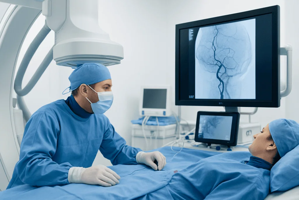
Interventional neurology uses image-guided, minimally invasive techniques to treat conditions affecting the brain and blood vessels. During thrombectomy, the specialist usually accesses the blood vessels through a small puncture in the groin or, in selected cases, the wrist. A thin tube called a catheter is then guided through the arteries toward the blocked vessel in the brain.
Once the catheter reaches the clot, the specialist may use aspiration, a device that applies suction to remove the clot, or a stent retriever, a tiny expandable mesh device that captures the clot so it can be withdrawn. In many procedures, both techniques are used together. X-ray imaging helps the team see the catheter, devices, and blood vessels in real time.
The goal is to achieve reperfusion, meaning blood flow returns to the affected part of the brain. The stroke team monitors the patient closely throughout the procedure, including blood pressure, oxygen levels, neurological status, and any signs of bleeding. After the procedure, the patient is cared for in a stroke unit or intensive care setting where recovery and complications can be monitored carefully.
Who May Be a Candidate

Mechanical thrombectomy is mainly used for people with an ischemic stroke caused by a large vessel occlusion. This means a clot is blocking one of the major arteries supplying the brain. Symptoms are often sudden and may include weakness on one side, difficulty speaking, facial drooping, vision loss, confusion, severe imbalance, or reduced alertness.
Time is an important factor. Many patients are assessed for thrombectomy within the first 6 hours after symptoms begin. However, some patients may still benefit up to 24 hours after they were last known to be well if advanced imaging shows that a significant area of brain tissue is at risk but not yet permanently damaged.
Doctors consider several factors before recommending thrombectomy, including:
- The time when symptoms started or when the person was last known to be well
- The location and size of the blocked artery
- Brain imaging results showing the balance between damaged tissue and potentially salvageable tissue
- The person’s neurological symptoms and general medical condition
- Whether the stroke is ischemic rather than hemorrhagic
- Potential risks such as bleeding tendency or severe underlying illness
Diagnosis and Emergency Assessment
Rapid diagnosis is essential because stroke treatment is highly time-sensitive. The first step is usually a neurological examination and urgent brain imaging. A non-contrast CT scan can quickly help distinguish an ischemic stroke from bleeding in the brain. This distinction matters because treatments for blocked arteries and bleeding are very different.
Additional imaging may include CT angiography or MR angiography to show whether a large artery is blocked. CT perfusion or MRI perfusion may be used in some hospitals to estimate how much brain tissue has already been injured and how much may still be saved if blood flow is restored. These scans help guide treatment decisions, especially when the exact time of stroke onset is unclear.
Blood tests, heart rhythm monitoring, blood pressure measurement, and assessment of other conditions are also part of the emergency evaluation. If the patient is eligible, intravenous clot-dissolving medication may be given before or during transfer for thrombectomy. Thrombectomy and intravenous thrombolysis are not necessarily competing treatments; for some patients, they are complementary parts of acute stroke care.
Treatment Options and the Procedure
For ischemic stroke, treatment aims to restore blood flow as quickly and safely as possible. Intravenous thrombolysis may be offered to eligible patients within the approved time window and when there are no contraindications. Mechanical thrombectomy is considered when imaging confirms a suitable large vessel blockage and the patient meets clinical and imaging criteria.
Before thrombectomy, the team explains the procedure when possible, reviews risks, and prepares the patient. Some procedures are performed with conscious sedation, while others require general anesthesia, depending on the patient’s condition, airway safety, movement, and hospital practice. The neurointerventional specialist then advances catheters through the blood vessels to the clot and removes it using specialized devices.
After the blocked artery is reopened, the team performs imaging to assess blood flow and check for complications. The puncture site in the groin or wrist is closed and monitored. Follow-up brain imaging is commonly performed later to check for bleeding or swelling. Rehabilitation planning may begin early, often involving physiotherapists, speech and language therapists, occupational therapists, nurses, dietitians, and physicians.
At Acibadem International, multidisciplinary stroke and neurointerventional teams in JCI-accredited hospitals evaluate and treat international patients with ischemic stroke, including appropriate candidates for thrombectomy, according to individual clinical needs and imaging findings.
Benefits, Risks, and Recovery
The main potential benefit of thrombectomy is restoring blood flow before more brain tissue is permanently damaged. For appropriately selected patients with large vessel occlusion, this can improve the chance of better functional recovery. Some patients show improvement soon after the artery is opened, while others recover gradually over days, weeks, or months with rehabilitation.
Like all emergency procedures, thrombectomy has risks. These may include bleeding in the brain, injury to a blood vessel, movement of clot fragments to another artery, reaction to contrast dye, kidney strain in vulnerable patients, infection or bleeding at the puncture site, and complications related to anesthesia or sedation. The stroke team weighs these risks against the expected benefit of reopening the artery.
Recovery depends on many factors, including the size and location of the stroke, how quickly blood flow was restored, age, other medical conditions, and the quality of rehabilitation. Thrombectomy can be a powerful treatment, but it cannot always reverse damage that has already occurred. Ongoing follow-up helps identify the cause of the stroke and reduce the risk of another event.
Prevention, Self-care, and When to Seek Help
After an ischemic stroke, prevention focuses on reducing the chance of another clot. Depending on the cause, doctors may recommend antiplatelet medication, anticoagulation for certain heart rhythm problems, cholesterol-lowering therapy, blood pressure control, diabetes management, smoking cessation, weight management, and treatment of carotid artery disease when appropriate. Medication decisions should always be individualized by a qualified physician.
Lifestyle measures can support medical care. A heart-healthy eating pattern, regular physical activity as advised by the rehabilitation team, limiting alcohol, good sleep, and adherence to follow-up appointments can all contribute to long-term vascular health. Patients and families may also benefit from learning how to monitor symptoms, manage swallowing or speech problems, prevent falls, and support emotional recovery.
Emergency help should be sought immediately for any sudden signs of stroke. These include face drooping, arm weakness, speech difficulty, sudden vision loss, severe dizziness or imbalance, confusion, numbness on one side, or a sudden severe headache. It is safer to call emergency services right away than to wait to see whether symptoms improve, because early treatment offers the best opportunity to protect brain tissue.
Frequently asked questions
What is thrombectomy for ischemic stroke?
Thrombectomy for ischemic stroke is a minimally invasive procedure that removes a clot from a blocked brain artery. A specialist guides a catheter through the blood vessels to reach the clot and restore blood flow. It is mainly used for large vessel occlusion strokes.
How quickly must thrombectomy be performed?
Many patients are assessed for thrombectomy within 6 hours of symptom onset. Some may still be eligible up to 24 hours after they were last known to be well if advanced imaging shows that brain tissue can still be saved. The sooner emergency care begins, the better the opportunity for effective treatment.
Is thrombectomy the same as clot-busting medicine?
No. Clot-busting medicine, also called intravenous thrombolysis, is given through a vein to help dissolve a clot. Thrombectomy physically removes the clot using a catheter and specialized devices. Some eligible patients may receive both treatments.
Is mechanical thrombectomy painful?
Patients receive anesthesia or sedation, so the procedure itself should not be painful. The medical team chooses conscious sedation or general anesthesia based on the patient’s condition and safety needs. Afterward, there may be mild soreness or bruising at the catheter entry site.
Can thrombectomy completely reverse a stroke?
Thrombectomy can restore blood flow and may significantly improve recovery in suitable patients, but it cannot guarantee full reversal of symptoms. The outcome depends on how much brain tissue was affected, how quickly treatment occurred, and the person’s overall health. Rehabilitation is often an important part of recovery.
What happens after the procedure?
After thrombectomy, the patient is monitored closely in a stroke unit or intensive care setting. Doctors check neurological function, blood pressure, the catheter site, and follow-up brain imaging. The team also investigates the cause of the stroke and starts prevention and rehabilitation planning.
When should someone call emergency services for stroke symptoms?
Emergency services should be called immediately for sudden facial drooping, arm weakness, speech difficulty, vision changes, severe imbalance, confusion, or one-sided numbness. Even if symptoms improve, urgent assessment is needed because a transient blockage can be a warning sign. Fast action can make more treatment options available.
References
- American Heart Association and American Stroke Association
- European Stroke Organisation
- World Stroke Organization
- National Institute of Neurological Disorders and Stroke
- Society of NeuroInterventional Surgery
This article is for general information only and is not a substitute for professional medical advice. Please consult a qualified doctor about your individual situation.
Interventional Neurology in Turkey — costs, top hospitals & a free quote
JCI-accredited · board-certified surgeons · reply within 24h
Check your numbers in seconds
BMI, calories, due date, blood pressure and 30+ more clinical calculators — free, instant, doctor-reviewed ranges.