

Choroid: A Complete Medical Overview

The choroid sits between the retina and the sclera and supports retinal health. Choroid disorders may cause blurred vision, flashes, floaters, dark spots, or no symptoms at first.
Key Takeaways
- The choroid sits between the retina and the sclera and supports retinal health.
- Choroid disorders may cause blurred vision, flashes, floaters, dark spots, or no symptoms at first.
- Common choroid-related problems include inflammation, fluid buildup, benign lesions, and cancerous tumors.
- Diagnosis often involves a dilated eye exam and imaging tests such as OCT, ultrasound, or angiography.
- Treatment depends on the cause and may include monitoring, medicines, laser-based therapies, or surgery.
- New visual symptoms should be evaluated promptly to protect sight and identify serious eye disease early.
The choroid is a thin, blood vessel-rich layer inside the eye that helps supply oxygen and nutrients to the retina. When the choroid becomes inflamed, swollen, or develops abnormal growths, it can affect vision and should be assessed by an eye specialist.
Overview: what the choroid is and why it matters
The choroid is the middle vascular layer of the eye, located between the white outer coat of the eye (the sclera) and the retina, which is the light-sensitive tissue lining the back of the eye. It contains many blood vessels and pigment cells. Its main job is to nourish the outer retina, especially the cells that help convert light into visual signals.
Because the choroid is so rich in blood supply, it plays an important role in keeping the retina healthy and functioning well. It also helps regulate temperature inside the eye and absorbs stray light, which may improve visual clarity. Even though most people have never heard of the choroid, it is essential to normal vision.
The term “choroid” may also come up when discussing eye diseases rather than normal anatomy. Doctors may identify choroidal inflammation, fluid leakage, thickening, moles, or tumors during an eye examination. Some choroid conditions are harmless and simply need observation, while others require prompt treatment to help preserve sight.
How the choroid supports vision
The retina has a high demand for oxygen and nutrients. The choroid helps meet that demand through an extensive network of blood vessels. This is especially important for the outer retinal layers, including the photoreceptors, which are responsible for detecting light and enabling detailed vision.
The choroid also contains dark pigment that reduces internal light scatter. In simple terms, this means it helps the eye manage incoming light more effectively, supporting sharper vision. This function is less noticeable in daily life, but it is part of the eye’s finely balanced visual system.
When the choroid is affected by disease, the retina may also suffer. For example, inflammation or abnormal leakage from choroidal blood vessels can distort the retina and blur vision. In other cases, growths in the choroid may lift or damage nearby retinal tissue. This close relationship is why choroid problems often become vision problems.
Some retinal disorders are strongly linked with the deeper layers of the eye, including the choroid. During an eye specialist evaluation, the doctor may assess both structures together, particularly when investigating retinal diseases.
Symptoms of choroid problems

Choroid conditions do not always cause symptoms in the early stages. Some are discovered during a routine dilated eye examination. When symptoms do occur, they usually relate to changes in the retina or to fluid, bleeding, inflammation, or a mass beneath it.
Possible symptoms include blurred or distorted vision, reduced central vision, a dark or empty spot in the field of view, flashes of light, floaters, or difficulty seeing fine detail. Some people notice straight lines appearing wavy, colors seeming duller, or one eye seeing less clearly than the other.
Less commonly, a person may develop eye discomfort, redness, or sensitivity to light if inflammation involves nearby eye structures. Sudden vision loss is not typical for every choroid condition, but it can occur in some urgent situations and should never be ignored.
- Blurred or distorted vision
- Dark spot or shadow in vision
- Flashes or new floaters
- Reduced central vision
- Wavy lines or altered color perception
- Occasionally redness, light sensitivity, or discomfort
Common choroid disorders and risk factors
Choroid disorders include a wide range of conditions. Inflammation of the choroid is often referred to as choroiditis and may occur as part of uveitis. Fluid-related disorders can involve abnormal leakage beneath the retina, while structural changes may include choroidal nevi, which are eye freckles or moles. Some lesions remain benign, but a small number can change over time and need closer monitoring.
Another important condition is choroidal melanoma, a rare cancer that begins in pigment-producing cells within the choroid. It may not cause symptoms early on and is often found during an eye exam. Other abnormalities include choroidal hemangioma, metastatic tumors that spread from other parts of the body, and thickening related to inflammatory or vascular disease.
Risk factors depend on the specific disorder. They can include increasing age, fair skin or light-colored eyes for some tumors, inflammatory or autoimmune disease, infection, high stress levels in some fluid-leak syndromes, and a history of cancer elsewhere in the body. Having symptoms does not necessarily mean a serious condition is present, but it does justify specialist assessment.
Some eye conditions affecting the choroid overlap with disorders of the retina and macula. For example, fluid under the retina may resemble or contribute to problems discussed in macular degeneration or other retinal disease patterns, which is why careful diagnosis matters.
How doctors diagnose choroid conditions
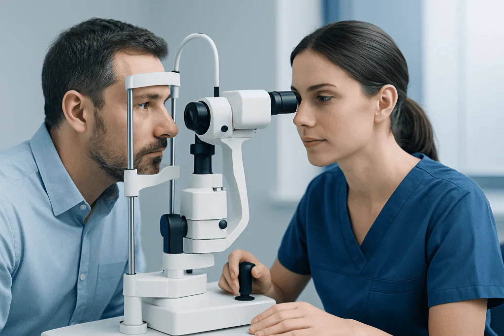
Diagnosis usually begins with a detailed eye history and a comprehensive eye examination. An ophthalmologist may ask about vision changes, timing of symptoms, past eye conditions, medical illnesses, medications, family history, and whether there has been a previous cancer diagnosis. A dilated eye exam allows the doctor to look at the back of the eye and inspect the retina and choroid more closely.
Imaging tests are often important. Optical coherence tomography, commonly called OCT, creates cross-sectional images of the retina and can show fluid, swelling, or changes related to underlying choroid disease. Fundus photography documents lesions over time, and ocular ultrasound can help measure deeper growths or distinguish between types of masses.
In some cases, angiography is used to study blood flow and leakage. This may involve fluorescein angiography or indocyanine green angiography, depending on what the doctor needs to assess. Additional tests, including blood work or body imaging, may be advised if inflammation, infection, or spread from another cancer is suspected.
When a deeper look is needed, the evaluation may include advanced eye examination techniques and imaging by a retina or ocular oncology team. Accurate diagnosis is the key step that guides whether the safest plan is simple monitoring or active treatment.
Treatment options for choroid disease
Treatment depends entirely on the cause. Many benign choroidal nevi do not need immediate treatment and can be monitored with regular photographs and scans. If inflammation is present, treatment may involve corticosteroids or other medicines to control the immune response. If an infection is responsible, the underlying infection must be treated appropriately.
Conditions that cause fluid leakage may be managed with observation, medicines, or targeted procedures, depending on severity and the diagnosis. Some choroidal vascular problems and retinal complications are treated using lasers, injections, or other retina-focused therapies. If the choroid problem is associated with a retinal detachment or severe structural change, surgery may be considered.
When a tumor is found, treatment may include close observation, radiation-based therapy, laser-based treatment, or surgery. The goal is to control the lesion while preserving as much vision and eye function as possible. Care is often individualized and may involve several specialists.
Depending on the specific problem, the care plan may overlap with retinal laser treatment, vitrectomy, or other retina and ocular oncology procedures. Near the end of the diagnostic process, some patients may also benefit from multidisciplinary input; Acibadem International’s specialists in JCI-accredited hospitals diagnose and treat eye conditions for international patients when coordinated care is needed.
Prevention, monitoring, and self-care
Not every choroid disorder can be prevented, but regular eye examinations are one of the best ways to protect vision. This is especially important for adults with changes in sight, a personal or family history of eye disease, inflammatory conditions, or past cancer. A dilated eye exam can reveal problems before symptoms become obvious.
General eye health habits may also help lower risk of complications. These include controlling blood pressure and blood sugar when relevant, not smoking, protecting the eyes from excessive ultraviolet exposure with appropriate sunglasses, and seeking medical care for new visual symptoms instead of waiting for them to pass.
People who have a known choroidal nevus or another monitored lesion should keep all follow-up appointments. Comparing new images with older ones helps doctors detect subtle changes. Self-care should not replace professional assessment, but paying attention to changes such as distortion, blind spots, flashes, or floaters can help patients seek care sooner.
- Attend routine dilated eye exams
- Report new or worsening visual symptoms promptly
- Manage chronic conditions such as diabetes or hypertension
- Avoid smoking and support overall vascular health
- Use UV-protective eyewear outdoors
- Keep follow-up visits for any known choroid lesion
When to seek medical care
Medical care should be sought promptly for new blurred vision, sudden distortion, a dark spot in central vision, flashes, a shower of new floaters, or any rapid change in sight. These symptoms do not always mean a serious choroid problem, but they can signal conditions that need urgent assessment to protect vision.
An earlier appointment is also advisable if a person has been told they have a choroidal nevus, has a history of uveitis, or has had cancer elsewhere in the body and develops visual changes. Regular follow-up is part of safe care, even when a lesion has been described as likely benign.
Emergency care may be needed if visual loss is sudden or severe, or if symptoms are accompanied by major eye pain, marked redness, or trauma. In any uncertain situation, the safest step is to contact an ophthalmologist or emergency service for guidance.
Frequently asked questions
What is the choroid in the eye?
The choroid is a layer of tissue filled with blood vessels that lies between the retina and the sclera. Its main role is to supply oxygen and nutrients to the outer retina and support healthy vision.
Can choroid problems cause vision loss?
Yes, some choroid conditions can affect vision, especially if they cause fluid buildup, inflammation, bleeding, or a growth beneath the retina. Early diagnosis can help guide treatment and may reduce the risk of lasting visual damage.
Are all choroidal lesions cancer?
No, many choroidal lesions are benign, such as a choroidal nevus, which is similar to a mole in the eye. However, some lesions need regular monitoring because a small number may show suspicious changes over time.
How are choroid disorders diagnosed?
Doctors usually diagnose choroid disorders with a dilated eye exam and imaging tests such as OCT, fundus photography, ultrasound, or angiography. The exact tests depend on whether the concern is inflammation, fluid leakage, or a possible tumor.
Can the choroid heal on its own?
Some mild or self-limited choroid-related changes may improve with monitoring, but this depends on the cause. Because different conditions can look similar at first, it is important not to assume symptoms will resolve without an eye specialist’s advice.
Who is more likely to develop choroid disease?
Risk varies by condition, but factors can include age, inflammatory or autoimmune disease, certain infections, prior cancer, and some inherited or pigment-related characteristics. An ophthalmologist can explain an individual’s risk based on the specific diagnosis.
References
- American Academy of Ophthalmology
- National Eye Institute
- Merck Manual Professional Edition
- Mayo Clinic
- National Cancer Institute
This article is for general information only and is not a substitute for professional medical advice. Please consult a qualified doctor about your individual situation.
Explore treatments in Turkey — costs, top hospitals & a free quote
JCI-accredited · board-certified surgeons · reply within 24h

More from the Health Library


Related Specialists

Prof. Dr. Mustafa Herdem
Orthopedic Surgery & Traumatology
Fzt. Semih Kızıltan
Physical Medicine and Rehabilitation
Prof. Dr. Arzu Akçay
Pathology
Dr. Burcu Sena Günaydın Şahin
Pediatrics