Uveitis
Uveitis is eye inflammation that can threaten vision. Learn symptoms, causes, diagnosis and treatment options, and when to see an eye specialist.
Quick answer
Uveitis is inflammation inside the eye that can affect the iris, ciliary body, or choroid and may cause pain, redness, blurred vision, and light sensitivity. Treatment depends on the cause and severity, and at Acibadem in Turkey it is evaluated with detailed eye examination and imaging, then managed with medicines such as anti-inflammatory or immune-targeting therapy, and sometimes surgery when…
What is uveitis?
Uveitis is inflammation of the uvea, the middle layer of the eye. The uvea sits between the white outer wall of the eye (the sclera) and the light-sensing layer at the back (the retina). It has three main parts: the iris (the colored ring around the pupil), the ciliary body (a ring of muscle and tissue behind the iris that helps the eye focus and produces fluid), and the choroid (a layer rich in blood vessels that nourishes the retina). When any of these structures becomes inflamed, doctors use the term uveitis.
Understanding what is uveitis begins with knowing that it is not a single disease. It is a group of inflammatory conditions that can arise from infections, autoimmune diseases (conditions in which the body’s immune system attacks its own tissues), eye injuries, or, in many cases, no identifiable cause at all. Doctors classify uveitis by the part of the eye involved:
- Anterior uveitis affects the front of the eye, mainly the iris. It is the most common form and is sometimes called iritis.
- Intermediate uveitis affects the middle of the eye, including the vitreous (the clear gel that fills the eyeball).
- Posterior uveitis affects the back of the eye, including the choroid and often the retina.
- Panuveitis means inflammation throughout all layers of the uvea.
Uveitis can affect people of any age, including children, though it is most often diagnosed in adults of working age. It may involve one eye or both, and it can be acute (appearing suddenly and lasting a limited time), recurrent (returning after quiet periods), or chronic (persisting for months). Although some episodes are mild, uveitis is a potentially serious condition. Untreated or poorly controlled inflammation inside the eye can lead to complications such as glaucoma (raised pressure inside the eye that damages the optic nerve), cataract (clouding of the eye’s natural lens), and permanent vision loss. Prompt evaluation and treatment are therefore important.
Symptoms of uveitis
Uveitis symptoms vary depending on which part of the eye is inflamed, how quickly the inflammation develops, and whether one or both eyes are involved. Common uveitis symptoms include:
- Eye redness, often most noticeable around the edge of the iris
- Eye pain, which may feel like a deep ache or soreness
- Sensitivity to light (photophobia), where normal light feels uncomfortably bright
- Blurred or hazy vision
- Floaterssmall dark spots, threads, or cobweb-like shapes drifting across your vision
- Decreased vision, ranging from mild dimming to significant loss
- A small or irregularly shaped pupil in some cases of anterior uveitis
How these symptoms appear often depends on the type of uveitis. Anterior uveitis, involving the front of the eye, typically comes on quickly and tends to cause redness, aching pain, and light sensitivity. Intermediate and posterior uveitis, which involve the middle and back of the eye, are often less painful. Instead, they may cause floaters and gradually blurring vision, sometimes without any redness at all. Because the eye can look normal from the outside in these forms, people may delay seeking care, which can allow damage to accumulate.
Chronic uveitis can be especially deceptive. Inflammation may smolder quietly with few obvious symptoms while slowly harming the structures of the eye. This is one reason regular follow-up with an eye specialist is important once uveitis has been diagnosed, even when the eye feels comfortable.
It is also worth noting that symptoms such as redness, pain, and light sensitivity overlap with many other eye problems, including infections of the eye’s surface and acute glaucoma. Only an eye examination can distinguish between these conditions, so new or worsening symptoms should always be assessed by a professional rather than self-diagnosed.
Causes and risk factors
Uveitis causes fall into several broad categories, and in a substantial share of cases, often called idiopathic uveitis, no specific cause is ever found even after thorough testing. Known contributors include:
- Autoimmune and inflammatory diseases. Conditions in which the immune system attacks the body’s own tissues are among the most common identifiable uveitis causes. Examples include ankylosing spondylitis (an inflammatory arthritis of the spine), sarcoidosis (a disease that causes clusters of inflammatory cells in organs), inflammatory bowel disease (Crohn’s disease and ulcerative colitis), Behçet’s disease (a condition causing blood vessel inflammation), juvenile idiopathic arthritis in children, and psoriatic arthritis.
- Infections. Certain infections can trigger inflammation inside the eye. These include herpes simplex and herpes zoster (shingles) viruses, toxoplasmosis (a parasitic infection often acquired from undercooked meat or contact with cat feces), tuberculosis, syphilis, and, less commonly, other bacterial, viral, or fungal infections.
- Eye injury or surgery. Trauma to the eye, or occasionally eye surgery, can provoke an inflammatory response in the uvea.
- Certain medications. A small number of drugs have been associated with uveitis as a side effect. Your doctor may review your medication list if the cause is unclear.
- Genetic factors. Some people carry a genetic marker called HLA-B27, which is linked to a higher risk of recurrent anterior uveitis, particularly in people who also have related inflammatory conditions.
Risk factors, then, include having an autoimmune or inflammatory disease, a history of certain infections, previous eye trauma or surgery, smoking (which has been associated with a higher risk of uveitis in some research), and a family or personal history of HLA-B27–related conditions. That said, uveitis can occur in people with none of these risk factors, and having a risk factor does not mean you will develop the condition.
Diagnosis
Uveitis diagnosis rests on a careful eye examination by an ophthalmologist, a medical doctor who specializes in eye disease, combined, where needed, with laboratory tests and imaging to look for an underlying cause. The main steps often include:
- Medical history. Your doctor will ask about your symptoms, past episodes, general health, joint pain, skin problems, bowel symptoms, infections, recent travel, medications, and family history. These clues help point toward possible causes.
- Visual acuity testing. A standard reading chart measures how well each eye sees.
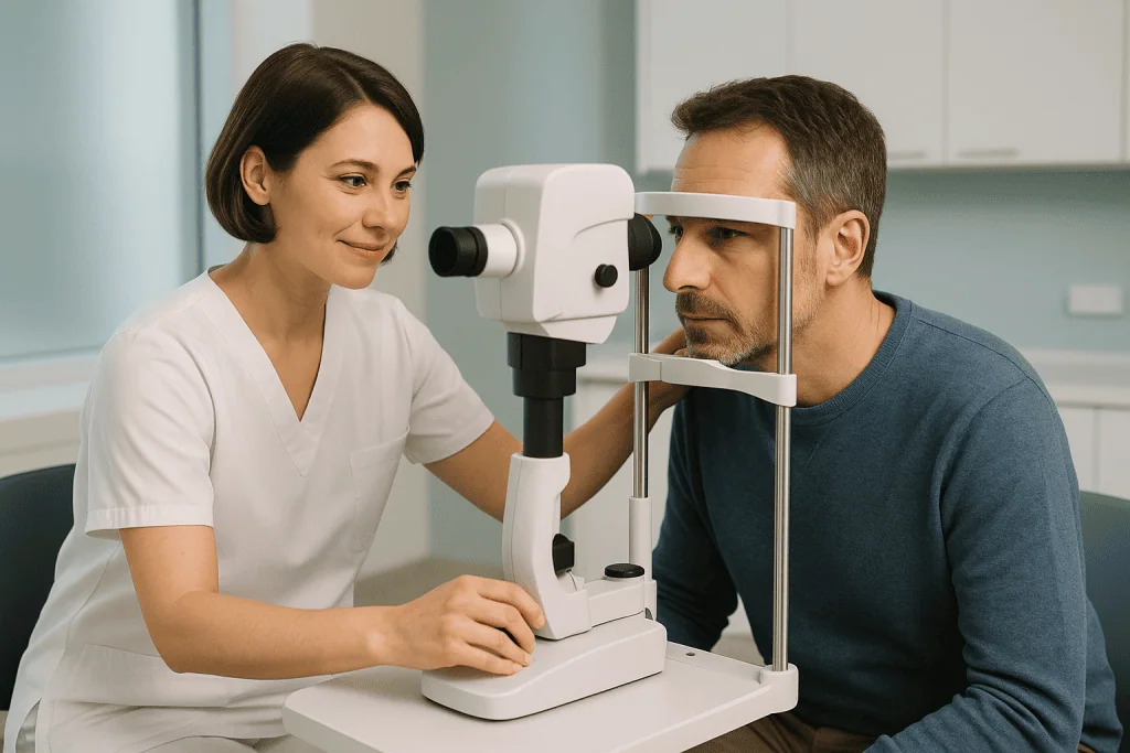
- Slit-lamp examination. This is the central test for uveitis. A slit lamp is a special microscope with a bright, narrow beam of light that lets the doctor see the front of the eye in fine detail. In active anterior uveitis, the doctor can often see inflammatory cells floating in the fluid at the front of the eye, along with protein haze known as flare.
- Eye pressure measurement (tonometry). Uveitis can raise or lower the pressure inside the eye, so this measurement is routinely checked.
- Dilated fundus examination. Drops are used to widen the pupil so the doctor can inspect the vitreous, retina, and choroid at the back of the eye for signs of intermediate or posterior inflammation.
- Imaging tests. Optical coherence tomography (OCT), a painless scan that produces cross-sectional pictures of the retina, can detect swelling of the central retina (macular edema), a common complication. Fluorescein angiography, in which a dye is injected into a vein and photographed as it passes through the eye’s blood vessels, may be used to assess inflammation of the retina and choroid. Ultrasound of the eye is sometimes helpful when the view inside is blocked.
- Blood tests and other investigations. Depending on the pattern of inflammation, your doctor may order blood tests for autoimmune markers and infections such as syphilis or tuberculosis, a chest X-ray or chest scan to look for sarcoidosis or tuberculosis, and occasionally genetic testing such as HLA-B27. Not every patient needs every test; investigations are tailored to the type of uveitis and the individual’s history.
Because uveitis can be the first visible sign of a disease affecting the whole body, uveitis diagnosis sometimes involves collaboration between ophthalmologists and other specialists, such as rheumatologists (doctors who treat autoimmune and joint diseases) or infectious disease physicians. Even so, in many patients no underlying disease is found, and the uveitis is managed on its own terms.
Treatment options
The goals of uveitis treatment are to control inflammation, relieve pain, prevent complications, and preserve vision. The right approach depends on the type of uveitis, its severity, its cause, and whether one or both eyes are affected. Specialized care for this condition is typically provided within an ophthalmology department, where at Acibadem, as at other centers, uveitis is managed by eye specialists, often together with other medical disciplines when a systemic disease is involved.
Common elements of uveitis treatment include:
- Corticosteroid eye drops. Steroids are anti-inflammatory medicines, and steroid drops are usually the first-line treatment for anterior uveitis. They are typically started at a frequent dosing schedule and then tapered slowly as the inflammation settles. Stopping them abruptly can cause the inflammation to rebound, so it is important to follow the tapering plan.
- Pupil-dilating (cycloplegic) drops. These drops relax the muscles inside the eye, which eases pain and helps prevent the iris from sticking to the lens: a complication called synechiae that can distort the pupil and raise eye pressure.
- Steroid injections or implants. When inflammation affects the middle or back of the eye, drops often cannot reach it effectively. Doctors may then inject steroid medication around or into the eye, or place a small slow-release steroid implant inside the eye, to deliver treatment where it is needed.
- Oral corticosteroids. Tablets such as prednisone may be used for more severe or widespread inflammation. Because long-term steroid use can cause side effects, including weight gain, high blood sugar, bone thinning, cataract, and raised eye pressure, doctors aim to use the lowest effective dose for the shortest necessary time.
- Immunosuppressive and biologic medications. For chronic or recurrent uveitis that cannot be controlled with steroids alone, or when long-term steroid use would be harmful, medicines that calm the immune system more broadly may be prescribed. These include conventional immunosuppressants such as methotrexate, azathioprine, or mycophenolate, and biologic agents such as adalimumab (an antibody that blocks a specific inflammatory signal). These treatments require regular monitoring with blood tests and are usually managed jointly by an ophthalmologist and a physician experienced in immune-modulating therapy.
- Treatment of an underlying infection. If uveitis is caused by an infection, the infection itself is treated first or alongside anti-inflammatory therapy, for example, antiviral medicines for herpes-related uveitis, or antiparasitic medicines for toxoplasmosis. Using steroids alone in an infectious uveitis can make matters worse, which is one more reason accurate diagnosis matters.
- Surgery. Surgery is not a routine treatment for uveitis itself, but it may be needed for its complications. Examples include cataract surgery when the lens has become cloudy, glaucoma surgery when eye pressure cannot be controlled with medication, and vitrectomy, removal of the vitreous gel, in selected cases to clear severe debris, treat complications, or obtain a sample for diagnosis.
Watchful waiting has only a very limited role. A few mild, clearly self-limited cases may be observed closely, but most uveitis needs active treatment, because ongoing inflammation inside the eye tends to cause cumulative damage. Your doctor will weigh the risks and benefits of each option with you and adjust treatment based on how your eye responds at follow-up visits.
Living with uveitis and outlook
The outlook for uveitis varies widely. Many people with a single episode of anterior uveitis respond well to steroid drops and recover good vision, though recurrences are possible. Chronic, intermediate, posterior, and panuveitis tend to require longer and more complex treatment, and they carry a higher risk of complications such as macular edema (swelling of the central retina), cataract, glaucoma, and, in severe untreated cases, permanent vision loss. With modern therapy and consistent follow-up, many patients maintain useful vision, but no outcome can be guaranteed, and some people live with reduced vision despite good care.
Practical points that often help people living with uveitis include:
- Attend every follow-up appointment, even when your eye feels normal. Inflammation and complications can develop silently.
- Take medications exactly as prescribed, including steroid tapering schedules, and never stop immune-modulating drugs without medical advice.
- Learn your early warning signs. Many people with recurrent uveitis come to recognize the first hints of a flare, mild ache, light sensitivity, or new floaters, and seeking care early can shorten and soften the episode.
- Manage any underlying condition. If your uveitis is linked to an autoimmune disease, keeping that disease controlled often helps protect your eyes.
- Protect your general health. Not smoking, attending regular checkups, and reporting medication side effects promptly all support long-term treatment.
Living with a recurrent or chronic eye condition can be stressful, and anxiety about vision is common and understandable. Discussing concerns openly with your care team, and asking what your specific type of uveitis is likely to mean for you, can make the condition easier to manage day to day.
Frequently asked questions
What is uveitis in simple terms?
Uveitis is inflammation of the middle layer of the eye, called the uvea, which includes the iris, the ciliary body, and the choroid. In simple terms, part of the inside of the eye becomes swollen and irritated, which can cause redness, pain, light sensitivity, floaters, and blurred vision. It can result from autoimmune disease, infection, or injury, and in many cases no cause is found.
Can uveitis go away on its own?
Some mild episodes may settle without treatment, but relying on this is risky. Untreated inflammation inside the eye can damage delicate structures and lead to complications such as glaucoma, cataract, or permanent vision loss. In most cases doctors recommend active treatment, so anyone with suspected uveitis symptoms should be examined rather than waiting to see whether the problem resolves.
How serious is uveitis?
Uveitis ranges from a single, easily treated episode to a chronic condition that threatens sight. It is considered a significant cause of preventable vision loss worldwide, mainly when it goes untreated or is poorly controlled. With prompt diagnosis and appropriate uveitis treatment, many people keep good vision, but the condition should always be taken seriously and monitored by an eye specialist.
What does uveitis pain feel like?
People with anterior uveitis often describe a deep, dull ache in or around the eye, frequently accompanied by discomfort in bright light. The pain typically differs from the surface irritation or gritty feeling of conditions like conjunctivitis. Intermediate and posterior uveitis, by contrast, are often painless, causing floaters and blurred vision instead. Because pain patterns vary, an eye examination is needed to identify the cause.
Will uveitis come back after treatment?
It can. Some people have a single episode and never experience another, while others have recurrent flares over months or years, particularly if the uveitis is linked to an underlying autoimmune condition or a genetic marker such as HLA-B27. Regular follow-up, prompt attention to early symptoms, and, in some cases, long-term preventive medication can help reduce the impact of recurrences, though they cannot always be prevented.
How is uveitis diagnosed?
Uveitis diagnosis is made by an ophthalmologist using a slit-lamp examination, which reveals inflammatory cells inside the eye, together with eye pressure measurement and a dilated examination of the back of the eye. Depending on the findings, doctors may add imaging such as optical coherence tomography, plus blood tests or chest imaging to search for underlying causes like autoimmune disease or infection. Testing is tailored to each patient.
Can uveitis cause blindness?
Severe or untreated uveitis can lead to permanent vision loss, usually through complications such as macular edema, glaucoma, cataract, or retinal damage rather than the inflammation alone. This outcome is far less likely when the condition is diagnosed early, treated appropriately, and followed up regularly. Prompt care is the most important step a patient can take to protect their sight.
When to see a doctor
Any new eye pain, redness, or change in vision deserves professional assessment, because uveitis and several other serious eye conditions can look similar in their early stages. Seek urgent medical attention, the same day if possible, if you experience any of the following red-flag warning signs:
- Sudden loss of vision or a rapid, noticeable drop in how well you see
- Severe eye pain, especially with headache, nausea, or vomiting, which can signal dangerously high eye pressure
- Marked sensitivity to light with a red, aching eye
- A sudden shower of new floaters, flashing lights, or a dark curtain or shadow moving across your vision, which can indicate a retinal problem
- A visibly changed pupilsmaller, larger, or irregular compared with the other eye
- Eye symptoms after an injury to the eye or face
- Worsening symptoms during uveitis treatment, or a flare of symptoms after stopping or tapering your eye drops
If you have already been diagnosed with uveitis, contact your eye care team promptly whenever familiar flare symptoms return, even if they seem mild. Early treatment of a recurrence is generally easier and safer than treating established inflammation, and timely care offers the best chance of protecting your vision over the long term.
Medically reviewed by the Acıbadem International Medical Board September 2, 2026
See our medical review board →
Update history
- PublishedJune 9, 2026
- Medical review approvedSeptember 2, 2026
- Last content updateSeptember 2, 2026