Conjunctival Nevus Removal It Is Complicated: Procedure, Recovery and Results

A conjunctival nevus is a usually benign pigmented spot on the clear membrane covering the white of the eye. Removal may be considered for diagnostic uncertainty, documented change, irritation, cosmetic concerns, or suspected malignancy.
Key Takeaways
- A conjunctival nevus is a usually benign pigmented spot on the clear membrane covering the white of the eye.
- Removal may be considered for diagnostic uncertainty, documented change, irritation, cosmetic concerns, or suspected malignancy.
- The procedure is commonly performed as day surgery using local anesthesia, with the lesion sent for laboratory examination when appropriate.
- Temporary redness, irritation, and a foreign-body sensation are common during healing; serious complications are uncommon but possible.
- A changing, newly appearing, raised, bleeding, or unusually vascular eye lesion should be assessed promptly by an ophthalmologist.
Conjunctival nevus removal is generally not a complicated operation when a lesion is clearly benign and performed by an experienced ophthalmic surgeon. The most important step is a careful eye examination, as monitoring may be safer than removal for many stable conjunctival nevi.
Overview: Is Conjunctival Nevus Removal Complicated?
Conjunctival nevus removal is usually a relatively small outpatient eye operation rather than a complex major surgery. A conjunctival nevus is a collection of pigment-producing cells that appears as a brown, tan, yellowish, or occasionally non-pigmented spot on the conjunctiva, the thin transparent tissue over the white part of the eye and inside the eyelids.
Whether removal is appropriate depends less on the size alone and more on the lesion’s appearance, location, documented changes, symptoms, and the person’s preferences. Many nevi remain harmless and can be observed with photographs and regular eye examinations. Removal becomes more important when the diagnosis is uncertain or there are features that require exclusion of a more serious condition.
The operation may involve simple excision, careful reconstruction of the conjunctiva, and occasionally a tissue graft when a larger area must be removed. A specialist may also recommend laboratory analysis of the removed tissue. This approach helps provide a clear diagnosis while protecting the eye’s surface and appearance.
How a Conjunctival Nevus Is Assessed and Who May Be a Candidate
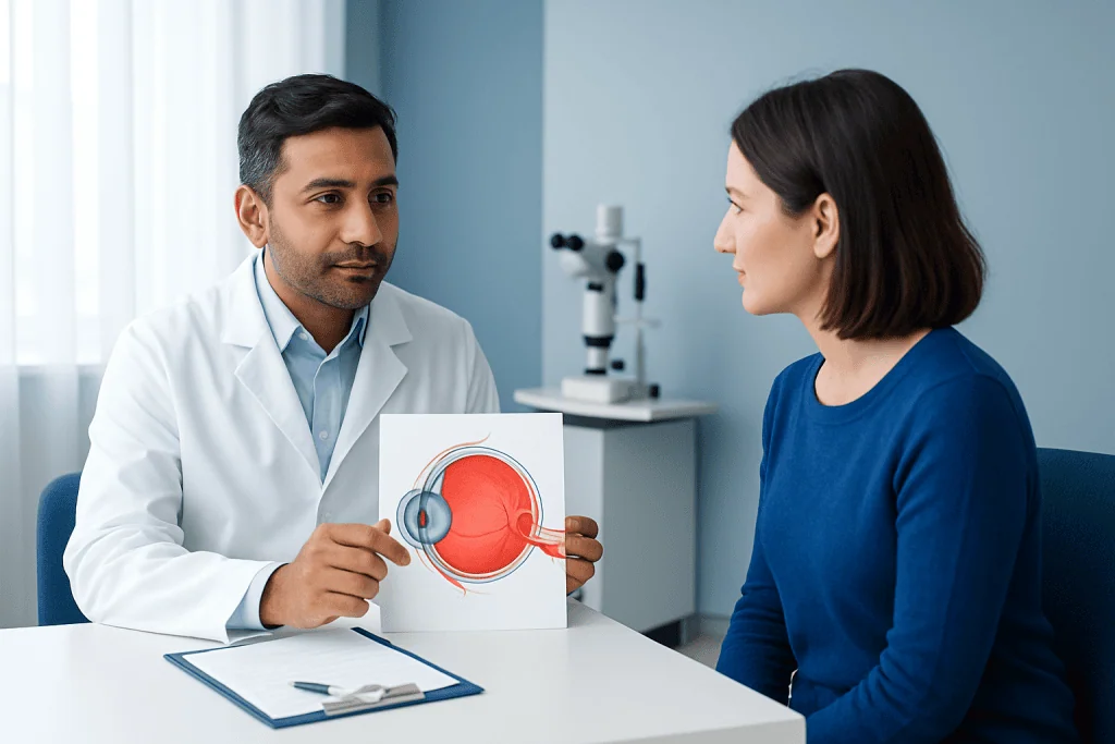
An ophthalmologist begins with a detailed history and slit-lamp examination. They may ask when the spot was first noticed, whether it has changed, and whether there is irritation, pain, recurrent redness, blurred vision, bleeding, or a sensation of a lump. High-quality clinical photographs can be useful for comparing the lesion over time.
Removal may be considered when a lesion enlarges after childhood, changes in colour or contour, develops new blood vessels, becomes symptomatic, repeatedly catches on the eyelid, or causes significant cosmetic concern. It may also be advised if the clinician cannot confidently distinguish a nevus from another pigmented conjunctival lesion.
Not every dark spot on the eye is a nevus. Conditions such as racial or complexion-associated pigmentation, primary acquired melanosis, conjunctival cysts, and rarely conjunctival melanoma can look similar. For this reason, self-diagnosis or trying to remove an eye lesion outside a specialist setting is unsafe.
- Stable, typical lesions may be monitored without surgery.
- Suspicious or changing lesions generally need specialist review.
- Removal may be diagnostic, therapeutic, cosmetic, or a combination of these reasons.
How Conjunctival Nevus Removal Works: Step by Step
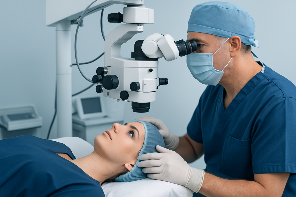
Conjunctival nevus removal is usually carried out by an ophthalmologist with experience in ocular surface surgery. Most procedures are performed on an outpatient basis under local anesthesia, often with anesthetic eye drops and, when needed, a small injection to numb the area. Sedation may be considered for selected patients, including some children or people who cannot comfortably remain still.
After the eye is cleaned and protected, the surgeon gently separates the lesion from the surrounding conjunctiva and underlying tissue. The goal is complete and careful removal while preserving as much healthy tissue as possible. The specimen is commonly sent for histopathology, where a laboratory specialist examines it under a microscope.
Small wounds may heal naturally or be closed with very fine dissolvable stitches. If the removed area is larger, the surgeon may use a conjunctival autograft, amniotic membrane, or another reconstructive technique to support healing and reduce scarring. These choices depend on the lesion’s size and location rather than indicating that every removal is complicated.
Patients considering conjunctival nevus removal should discuss the intended surgical technique, need for tissue analysis, likely cosmetic result, and follow-up plan with their eye surgeon before consenting to treatment.
Benefits, Risks and Expected Results
The possible benefits of removal include obtaining a definite tissue diagnosis, treating irritation caused by a raised lesion, addressing a suspicious change, and improving appearance when the nevus is noticeable. If pathology confirms a benign nevus that has been completely removed, no further treatment is often needed beyond routine eye care and the surgeon’s recommended review.
As with any eye procedure, there are risks. These can include temporary discomfort, redness, swelling, bleeding, infection, delayed surface healing, scarring, dry-eye symptoms, visible blood vessels, pigment remaining or returning, and an unsatisfactory cosmetic result. Rarely, surgery may affect nearby structures or require additional treatment.
Removing a nevus does not guarantee that all future pigmented lesions will be prevented. It is also important to understand that pathology results may occasionally identify a different diagnosis than expected, which could change the follow-up plan. An ophthalmologist can explain individual risks based on the lesion and the person’s general eye health.
For lesions with concerning features, treatment planning may involve ophthalmic oncology, pathology, reconstructive eye-surface specialists, and other relevant teams. This multidisciplinary approach is particularly valuable when malignancy must be excluded or confirmed.
Recovery Timeline and Aftercare
Most people go home on the day of surgery. The eye may feel gritty, watery, sensitive to light, or mildly sore during the first few days. Redness can be noticeable and may take several weeks to fade, particularly when stitches, a graft, or a larger area of conjunctival healing is involved.
The surgeon usually prescribes medicated eye drops or ointment to reduce inflammation and prevent infection. It is important to use these exactly as directed, avoid rubbing the eye, wash hands before touching the face or applying medication, and attend scheduled follow-up visits. Contact lenses should not be worn until the eye specialist confirms that the surface has healed sufficiently.
Many people can resume gentle daily activities soon after the procedure, but swimming, dusty environments, heavy exertion, and eye makeup may need to be avoided temporarily. Recovery times vary; a small superficial excision may settle quickly, while healing after grafting or more extensive surgery can take longer.
Pathology results are typically discussed at a follow-up appointment. If the result is benign, the doctor may recommend periodic eye examinations and photographs. If there is an atypical or malignant finding, the ophthalmology team will explain the next diagnostic and treatment steps clearly.
Is It Safe to Remove Conjunctival Nevus?
Conjunctival nevus removal is generally considered safe when it is appropriately indicated and performed by a qualified ophthalmic surgeon in a suitable clinical setting. The eye surface usually heals well, and serious complications are uncommon. However, safe care starts with confirming whether surgery is necessary and selecting a technique that matches the lesion’s location and size.
Monitoring can be the safer choice for a typical, stable nevus with no concerning features. Unnecessary surgery can expose a person to avoidable discomfort, scarring, and other procedural risks. Conversely, a lesion that is changing or difficult to diagnose should not simply be ignored because it looks like a harmless mole.
Before surgery, patients should disclose prior eye operations, dry eye disease, bleeding conditions, medication use, allergies, contact lens use, and any history of skin or eye cancer. These details help the team plan anesthesia, aftercare, and follow-up safely.
Can a Nevus in the Eye Become Malignant?
Most conjunctival nevi are benign and do not become malignant. A nevus that has been present for years and remains stable is usually reassuring, but it should still be assessed if its appearance changes. The risk is not the same for every pigmented lesion, which is why an ophthalmologist’s examination is essential.
Features that deserve prompt review include new growth in adulthood, a new raised area, increasing thickness, colour variation, persistent inflammation, bleeding, newly prominent feeder vessels, or extension onto the cornea. These signs do not prove cancer, but they can indicate that closer assessment is needed.
Some lesions initially thought to be nevi may instead represent primary acquired melanosis or another condition with a different level of risk. When clinical assessment cannot provide a confident answer, excision and histopathological examination may be the most reliable way to establish the diagnosis.
What Is the Survival Rate for Conjunctival Melanoma?
Conjunctival melanoma is rare and differs substantially from a benign conjunctival nevus. Survival varies widely because prognosis depends on factors such as the tumour’s location, size, thickness, cell characteristics, whether it has recurred, and whether it has spread to lymph nodes or distant organs. For this reason, an individual survival estimate should come from the specialist team after complete assessment.
Published outcomes differ between studies and healthcare settings, so a single percentage can be misleading. In general, earlier diagnosis and treatment before spread are associated with better outcomes. Ongoing follow-up is important because conjunctival melanoma can recur locally or spread even after initial treatment.
People should not assume that a common benign eye spot is melanoma. At the same time, any suspicious or changing pigmented conjunctival lesion deserves timely evaluation by an ophthalmologist, ideally one experienced in ocular surface tumours or ocular oncology.
How Much Does Conjunctival Nevus Removal Cost?
The cost of conjunctival nevus removal varies considerably by country, hospital, surgeon expertise, anesthesia needs, operating-room setting, laboratory pathology, use of graft material, insurance coverage, and the number of follow-up visits required. It is therefore not possible to give one accurate cost figure without an individual clinical and administrative assessment.
Patients can request a written estimate that separates the consultation, preoperative tests if needed, surgeon and facility charges, anesthesia, pathology, medications, graft materials, and planned follow-up. They should also ask what may create additional costs, such as more extensive reconstruction or further treatment following an unexpected pathology result.
At Acibadem International, multidisciplinary specialists and JCI-accredited hospitals can assess and treat conjunctival lesions for international patients. A personalised plan should be based on clinical examination first, rather than cost alone.
When to Seek Medical Care
An eye doctor should assess any new pigmented spot on the eye, especially if it develops in adulthood or its history is unclear. Prompt medical review is also appropriate for a known nevus that changes in size, shape, elevation, or colour, or develops new visible blood vessels.
Urgent assessment is sensible if there is persistent pain, significant redness, bleeding, a rapidly growing mass, new visual disturbance, or difficulty closing the eyelid. These symptoms can have many causes, and they do not necessarily mean cancer, but they should not be managed with over-the-counter products alone.
Regular comprehensive eye examinations support early identification of changes on the ocular surface. Patients who have had a lesion removed should keep follow-up appointments and contact their surgeon if pain, discharge, worsening redness, or reduced vision occurs during recovery.
Frequently asked questions
Is conjunctival nevus removal painful?
The procedure is usually performed with local anesthesia, so sharp pain should not be felt during the operation. Some pressure or awareness of movement may occur. Mild soreness, grittiness, tearing, and light sensitivity are common for a short time afterwards and can usually be managed with the prescribed treatment.
How long does conjunctival nevus removal take?
The surgical time depends on the lesion’s size, position, and whether reconstruction or grafting is required. A straightforward removal is often completed during a short outpatient procedure, while more extensive cases take longer. The eye surgeon can give a more specific estimate after examining the lesion.
Will the eye stay red after conjunctival nevus removal?
Redness is expected after surgery and may remain visible for several weeks while the conjunctiva heals. It can last longer when stitches or a graft are used. Worsening redness, increasing pain, discharge, or reduced vision should be reported to the treating eye team.
Is a biopsy always needed for a conjunctival nevus?
A biopsy is not always needed for a typical, stable lesion that an ophthalmologist can confidently diagnose and monitor. If the lesion is removed, it is often sent for histopathology to confirm the diagnosis. Biopsy or excision is more strongly considered when there are suspicious changes or diagnostic uncertainty.
Can a conjunctival nevus grow back after removal?
Pigment may remain, recur, or become visible again after removal, particularly if cells extend beyond the visibly pigmented area. The chance depends on the lesion and the surgical technique. Follow-up examinations help distinguish expected healing changes from recurrence or a new lesion.
Can I wear contact lenses after conjunctival nevus surgery?
Contact lenses should be avoided until the surgeon confirms that the conjunctiva has healed. Wearing them too early may irritate the surgical area and increase the risk of complications. The timing varies according to the procedure and the individual’s recovery.
References
- American Academy of Ophthalmology
- American Academy of Ophthalmology EyeWiki
- National Cancer Institute
- American Cancer Society
- World Health Organization
This article is for general information only and is not a substitute for professional medical advice. Please consult a qualified doctor about your individual situation.
Explore treatments in Turkey — costs, top hospitals & a free quote
JCI-accredited · board-certified surgeons · reply within 24h