CT Scan for Prostate Cancer: Preparation, Procedure and Results
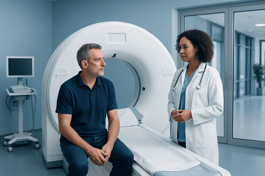
CT is usually a staging test rather than the main test used to diagnose prostate cancer. A contrast-enhanced CT scan may require temporary fasting and a review of kidney function, allergies and medicines.
Key Takeaways
- CT is usually a staging test rather than the main test used to diagnose prostate cancer.
- A contrast-enhanced CT scan may require temporary fasting and a review of kidney function, allergies and medicines.
- CT can identify enlarged lymph nodes or spread to organs, but small or early prostate tumors may not be visible.
- The scan itself commonly takes only minutes, while the full appointment may be longer if contrast is used.
- Results are interpreted alongside PSA testing, examination findings, biopsy results and other imaging studies.
A CT scan for prostate cancer is most often used after diagnosis to check lymph nodes, nearby organs and other areas for signs that cancer may have spread. It is quick and painless, and preparation mainly depends on whether contrast dye is needed.
CT Scan for Prostate Cancer: What It Shows
A CT scan for prostate cancer uses X-rays and computer processing to create detailed cross-sectional images of the body. It is generally used to help determine the extent of a known or strongly suspected cancer, particularly whether there are enlarged lymph nodes or changes in nearby organs. It may also be used when symptoms or test results raise concern that cancer has spread beyond the prostate.
CT is not usually the first or most accurate test for finding cancer confined within the prostate gland. Blood testing for prostate-specific antigen (PSA), a digital rectal examination, prostate MRI and biopsy are more commonly used to investigate possible prostate cancer. Once cancer is confirmed, imaging contributes to the overall assessment of prostate cancer and supports treatment planning.
Depending on a person’s situation, clinicians may recommend CT alongside bone imaging, MRI or more specialized molecular imaging. The most appropriate approach depends on biopsy grade, PSA level, symptoms, previous treatment and the estimated likelihood of disease outside the prostate.
How a CT Scan Works and Who May Need One
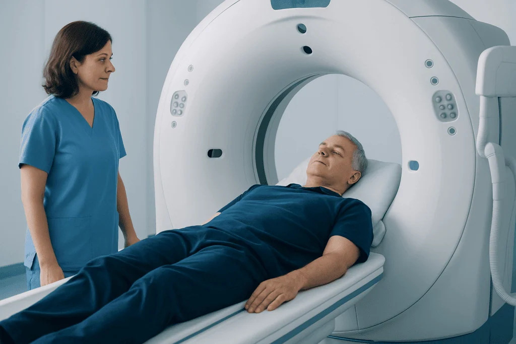
During CT scanning, the patient lies on a motorized table that moves slowly through a doughnut-shaped scanner. The machine takes many X-ray images from different angles. A computer combines them into detailed pictures of the pelvis, abdomen and, when appropriate, the chest or other body areas.
For prostate cancer staging, an intravenous contrast agent may be used. This iodine-containing dye makes blood vessels and some organs easier to distinguish on the images. It does not treat cancer; it simply improves image detail. Some people notice a brief warm sensation, a metallic taste or a feeling of needing to urinate when contrast is injected. These sensations usually pass quickly.
A CT scan may be considered for people with higher-risk disease, concerning symptoms, rising PSA after treatment or clinical findings that suggest possible spread. For people with low-risk, localized cancer, CT may not add useful information. The urologist and oncology team can explain whether CT, MRI, PSMA PET imaging or another test is most suitable.
Preparing for the Scan

Preparation instructions vary by imaging center and whether contrast will be used. Patients should follow the instructions from their radiology department rather than making changes independently. They may be asked to avoid food for several hours before a contrast-enhanced scan, while clear fluids may still be permitted.
Before the appointment, it is important to tell the care team about previous reactions to contrast dye, asthma or significant allergies, kidney disease, diabetes, thyroid conditions, pregnancy, and all prescribed medicines or supplements. A blood test may be arranged to assess kidney function before iodinated contrast is given. The team may give individualized instructions about diabetes medicines or other treatments.
Patients should bring relevant medical information, including biopsy reports, PSA results, previous scan reports and a current medication list if these are not already available to the hospital. Wearing loose, comfortable clothing can help. Metal items such as belts, jewelry, hearing aids and some removable dental items may need to be removed before imaging.
Is there anything I shouldn’t do before a CT scan?
Patients should not eat or drink contrary to the imaging center’s instructions, particularly when contrast is planned. They should also not withhold regular medicines, stop blood-thinning medicine or change diabetes treatment unless their doctor specifically advises this. It is important not to overlook a history of contrast reaction or kidney problems, as the radiology team can take precautions or consider an alternative test.
What should you not do before a prostate exam?
Preparation depends on the type of prostate exam. Before a digital rectal examination, there is usually little preparation, but patients should tell the clinician about rectal pain, bleeding, hemorrhoids, recent rectal surgery or severe bowel symptoms. Before a PSA blood test, clinicians may advise avoiding ejaculation and vigorous cycling for a short period because these can temporarily affect PSA levels; instructions can vary, so the testing provider should be asked.
What Happens During the Procedure
On arrival, a radiographer will confirm the patient’s identity, review safety questions and explain the scan. If contrast is needed, a small intravenous cannula is placed in a vein, usually in the hand or arm. The patient then lies on the scanning table, typically on their back, and may be positioned with cushions for comfort.
The radiographer operates the scanner from a nearby room but can see, hear and speak with the patient throughout the examination. The patient needs to remain still, and may occasionally be asked to hold their breath briefly. The CT scanner is open at both ends and is generally less confining than an MRI scanner.
The image acquisition itself often takes only a few minutes. Allow extra time for registration, safety checks, contrast preparation and observation if needed. Afterward, a radiologist analyzes the images and sends a report to the doctor who requested the examination.
Can they see prostate cancer on a CT scan?
CT may show a large prostate mass or signs that cancer has spread, such as enlarged lymph nodes or abnormalities in certain organs. However, it often cannot distinguish early prostate cancer from normal prostate tissue or non-cancerous enlargement. A normal CT scan therefore does not rule out prostate cancer, and biopsy remains the test that confirms the diagnosis.
Results, Benefits and Limits of CT Staging
CT results are considered together with PSA trends, examination findings, biopsy results and other scans. A report may describe the prostate, pelvic lymph nodes, bladder, rectum, bones and any other body areas included in the study. Enlarged lymph nodes can be suspicious, but enlargement is not always caused by cancer, and microscopic cancer deposits may not enlarge a node enough to be seen.
The main benefit of CT is that it is widely available, fast and useful for reviewing several anatomical areas in one examination. It can help clinicians select the next investigations and discuss suitable management options. Treatment may include active surveillance, surgery, radiation therapy, hormone therapy, systemic therapy or a combination, depending on the individual diagnosis and stage.
CT uses ionizing radiation, so it is ordered when the expected clinical value outweighs the small radiation risk. If contrast is used, uncommon risks include an allergic-like reaction or kidney-related complications in people with significant kidney impairment. Radiology teams screen carefully and are trained to manage reactions promptly. Where appropriate, multidisciplinary assessment can help coordinate prostate cancer treatment decisions.
What is the 2 week rule for prostate cancer?
The term “2 week rule” is used in some health systems to describe an urgent referral pathway: a person with symptoms or test findings that may suggest cancer is offered specialist assessment within approximately two weeks. It is not a rule that prostate cancer must be diagnosed, scanned or treated within two weeks. Referral urgency is based on the individual’s symptoms, PSA result, examination findings and overall health.
Recovery Timeline and Aftercare
Most people can return to normal daily activities immediately after a CT scan. If no sedative has been given, which is typical for CT, they can usually travel home and resume work or other routine plans. The scan does not leave radiation in the body.
After contrast, drinking fluids may be encouraged unless a clinician has advised fluid restriction because of heart, kidney or other health conditions. This supports normal elimination of the contrast agent. The cannula site may be mildly tender or bruised for a short time, but this usually settles without treatment.
Patients should seek prompt medical advice after leaving the department if they develop hives, swelling of the face or throat, trouble breathing, persistent vomiting, marked dizziness, or worsening pain and redness around the injection site. Delayed reactions are uncommon but should be discussed with a healthcare professional. The referring doctor will explain when and how results will be communicated.
When to Seek Medical Care
A person should arrange medical assessment for urinary symptoms that persist or worsen, such as difficulty starting urination, a weak stream, frequent urination, waking repeatedly at night to urinate, blood in urine or semen, or new pelvic discomfort. These symptoms are often caused by non-cancerous conditions, including prostate enlargement or infection, but they deserve proper evaluation.
Urgent medical care is appropriate for an inability to pass urine, heavy bleeding in urine, fever with urinary symptoms, severe back or bone pain with weakness or numbness, or unexplained symptoms that are rapidly worsening. These symptoms do not necessarily indicate prostate cancer, but timely assessment is important.
People with a raised PSA, an abnormal prostate examination, a strong family history of prostate cancer or known inherited cancer-risk variants should discuss individualized assessment with a qualified clinician. Acibadem International’s multidisciplinary specialists in JCI-accredited hospitals diagnose and treat prostate conditions for international patients, coordinating radiology, urology, pathology and oncology care where needed.
Frequently asked questions
Is a CT scan painful?
A CT scan is painless. The patient needs to lie still on a moving table while the scanner takes images. If contrast is injected, there may be a brief warm feeling or metallic taste, which usually fades quickly.
How long does a CT scan for prostate cancer take?
The scanning portion may take only a few minutes. The complete appointment can be longer because of check-in, safety screening, placement of an intravenous line and contrast preparation. The imaging department can provide an estimated appointment length in advance.
Do I need contrast dye for a prostate cancer CT scan?
Many CT staging examinations use intravenous contrast because it can improve visibility of blood vessels, lymph nodes and abdominal organs. However, contrast is not appropriate for everyone, such as some people with certain kidney conditions or previous severe reactions. The radiology team will decide based on the clinical question and safety assessment.
Can a CT scan tell whether prostate cancer has spread?
CT can help identify signs of spread to lymph nodes, organs or other areas included in the scan. It cannot detect every small area of cancer, particularly microscopic disease. Other imaging tests may be recommended when more sensitive staging information is needed.
When will CT scan results be available?
A radiologist reviews the images and prepares a report for the requesting doctor. Timing differs between healthcare settings and may depend on whether the scan is routine or urgent. The doctor who requested the scan should explain the results and what they mean in the context of other test findings.
Can I drive after a CT scan?
Most people can drive after a routine CT scan because sedation is not normally used. If a person feels unwell after contrast or has received medicine that affects alertness for another reason, they should not drive. It is sensible to follow the instructions given by the imaging team.
References
- American Cancer Society
- European Association of Urology
- National Cancer Institute
- Radiological Society of North America
- National Health Service
This article is for general information only and is not a substitute for professional medical advice. Please consult a qualified doctor about your individual situation.
Prostate Cancer in Turkey — costs, top hospitals & a free quote
JCI-accredited · board-certified surgeons · reply within 24h
Add us as a Preferred Source to see more of our trusted health content across Google Search, AI Overviews and Discover.

More from the Health Library



Related Specialists

Dr. Ezgi Gülüm
Oral Dental and Maxillofacial Surgery
Dr. Recep Özgür
Interventional Radiology
Dyt. Burcu Akbeyaz
Nutrition & Diet
Prof. Dr. Barış Kocaoğlu
Orthopedic Surgery & Traumatology