Life after Benign Meningioma Surgery: Procedure, Recovery and Results

A benign meningioma is usually slow-growing, but its location can still affect important brain, nerve or spinal functions. Surgery aims to remove the tumour safely while protecting nearby brain tissue, blood vessels and nerves.
Key Takeaways
- A benign meningioma is usually slow-growing, but its location can still affect important brain, nerve or spinal functions.
- Surgery aims to remove the tumour safely while protecting nearby brain tissue, blood vessels and nerves.
- Hospital recovery commonly takes several days, while fuller physical and cognitive recovery may take weeks to months.
- Possible postoperative symptoms include fatigue, headaches, scalp numbness and temporary changes in strength, speech, vision or concentration.
- Follow-up appointments and MRI scans are important because some meningiomas can regrow, particularly if complete removal was not safe or possible.
- Urgent medical assessment is needed for seizures, worsening neurological symptoms, fever, wound concerns or a sudden severe headache.
Life after benign meningioma surgery is often marked by a gradual return to daily activities, with recovery shaped by the tumour’s location, its size, the extent of removal and a person’s health before surgery. Many people need ongoing clinical review and MRI monitoring, even when the tumour is benign and has been completely removed.
Overview: What Life Is Like After Benign Meningioma Surgery
Life after benign meningioma surgery usually involves a period of healing, symptom monitoring and gradual rehabilitation. Many people resume valued daily activities over time, but the pace is individual. Recovery depends not only on whether the tumour was benign, but also on where it was located, whether it affected nerves or brain tissue, and how much surgery was needed.
Meningiomas develop from the meninges, the protective layers surrounding the brain and spinal cord. Most are classified as benign, also called World Health Organization grade 1. Benign describes the tumour’s usual growth pattern under the microscope; it does not mean that it is always harmless. A tumour can cause symptoms when it presses on nearby structures, and surgery in delicate areas may require careful balancing of tumour removal and function preservation.
For many people, surgery provides relief from symptoms caused by pressure from the tumour and offers a tissue diagnosis. A pathology report confirms the meningioma type and grade, which helps the neurosurgical team plan surveillance. Information about the condition is also available in meningioma guidance.
Who May Benefit From Surgery and How Planning Works
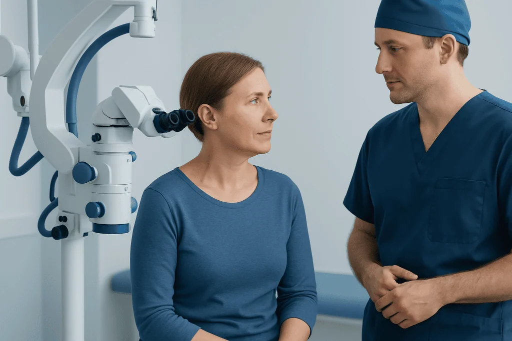
Not every benign meningioma needs immediate surgery. Small tumours that are not causing symptoms may be monitored with periodic MRI scans, especially when they are found incidentally. Surgery is more often considered when a tumour is growing, causing seizures, headaches, weakness, vision changes, balance problems or other neurological symptoms, or when imaging suggests pressure on important structures.
Before surgery, the team reviews MRI or CT images to understand the tumour’s location, relationship to blood vessels and nerves, and possible surgical route. Depending on the case, assessment may also include neurological examination, blood tests, vision testing, hearing tests or vascular imaging. The person’s age, general health, medicines and personal priorities are part of the decision-making process.
Some tumours cannot be fully removed without an unacceptable risk to nearby structures. In these situations, the safest plan may be partial removal followed by observation or focused radiation treatment when appropriate. This is one reason outcomes should be discussed in terms of long-term function and tumour control, rather than complete removal alone.
The Meningioma Surgery Procedure: Step by Step

Meningioma surgery is performed by a neurosurgeon under general anaesthesia. The most common approach is a craniotomy, in which an opening is made in the skull to reach the tumour. For selected tumours in particular locations, surgeons may use minimally invasive or endoscopic approaches through smaller openings or, occasionally, through the nose. The approach is chosen according to anatomy and safety rather than scar size alone.
During the operation, the surgical team uses detailed imaging, magnification and monitoring techniques to identify the tumour and protect critical structures. Depending on tumour location, monitoring may help assess functions such as movement, sensation, hearing or vision pathways. The surgeon carefully separates and removes tumour tissue where it can be done safely, controls bleeding and may remove or treat involved tissue around the tumour when appropriate.
After removal, the bone is secured and the incision is closed. Removed tissue is examined by a pathologist to confirm the diagnosis and grade. Patients and families can learn more about meningioma surgery and the factors that guide an individual surgical plan.
In some cases, surgery is not the only treatment considered. If a small tumour remnant remains near sensitive nerves or blood vessels, or if there is later growth, a specialist may discuss carefully targeted radiation. This is planned individually and is not necessary after every benign meningioma operation.
Recovery Timeline: Hospital Stay to Returning Home
Immediately after surgery, patients are monitored closely in a recovery area or intensive care setting. Nurses and doctors regularly check alertness, speech, limb movement, pupils, pain and the surgical wound. A brain scan is often performed soon after the operation to assess the surgical area. Temporary swelling, fatigue and headache are common after brain surgery and are managed by the clinical team.
The hospital stay varies, but many people remain in hospital for several days. Physical therapy, occupational therapy, speech and language therapy or swallowing assessment may begin early when needed. Before discharge, the team reviews medicines, wound care, warning signs, activity guidance and follow-up arrangements.
At home, energy levels may fluctuate and rest is an important part of recovery. Light activity, such as short walks, is often encouraged as advised by the care team. Strenuous exercise, heavy lifting, driving and returning to work should wait until the neurosurgeon confirms that they are safe. Return to work may be relatively early for some people, while others need several weeks or months, especially when symptoms or rehabilitation needs continue.
Concentration, memory, mood and sleep can also be affected during recovery. These changes may improve gradually, but they deserve attention rather than dismissal. Keeping follow-up appointments and sharing any concerns allows the team to arrange rehabilitation, medication review or further assessment when needed.
Expected Results, Benefits and Possible Risks
The potential benefit of surgery is removal or reduction of tumour pressure, which may improve symptoms such as headaches, seizures or focal neurological problems. However, improvement can be gradual, and symptoms related to long-standing nerve or brain compression may not resolve completely. The pathology result and postoperative scan help clarify the expected outlook and future monitoring plan.
All brain surgery carries risks. These can include bleeding, infection, fluid leakage, blood clots, stroke, seizures and reactions to anaesthesia. There can also be location-specific effects, such as weakness, numbness, speech difficulty, visual changes, hearing changes, balance problems, swallowing difficulty or changes in thinking and behaviour. Some effects are temporary, while others may persist.
A benign meningioma can recur or regrow, especially when a small portion is left behind for safety reasons. This does not necessarily mean that immediate further treatment is needed. Regular MRI surveillance helps detect changes early so that observation, further surgery or radiation can be considered if required.
Open discussion of likely benefits and risks is central to informed consent. The neurosurgical team can explain how the person’s tumour location, imaging features and medical history affect the likely results more accurately than general timelines can.
Follow-Up, Self-Care and Rehabilitation
Follow-up care commonly includes a postoperative visit to review wound healing, symptoms and pathology findings, followed by MRI scans at intervals recommended by the neurosurgeon. The schedule varies according to the grade of meningioma, completeness of removal, location and scan findings. It is helpful to bring a list of symptoms, questions and current medicines to each appointment.
Good recovery habits include taking prescribed medicines exactly as directed, attending therapy sessions, maintaining hydration and regular meals, and increasing activity gradually. Avoid smoking and limit alcohol, particularly while taking pain medicines, anti-seizure medicines or other drugs that affect alertness. A balanced routine that supports sleep and gentle movement can help with fatigue and emotional adjustment.
If seizures occurred before or after surgery, the clinical team will advise on medication, safety precautions and local rules about driving. People should not restart driving based on how they feel alone; clearance may depend on recovery, seizure history and legal requirements. Family members can be valuable partners in noticing subtle changes in speech, memory, mood or function.
Acibadem International’s multidisciplinary specialists and JCI-accredited hospitals support diagnosis, surgery, rehabilitation and follow-up planning for international patients with meningioma.
When to Seek Medical Care
Contact the surgical team promptly for increasing redness, swelling, drainage or separation of the wound; fever; persistent vomiting; worsening headache; new or worsening weakness; confusion; speech problems; vision changes; or a seizure. These symptoms do not always indicate a serious complication, but they need timely clinical advice after brain surgery.
Emergency medical care is needed for a sudden severe headache, loss of consciousness, a seizure that does not stop promptly, severe breathing difficulty, sudden one-sided weakness, new facial drooping or sudden trouble speaking. It is safer to seek urgent help rather than wait for a scheduled appointment when a neurological change is abrupt or worsening.
Emotional symptoms also warrant support. Persistent low mood, overwhelming anxiety, major personality changes or difficulty coping with recovery should be discussed with a doctor. Psychological support, rehabilitation services and practical help at home can be important parts of recovery.
Frequently asked questions
How long does it take to recover from benign meningioma surgery?
Early recovery often takes several weeks, but full recovery can take months. The timeline depends on the tumour location, surgical approach, any neurological symptoms and the need for rehabilitation. Fatigue and reduced stamina are common during the first part of recovery.
Can a benign meningioma come back after surgery?
Yes, a benign meningioma can regrow, although the likelihood varies. Regrowth is more likely when complete removal was not safe or possible, but it can also occur after apparently complete removal. Follow-up MRI scans are the most reliable way to monitor for change.
Will symptoms improve after meningioma surgery?
Symptoms caused by pressure from the tumour may improve after surgery, but the degree and speed of improvement vary. Some symptoms settle quickly, while nerve-related, visual, cognitive or movement symptoms may recover more gradually. In some cases, symptoms may persist despite successful tumour treatment.
When can a person drive after meningioma surgery?
Driving should only restart after the treating clinician confirms it is safe. Decisions may be affected by visual or cognitive recovery, weakness, medication effects and whether the person has had seizures. Local driving regulations may also apply.
Is rehabilitation needed after benign meningioma surgery?
Not everyone needs formal rehabilitation, but many people benefit from targeted support. Physiotherapy, occupational therapy, speech and language therapy, cognitive rehabilitation or psychological support may be recommended according to individual needs. Starting rehabilitation early can help restore independence and confidence.
What follow-up is needed after a benign meningioma is removed?
Follow-up usually includes a clinical review and scheduled MRI scans. The frequency is individualized based on pathology, the extent of removal, tumour location and scan results. The neurosurgeon will explain the recommended surveillance plan after surgery.
References
- World Health Organization Classification of Tumours Editorial Board
- National Institute of Neurological Disorders and Stroke
- National Cancer Institute
- American Association of Neurological Surgeons
- European Association of Neuro-Oncology
This article is for general information only and is not a substitute for professional medical advice. Please consult a qualified doctor about your individual situation.
Joint and spine care in Turkey — expert assessment & treatment
JCI-accredited · board-certified surgeons · reply within 24h
Add us as a Preferred Source to see more of our trusted health content across Google Search, AI Overviews and Discover.