Gamma Knife Radiosurgery: Focused Treatment for Brain Conditions

Gamma Knife radiosurgery delivers many small radiation beams that meet precisely at a target inside the brain. It may be used for selected benign and malignant brain tumors, arteriovenous malformations, vestibular schwannomas, pituitary tumors, and trigeminal neuralgia.
Key Takeaways
- Gamma Knife radiosurgery delivers many small radiation beams that meet precisely at a target inside the brain.
- It may be used for selected benign and malignant brain tumors, arteriovenous malformations, vestibular schwannomas, pituitary tumors, and trigeminal neuralgia.
- Treatment is usually completed in one session, although some conditions may require staged or repeat treatment.
- Most people go home the same day, but follow-up imaging is important because results often develop gradually over weeks, months, or years.
- Suitability depends on the diagnosis, size and location of the target, symptoms, previous treatments, and overall health.
Gamma Knife radiosurgery is a highly focused form of radiation treatment used for selected brain tumors, blood vessel abnormalities, and certain nerve-related conditions. It is called radiosurgery because of its precision, but it does not involve an incision or removal of tissue.
Overview
Gamma Knife radiosurgery is a type of stereotactic radiosurgery, a treatment that uses advanced imaging and computer-guided planning to deliver radiation with very high accuracy. Despite the word surgery, it does not involve a scalpel, an incision, or opening the skull. Instead, multiple low-dose radiation beams are directed from different angles and converge on a carefully defined target, where the combined dose has a therapeutic effect.
The treatment is designed for conditions inside the head, including some brain tumors, abnormal blood vessels, and certain functional nerve disorders. Because the beams are highly focused, the surrounding healthy brain tissue receives much less radiation than the target area. This precision is one reason Gamma Knife radiosurgery may be considered when conventional surgery is difficult, carries higher risk, or is not the preferred first option.
Gamma Knife treatment is typically planned and delivered by a multidisciplinary team. This may include neurosurgeons, radiation oncologists, neuroradiologists, medical physicists, nurses, and radiation therapists. The goal is to choose the safest and most effective approach for the individual condition, rather than applying one standard plan to every patient.
How Gamma Knife Radiosurgery Works
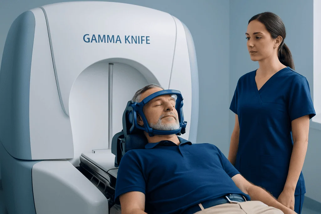
Gamma Knife systems use many precisely aimed beams of gamma radiation, commonly produced by cobalt sources, to treat a small or clearly defined area within the brain. Each individual beam is relatively weak as it passes through healthy tissue. At the target, however, the beams meet and deliver a concentrated dose that can damage the DNA of tumor cells, close abnormal blood vessels over time, or interrupt pain pathways involved in certain nerve conditions.
Before treatment, high-resolution imaging is used to map the target and nearby sensitive structures such as the optic nerves, brainstem, hearing pathways, and normal blood vessels. Planning software allows the team to shape the radiation dose to the target as closely as possible. This planning step is a major part of the treatment and helps balance effectiveness with safety.
Depending on the equipment and the patient’s condition, the head may be kept still with a lightweight frame attached to the skull after local anesthesia, or with a custom mask system. Immobilization is essential because even small movements can affect accuracy. The medical team explains the method in advance and monitors the patient throughout the procedure.
Conditions Treated with Gamma Knife

Gamma Knife radiosurgery may be used for both non-cancerous and cancerous conditions, but it is not suitable for every brain problem. It is often considered when the target is small to moderate in size, well defined on imaging, and located in an area where focused radiation can be delivered safely. The expected benefit, possible risks, and available alternatives are reviewed before a treatment recommendation is made.
Commonly treated conditions include:
- Brain metastases, which are tumors that have spread to the brain from cancers elsewhere in the body
- Benign tumors such as meningiomas and vestibular schwannomas
- Pituitary adenomas in selected cases
- Arteriovenous malformations, also called AVMs, which are abnormal connections between arteries and veins
- Trigeminal neuralgia, a condition that causes severe facial pain
- Selected recurrent or residual tumors after surgery or previous radiation
The way Gamma Knife works depends on the condition being treated. Tumors may stop growing, shrink gradually, or become controlled over time. AVMs may close slowly as the treated blood vessels scar and seal. For trigeminal neuralgia, the treatment aims to reduce abnormal pain signaling, although pain relief may take time and may vary from person to person.
Who May Be a Candidate
A person may be considered for Gamma Knife radiosurgery after a detailed review of the diagnosis, MRI or CT findings, symptoms, neurological examination, and previous treatments. The size, shape, and location of the target are especially important. Small, clearly outlined targets are generally more suitable than very large, diffuse, or poorly defined areas.
Gamma Knife may be recommended as a primary treatment, as an additional treatment after surgery, or as a treatment for a condition that has returned. It may also be considered for patients who are not ideal candidates for open surgery because of medical risk, age, tumor location, or personal treatment goals. However, open surgery may still be the better option when a large mass is causing pressure, when tissue diagnosis is needed, or when immediate decompression is required.
Some people need a different form of radiation therapy, surgery, medication, observation with regular scans, or a combination of approaches. For example, a very large tumor may be treated more safely with surgery first, while multiple brain metastases may require a personalized plan that considers the number of lesions, overall cancer treatment, and neurological symptoms. The decision should be made with specialists experienced in brain imaging, neurosurgery, and radiation treatment.
What to Expect Before and During Treatment
Preparation begins with a consultation and review of medical records, imaging studies, medications, allergies, and general health. Patients are usually advised about eating, drinking, and regular medications before the procedure according to the treatment center’s protocol. If a head frame is used, local anesthesia is given to numb the pin sites, and some patients may receive mild sedation to help them relax.
On the day of treatment, detailed imaging is performed after the frame or mask is in place. MRI is commonly used, and CT or angiography may be added for certain conditions such as AVMs. The patient then rests while the team designs and checks the treatment plan. This planning time varies depending on the complexity of the target and the number of areas being treated.
During treatment, the patient lies on a treatment couch while the head is kept still. The procedure itself is painless; patients do not feel the radiation. The team monitors the patient by camera and audio connection, and treatment may last from minutes to a few hours depending on the plan. Afterward, the frame or mask is removed, and the patient is observed for a period before going home in most cases.
Benefits, Risks, and Recovery
Potential benefits of Gamma Knife radiosurgery include high precision, no surgical incision, no general anesthesia in many cases, and a relatively short recovery time. Many patients return home the same day and resume light activities soon afterward, depending on how they feel and their doctor’s instructions. It may also be useful for targets in deep or delicate brain areas where open surgery would be more complex.
Like all medical treatments, Gamma Knife radiosurgery has possible side effects. Short-term effects may include headache, nausea, fatigue, scalp tenderness, or swelling at frame pin sites. Some patients may need medication to reduce inflammation or manage symptoms after treatment. Temporary or longer-lasting neurological symptoms can occur if swelling affects nearby brain structures, although the treatment team plans carefully to reduce this risk.
Results are not always immediate. Tumor control or shrinkage may be seen gradually on follow-up scans, AVM closure can take years, and pain relief in trigeminal neuralgia may develop over weeks or months. Regular follow-up is essential so the medical team can evaluate response, manage side effects, and decide whether further treatment is needed.
Follow-up, Self-care, and When to See a Doctor
After Gamma Knife radiosurgery, patients receive individualized instructions about activity, wound care if a frame was used, medications, and follow-up appointments. It is important to attend scheduled MRI, CT, or other imaging visits even if symptoms improve. These scans help the care team assess whether the treated area is stable, shrinking, or requires additional attention.
Patients should contact their doctor promptly if they develop new or worsening headaches, seizures, weakness, numbness, vision changes, balance problems, persistent vomiting, fever, drainage or increasing redness at pin sites, or any sudden neurological symptom. These symptoms do not always mean something serious has happened, but they should be assessed by a qualified medical professional. People being treated for cancer should also keep their oncology team informed, because brain treatment may need to be coordinated with systemic therapy.
General self-care includes getting adequate rest, following medication instructions, avoiding driving or strenuous activity until cleared by the doctor, and asking the care team which symptoms should prompt urgent evaluation. International patients may also need a clear plan for follow-up imaging after returning home. Acibadem International’s multidisciplinary specialists and JCI-accredited hospitals provide diagnosis and treatment planning for brain conditions, including Gamma Knife radiosurgery, for international patients when medically appropriate.
Frequently asked questions
Is Gamma Knife radiosurgery the same as brain surgery?
No. Gamma Knife radiosurgery is called surgery because it treats a precise target, but it does not involve an incision or opening the skull. It uses focused radiation beams to affect tissue inside the brain.
Does Gamma Knife treatment hurt?
The radiation itself is painless and cannot be felt during treatment. If a head frame is used, patients may feel pressure during placement, but local anesthesia helps numb the area. Some people have a mild headache or scalp tenderness afterward.
How long does Gamma Knife radiosurgery take?
The treatment session may take from minutes to a few hours, depending on the number, size, and complexity of the targets. The full visit is usually longer because it includes preparation, imaging, treatment planning, the procedure, and observation afterward.
When will results be visible?
Results depend on the condition being treated. Some symptoms may improve within weeks, while tumor changes may take months and AVM closure may take years. Follow-up imaging is needed to measure the response safely.
Can Gamma Knife be used for all brain tumors?
No. Suitability depends on the tumor type, size, location, symptoms, and previous treatments. Some tumors are better treated with surgery, conventional radiation therapy, medication, observation, or a combination of approaches.
Will hair fall out after Gamma Knife radiosurgery?
Most patients do not have widespread hair loss because the radiation is highly focused. A small patch of temporary hair thinning can occur if the treated area is close to the scalp. The care team can explain the likelihood based on the treatment plan.
Is follow-up necessary if the patient feels well?
Yes. Many treatment effects are visible on imaging before symptoms change, and some conditions require long-term monitoring. Follow-up scans help doctors confirm treatment response and identify any delayed side effects early.
References
- American Society for Radiation Oncology
- American Association of Neurological Surgeons
- National Cancer Institute
- Mayo Clinic
- International Stereotactic Radiosurgery Society
This article is for general information only and is not a substitute for professional medical advice. Please consult a qualified doctor about your individual situation.
Gamma Knife in Turkey — costs, top hospitals & a free quote
JCI-accredited · board-certified surgeons · reply within 24h