

Mammogram: What It Is, Whether It Hurts, How Long It Takes, and the Deodorant Rule
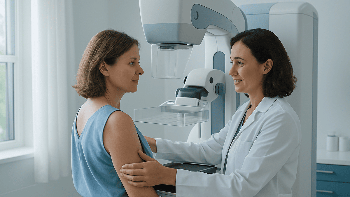
Key Takeaways
- Compression lasts only a few seconds per image, and a standard screening mammogram takes just four images — two views of each breast.
- Aluminum in many antiperspirants shows up as white specks that can mimic breast calcifications on the X-ray, which is why deodorant, powders, and lotions are off-limits on exam day.
- A screening appointment runs about 30 minutes door to door, but the actual imaging takes only a few minutes.
- You undress from the waist up only, so a two-piece outfit means your pants or skirt never come off.
- Among women screened annually for ten years, roughly half will experience at least one false-positive result — so a callback usually signals a question, not a cancer.
- Nearly half of women over 40 have dense breast tissue, which can hide tumors on a mammogram, and U.S. facilities are now required to tell you your density with your results.
A mammogram is a low-dose X-ray of the breast, used either to screen for cancer before symptoms appear or to investigate a lump or other change. Each breast is compressed between two plates for a few seconds per image; most people describe it as uncomfortable rather than painful. The full appointment usually takes about 30 minutes, and you should skip deodorant that day because it can blur the images.
There’s a sign taped to the mirror in nearly every mammography changing room in America, and it never mentions cancer. It says something like: Please tell the technologist if you are wearing deodorant. First-timers read it twice, gown half-tied, and wonder what antiperspirant could possibly have to do with an X-ray.
Quite a lot, it turns out — and that small mystery is a good doorway into the bigger ones. Roughly 40 million mammograms are performed in the U.S. each year, yet the same questions keep landing in search bars the night before an appointment: Will it hurt? How long am I in there? What are they actually doing behind that glass?
Here are the honest answers — including the ones about squeezing, waiting for results, and why your pants get to stay exactly where they are.
What is a mammogram, exactly?
A mammogram is an X-ray picture of the breast taken with a machine designed for one job only. Unlike a chest X-ray, which shoots through bone and lung, a mammography unit uses a low radiation dose tuned to soft tissue, so a radiologist can distinguish fat, glandular tissue, cysts, calcium deposits, and masses from one another, according to the National Library of Medicine.
The machine itself looks nothing like the tunnel scanners people picture. It’s an upright unit, roughly the size of a tall filing cabinet, with two flat plates that gently flatten the breast for a few seconds while the image is captured. Flattening matters for two reasons: it spreads overlapping tissue so small findings aren’t hidden, and it thins the breast so less radiation is needed for a sharp picture, as the Mayo Clinic explains.
What makes the test valuable is timing. The CDC notes that a screening mammogram can find breast cancer years before a lump is large enough to feel — and cancers found at that stage are generally easier to treat. That head start, not the technology itself, is the whole point of the exam. A mammogram doesn’t diagnose cancer on its own; it flags areas that deserve a closer look, which is a distinction worth keeping in mind when results arrive.
Screening vs. diagnostic mammogram: which one am I getting?
The two exams use the same machine but answer different questions. A screening mammogram asks, “Is anything here that shouldn’t be?” in a person with no symptoms. A diagnostic mammogram asks, “What exactly is that?” — it’s ordered when you or your clinician has found a lump, nipple discharge, skin change, or when a screening exam turned up something that needs a second look, per the National Cancer Institute.
| Screening mammogram | Diagnostic mammogram | |
|---|---|---|
| Who it’s for | People with no breast symptoms | People with a symptom or an abnormal screening result |
| Typical images | Two views per breast (four total) | Extra angles, magnified or spot-compression views as needed |
| Time in the room | Imaging takes just a few minutes | Often longer; images are reviewed while you wait |
| Who reviews it | Radiologist reads it afterward | Radiologist often checks images during the visit |
| What it answers | “Does anything need a closer look?” | “What is this specific finding?” |
One practical consequence: being sent for a diagnostic mammogram is not a diagnosis. It simply means the first set of pictures raised a question — often about overlapping tissue rather than anything worrisome. The Cleveland Clinic notes that diagnostic visits frequently pair the mammogram with an ultrasound to settle the question the same day.
What is done during a mammogram, step by step
Here’s the sequence, minute by minute, drawn from the Mayo Clinic’s description of the exam:
- Check-in and changing. You undress from the waist up, remove necklaces, and put on a gown that opens in front.
- Positioning. A mammography technologist — nearly always a woman, and you can request one — places one breast on the machine’s lower plate and adjusts your shoulder, arm, and chin so nothing else drifts into the image.
- Compression. The upper plate lowers and presses the breast flat for a few seconds while the X-ray is taken. You’ll be asked to hold still and briefly hold your breath.
- Repeat. A standard screening exam captures two views of each breast — one from above, one from the side at an angle — four images in all.
- Quick review. The technologist checks that each picture is sharp and complete, and retakes any that aren’t. That’s a quality step, not a red flag.
Then you dress and leave. There are no needles, no dye, no fasting, and no recovery time. The technologist can’t tell you results — interpreting the images is the radiologist’s job, done afterward with your prior mammograms alongside for comparison. Bringing or transferring old images, the Mayo Clinic advises, is one of the most useful things you can do, because change over time is often the most telling clue.
Is a mammography test painful? An honest answer
For most people, no — but it’s not nothing, either. Compression feels like firm, sustained pressure, and the honest range of experience runs from “mildly odd” to “genuinely painful for a few seconds.” The NHS puts it plainly: some find it uncomfortable, some find it painful, and any discomfort typically passes quickly once the plate lifts.
Several factors shift where you land on that spectrum. Breast tenderness is highest the week before and during a period, so scheduling for the week after often helps, per the Cleveland Clinic. Smaller breasts sometimes require more maneuvering into position. Caffeine makes some people’s breasts more tender. And anxiety tightens the chest and shoulder muscles, which can make positioning harder — slow breathing genuinely helps here.
Two things are worth saying clearly. First, the squeeze lasts seconds per image, not minutes; nobody stands in compression for long. Second, you have a voice in the room. Tell the technologist if it hurts — compression can often be adjusted slightly without ruining the image, and a skilled technologist would rather reposition than have you dread the next visit. If a past mammogram was painful, say so at booking; that single sentence changes how the appointment is handled.
The trade is a few seconds of pressure for an image that can catch cancer before it’s palpable. Most people who’ve had one say the anticipation was worse than the exam.
How long does a mammogram take?
Budget about 30 minutes door to door for a screening mammogram, according to the Mayo Clinic — and most of that is changing, paperwork, and positioning. The actual imaging is startlingly brief: the NHS notes the X-rays themselves take only a few minutes, with each compression lasting a matter of seconds.
A diagnostic mammogram runs longer, and for a reassuring reason rather than an ominous one: the radiologist often reviews images while you wait, deciding in real time whether an extra angle or an ultrasound would answer the question on the spot. That can stretch the visit toward an hour, but it also means you frequently leave a diagnostic appointment with an answer instead of another wait.
A few timing tips from people who do this every day: morning slots tend to run closer to schedule; wearing a two-piece outfit shaves minutes off changing; and arriving with prior mammogram records already transferred prevents the most common delay of all. If you’re squeezing the appointment into a workday lunch hour, a screening mammogram fits — many people are back at their desk before anyone notices they left. The exam’s brevity is one of its most underrated features: for a test capable of finding cancer years early, the time cost is roughly that of waiting in line for coffee on a bad morning.
The deodorant rule: why the changing room has that sign
Now, the mystery from the changing-room mirror. Many antiperspirants contain aluminum-based compounds, and metallic particles show up on an X-ray as tiny bright white specks. The trouble is that certain breast cancers announce themselves the same way — as clusters of small white calcifications. The Mayo Clinic and the National Cancer Institute both advise skipping deodorant, antiperspirant, powders, lotions, and creams on the breasts and underarms on exam day, because residue can mimic exactly the finding radiologists are trained to chase.
The consequences of forgetting are inconvenient rather than dangerous. At best, the technologist hands you a wipe and you clean up before the images are taken — most centers keep wipes on hand for precisely this reason. At worst, residue slips through, the radiologist sees ambiguous white dots, and you get called back for repeat images and a week of unnecessary worry over what was, chemically speaking, your morning routine.
Practical workarounds: shower that morning and apply nothing from the waist up; toss a travel-size deodorant in your bag to apply after the exam; and if you use medicated creams on the chest area, mention it at check-in. Perfume and body glitter fall under the same logic — anything that sits on the skin can end up in the picture. It is likely the only medical instruction you’ll ever receive that’s entirely about your armpits, and it’s worth following to the letter.
Do I have to take off my pants for a mammogram?
No. A mammogram involves the chest only, so you undress from the waist up — pants, skirt, shoes, and socks all stay on, as the Cleveland Clinic confirms. You’ll be given a gown or cape that opens at the front, and the technologist uncovers one breast at a time during positioning.
This is why nearly every guide, including the NHS, suggests wearing separates rather than a dress: a two-piece outfit means you’re changed and ready in under a minute, and you never stand around feeling more exposed than necessary. Leave necklaces at home, since anything metal near the chest has to come off anyway.
A word on the dignity of the thing, because it’s a real concern that keeps some people from booking: you are never fully undressed, the room is private, only the technologist is present, and each breast is covered again the moment its images are done. If having a female technologist matters to you, say so when scheduling — it’s a routine request, honored without fuss. People who’ve delayed a first mammogram over modesty worries often report afterward that the exam felt closer to a bra fitting than a medical procedure: brief, businesslike, and considerably less awkward than imagination suggested.
Can a mammogram see cancer?
It can — and often before anything can be felt. The CDC states that mammograms can detect breast cancer up to three years before a tumor grows large enough to notice by touch. On the image, cancer may appear as a mass with irregular edges, a cluster of fine calcifications, a distortion in the tissue’s architecture, or a change from previous years’ pictures.
But precision matters here: a mammogram finds suspicious areas; it does not diagnose cancer. Plenty of benign things — cysts, fibroadenomas, overlapping dense tissue, even that deodorant residue — can look questionable on a first pass. Confirming or ruling out cancer takes additional steps: more detailed imaging, ultrasound, sometimes MRI, and ultimately a biopsy, in which a small tissue sample is examined under a microscope, per the National Cancer Institute.
The reverse is also true, and it’s the part too many articles soften: mammograms miss some cancers. Dense breast tissue can hide tumors, because both appear white on the image — radiologists describe it as looking for a snowball in a snowstorm. That’s why a normal mammogram never overrides a real symptom. If you can feel a lump but your mammogram was clear, the lump still needs evaluation. The evidence supports mammography as the best-studied screening tool we have for breast cancer — and supports being honest that no screening test catches everything.
What's the difference between a 2D and 3D mammogram?
If your imaging center offers “3D mammography,” the formal name is digital breast tomosynthesis. Instead of taking one flat picture per view, the X-ray arm sweeps in an arc over the breast, capturing a series of thin image “slices” that a computer assembles into a layered view, as described by the Mayo Clinic.
The advantage is unstacking. In a standard 2D image, tissue layers pile on top of one another like pages photographed through a closed book; normal tissue can overlap in ways that either hides a small mass or imitates one. Tomosynthesis lets the radiologist page through the breast slice by slice. The National Cancer Institute notes that studies associate 3D mammography with fewer callbacks for additional imaging and improved cancer detection compared with 2D alone — though research continues on whether that translates into fewer deaths, which is the outcome that matters most and takes decades to measure.
From where you’re standing, the two exams feel nearly identical: same machine silhouette, same compression, a sweep lasting a few seconds longer. Many centers now perform both together by default. Whether 3D is available — and whether insurance covers any difference in cost — varies by facility and plan, so it’s a fair question to ask when booking. If only 2D is offered, that is still a validated, effective screening exam; don’t postpone a mammogram waiting for a particular machine.
When should I start getting mammograms, and how often?
For people at average risk, the U.S. Preventive Services Task Force now recommends screening every two years from age 40 through 74 — guidance the CDC reflects, and a notable shift from earlier advice that started routine screening at 50. Other respected bodies differ at the margins: some support the option of annual screening, and the UK’s NHS invites women every three years, beginning with a first invitation between ages 50 and 53 and continuing to 71.
Those differences aren’t evidence of chaos; they’re evidence of trade-offs. Screening more often and starting younger catches some cancers earlier but produces more false alarms, more callbacks, and more biopsies of things that were never going to cause harm. Screening less often reduces those burdens but risks a later diagnosis for some. Reasonable experts weigh that balance differently, which is why the guidelines don’t perfectly align.
Two situations change the math entirely. If you have a strong family history of breast or ovarian cancer, a known genetic variant such as BRCA1 or BRCA2, or prior chest radiation, screening may need to start earlier and include MRI — a conversation to have with your clinician well before 40, per the National Cancer Institute. And if you have symptoms at any age, you don’t wait for a screening milestone; you get evaluated now. Screening schedules are for people with nothing to report.
What happens after: results, callbacks, and why most are fine
A radiologist reads your images — comparing them against prior years when available — and the results reach you afterward. In the UK, the NHS sends a letter within about two weeks. In the U.S., many facilities post results to a patient portal within days; ask at checkout when to expect yours, and call if nothing arrives.
Then there’s the callback — the phone message that quickens every pulse. Keep two facts within reach. First, being asked to return for more images is common and usually reflects a technical question: overlapping tissue, an unclear area, a picture that needs a different angle. Second, the overwhelming majority of callbacks end in reassurance, not diagnosis. The National Cancer Institute reports that among women screened annually for ten years, roughly half will experience at least one false-positive result — a statistic that reframes the callback from “something is wrong” to “this happens to almost everyone eventually.”
First mammograms generate more callbacks than later ones for a simple reason: there’s nothing to compare against. Every breast has its own baseline quirks, and without prior images, a radiologist can’t yet tell your normal from a change. This is the strongest argument for staying with one imaging center when you can, or having old images transferred when you can’t. If a callback does lead to a biopsy, remember that most breast biopsies also come back benign — the funnel narrows dramatically at every step.
What if I have dense breasts?
Breast density is about tissue composition, not size or firmness — and you can’t feel it. Dense breasts have more glandular and fibrous tissue relative to fat, and that tissue appears white on a mammogram. So do tumors. The CDC reports that nearly half of women over 40 have dense breasts, making this one of the most common findings in all of screening.
Density matters for two independent reasons. It can mask cancers on a mammogram, lowering the exam’s sensitivity. And density itself is associated with a somewhat higher risk of developing breast cancer — a separate effect, not merely a visibility problem. Neither fact means a mammogram is useless for dense breasts; it remains the foundation of screening. It means the picture may be incomplete.
You no longer have to guess where you stand. U.S. mammography facilities are now required to tell every patient whether their breasts are dense as part of the results report, following a federal rule that took effect in September 2024, per the CDC. If yours are, that’s the opening line of a conversation with your clinician — not a cause for alarm. Depending on your overall risk, supplemental imaging such as ultrasound or MRI may be worth discussing, though the evidence on who benefits most from added screening is still evolving, and honest clinicians will say so. Density can also decrease with age, so your status may change over the years.
How much radiation does a mammogram involve — is it safe?
Very little, and yes — the numbers hold up to scrutiny. According to the National Cancer Institute, the radiation dose from a standard screening mammogram is roughly what a person absorbs from natural background sources — soil, building materials, cosmic rays — over about seven weeks of ordinary life. Modern digital units are engineered to use the minimum dose that still yields a diagnostic-quality image, and compression itself reduces the dose needed by thinning the tissue.
Could decades of repeated low-dose exposure carry any theoretical risk? Researchers have modeled exactly that question, and the NCI’s assessment is that the potential harm from screening-level radiation is extremely small — far outweighed, for people in recommended age ranges, by the benefit of finding cancers early. This is not a case where the risks are being waved away; they’ve been counted, and the arithmetic favors screening.
One genuine caveat: pregnancy. Tell the technologist if you are or might be pregnant before the exam begins. Routine screening is typically postponed during pregnancy, and if breast imaging is needed for a symptom, clinicians can choose the approach and shielding accordingly, per the Cleveland Clinic. For everyone else, the radiation question deserves a place on the list of reasonable things to ask — and then, on the evidence, a place off the list of reasons to skip the exam.
When to see a doctor — with or without a mammogram on the calendar
Screening is for people with nothing to report. Symptoms follow a different rule: they get evaluated promptly, regardless of your age, your last mammogram’s result, or how recently it was done. Contact your clinician without waiting if you notice any of the following, which the NHS and CDC flag as changes that need assessment:
- A new lump or thickened area in the breast or armpit
- Nipple discharge — especially if bloody or from one side only
- A nipple that has newly turned inward or changed shape
- Skin changes: dimpling, puckering, redness, scaling, or a texture like orange peel
- A change in the size or shape of one breast
- Persistent pain in one spot that doesn’t follow your cycle
Most of these turn out to have benign explanations — cysts, hormonal shifts, infections. But “probably benign” is a conclusion a clinician reaches after examination and appropriate imaging, not one to reach alone at the bathroom mirror. And it bears repeating: a recent normal mammogram does not cancel out a new symptom. If you feel something the pictures didn’t show, the symptom wins, and it deserves its own workup. The people who fare best with breast cancer are, overwhelmingly, the ones whose disease was found early — either by a screening exam they didn’t postpone or a symptom they didn’t talk themselves out of.
Frequently asked questions
Is a mammography test painful?
For most people it’s uncomfortable rather than painful — firm pressure for a few seconds per image, gone the moment the plate lifts. Sensitivity varies: breasts are most tender the week before and during a period, so scheduling the week after often helps. Tell the technologist if it genuinely hurts; compression can often be adjusted slightly. Most people report the anticipation was worse than the exam itself.
What is done during a mammogram?
You undress from the waist up, and a technologist positions one breast at a time between two plates on an upright X-ray machine. Each breast is compressed for a few seconds while an image is taken — two views per breast, four images total for a standard screening exam. The technologist checks image quality, and a radiologist interprets the pictures afterward. There are no needles, no dye, and no recovery time.
Can a mammogram see cancer?
Yes — a mammogram can detect breast cancer up to three years before a tumor is large enough to feel, according to the CDC. However, it flags suspicious areas rather than diagnosing cancer; confirmation requires further imaging and usually a biopsy. Mammograms can also miss some cancers, particularly in dense breast tissue, which is why a new lump always deserves evaluation even after a normal result.
Do I have to take off my pants for a mammogram?
No. A mammogram images the chest only, so you undress from the waist up and wear a front-opening gown; pants, skirts, shoes, and socks stay on. That’s why wearing separates instead of a dress makes the appointment quicker and more comfortable. Only one breast is uncovered at a time during positioning, and you can request a female technologist when you book.
Why can't I wear deodorant to a mammogram?
Many antiperspirants contain aluminum compounds that appear on the X-ray as tiny white specks — the same appearance as the breast calcifications radiologists look for as a possible sign of cancer. Residue can therefore cause a false alarm and an unnecessary callback. Skip deodorant, powders, lotions, and creams on the breasts and underarms that day; most centers provide wipes if you forget, and you can reapply right after.
How long does a mammogram take?
Plan on about 30 minutes for the whole screening appointment, most of which is check-in, changing, and positioning. The imaging itself takes only a few minutes, and each compression lasts a matter of seconds. Diagnostic mammograms run longer — sometimes closer to an hour — because the radiologist often reviews images during the visit and may add extra views or an ultrasound on the spot.
What is the difference between a screening and a diagnostic mammogram?
A screening mammogram is a routine check for people with no symptoms, typically four images read by a radiologist afterward. A diagnostic mammogram investigates something specific — a lump, discharge, skin change, or an abnormal screening result — using additional angles and magnified views, often reviewed while you wait. Being sent for a diagnostic exam is not a diagnosis; it means the first images raised a question that needs a closer look.
How soon do mammogram results come back?
It varies by country and facility. In the UK, the NHS mails results within about two weeks. In the U.S., many centers post results to an online patient portal within days, and you should receive a written summary as well. Ask at checkout when to expect yours, and call the facility if nothing arrives — no news should never be assumed to mean good news.
Is the radiation from a mammogram dangerous?
No — the dose is very low. The National Cancer Institute compares the radiation from a standard mammogram to what a person absorbs from natural background sources over about seven weeks of everyday life. Researchers have modeled the theoretical risk of repeated screening and found it extremely small, far outweighed by the benefit of early detection for people in recommended age ranges. Do tell the technologist if you are or might be pregnant.
Should I schedule my mammogram around my period?
If you menstruate, aim for the week after your period ends, when breasts are typically least tender — this can make compression noticeably more comfortable. Avoid the week just before and during your period, when hormonal changes increase sensitivity. It’s a preference, not a requirement: the images are accurate at any point in the cycle, so don’t cancel an appointment you can’t reschedule easily just for timing.
References
- CDC — Screening for Breast Cancer
- NHS — Breast screening (mammogram)
- Cleveland Clinic — Mammogram
- MedlinePlus — Mammography
This article is for general information only and is not a substitute for professional medical advice. Please consult a qualified doctor about your individual situation.

More from the Blog

Functional, Dermoid or Endometrioma: How the Type of Ovarian Cyst Guides Treatment
Ovarian cysts fall into distinct types, and the type largely decides the plan. Functional cysts arise from normal ovulation and usually disappear on their…

Can You Prevent Gestational Diabetes, and Does It Go Away?
You cannot guarantee prevention, but you can meaningfully lower your risk of gestational diabetes by entering pregnancy near a healthy weight, staying physically active…

The Placenta: When It Forms, When It Takes Over and What It Does
The placenta starts forming at implantation, roughly a week after fertilization, and gradually takes over the pregnancy-sustaining hormone work of the corpus luteum by…

Does Endometriosis Go Away? What Happens Over Time, After Menopause and with Treatment
Endometriosis rarely goes away on its own. It is a long-term condition driven largely by estrogen, so symptoms often settle after menopause, when estrogen…

Why Mammogram Results Can Take Two Weeks: What Happens Between the Scan and the Letter
Mammogram results often take up to two weeks because screening images are interpreted after your visit by a radiologist — sometimes two, working independently…

Fibroids, Weight Gain and Bloating: Why It Happens and What Treatment Can Change
Fibroids can add weight directly, but usually only when they are large. Most fibroids are small and add very little, while a very large…