

Ankle Impingement Explained: Why the Joint Pinches and When Ankle Arthroscopy Is Considered

Key Takeaways
- Ankle impingement is pain from soft tissue or a bone spur being pinched at the end of the ankle's range, so it is typically felt at one spot in one position rather than constantly.
- Anterior impingement is provoked by bending the foot upward, as in a deep squat, while posterior impingement is provoked by pointing the foot hard, as in dance or kicking sports.
- Bone spurs seen on X-ray are common in active people and frequently painless; the spur matters only when tissue is caught against it.
- Physical therapy for impingement aims to restore how the talus glides and to rebuild stabilizing strength, not to stretch the ankle further into the painful position.
- A corticosteroid injection reduces inflammation in the joint lining and can help confirm the pain source, but it cannot shrink bone and is usually limited in how often it is repeated.
- Ankle arthroscopy typically takes 30 minutes to 2 hours and is usually a same-day procedure, with the NHS describing light activity returning within days to weeks and full recovery over weeks to months.
Ankle impingement is pain caused when soft tissue or a small bone spur is pinched at the front or back of the ankle joint, often after sprains or repeated deep ankle bending. Treatment usually begins with activity changes, physical therapy and sometimes an injection. Ankle arthroscopy, a keyhole procedure to remove the pinching tissue, is considered when months of nonsurgical care have not settled the pain.
She can run five miles without trouble. What she cannot do is the last two feet of a lunge, the deep squat at the end of a class, or the step off a curb that lands slightly wrong. Every time the ankle bends fully forward, a sharp pinch fires across the front of the joint, then disappears. Her doctor called it anterior ankle impingement and started talking about ankle impingement treatment in stages, with arthroscopy sitting at the far end of the list.
That staged approach is the story of this condition. Impingement is rarely an emergency, rarely mysterious once examined properly, and rarely the first thing a surgeon wants to operate on. It is, however, genuinely frustrating, because the ankle feels fine most of the day and then betrays you at one specific angle.
This explainer walks through why the joint pinches, what the early treatments are actually doing, and how surgeons decide whether a keyhole operation is worth discussing.
What is ankle impingement, in plain language?
Impingement simply means something is being squeezed where it should have room. In the ankle, that something is usually a fold of joint lining, a thickened scar band, or a small bony bump that has grown on the rim of the joint. When the foot moves to the end of its range, the bones close down on that tissue and the person feels a sharp, localized pinch.
The ankle is a hinge formed by the shin bone (the tibia), the smaller outer leg bone (the fibula) and the talus, the dome-shaped bone that sits on top of the heel. In a healthy joint, the front lip of the tibia and the neck of the talus glide past each other with a comfortable gap when the foot bends upward. At the back, the heel bone and a small bony prominence on the talus clear each other when the foot points downward. Impingement happens when those gaps have become crowded.
Two features separate impingement from many other ankle complaints. The pain is position-dependent rather than constant, and it is usually felt at a specific spot that the person can point to with one finger. Someone with anterior impingement often describes pain deep in the front crease of the ankle when squatting, climbing stairs or landing from a jump. Someone with posterior impingement tends to feel it behind the ankle when pointing the toes hard, as dancers and kicking athletes do repeatedly.
Neither pattern is a diagnosis you can make from a description alone. Ankle pain has many overlapping causes, from ligament injury to cartilage damage to tendon irritation, and MedlinePlus notes that sprains are the most common ankle injury of all. What the pattern does is give a clinician a strong lead, which they then confirm with examination and imaging.
Why does the ankle joint pinch? The mechanics behind it
The pinch has two ingredients: something extra inside the joint, and a movement that closes the joint down onto it. Understanding both explains why treatment starts where it does.

The extra tissue most often comes from injury. A sprain stretches or tears the ligaments that hold the ankle together. As the body repairs those fibers, it can lay down thickened scar tissue, and the joint lining, called the synovium, may stay inflamed and swollen long after the ligament itself has knitted. Swollen lining is bulkier than healthy lining, so it gets caught more easily. Mayo Clinic describes this kind of lingering pain and instability after sprains as one of the reasons a sprained ankle deserves proper rehabilitation rather than a shrug.
Bone spurs are the second common ingredient. When the front rim of the tibia and the talus repeatedly bump against each other, the bone responds the way bone tends to respond to repeated stress: it builds more of itself. Over years, a small ridge forms. Footballers and others who kick with a forced upward bend of the foot are classic examples, which is why anterior spurs were historically described in athletes. The ridge itself does not always hurt. Pain arrives when soft tissue becomes trapped between the ridge and the opposing bone.
At the back of the ankle, anatomy plays a larger role. Some people are born with an extra small bone behind the talus, or with an unusually long bony process there. Nothing is wrong with that variation until the foot is pointed hard and often enough, at which point the surrounding tissue is compressed between heel bone and shin bone.
The movement ingredient matters just as much. If the joint were never pushed to the end of its range, the extra tissue would sit quietly. That is why the earliest ankle impingement treatment steps focus on movement patterns, not just on the tissue itself.
Anterior ankle impingement versus posterior ankle impingement
Front and back impingement share a mechanism but behave like different conditions in practice, and the distinction changes both the examination and the surgical approach.
Anterior ankle impingement is the more familiar of the two. Pain sits across the front of the ankle, sometimes toward the outer side, and is provoked by dorsiflexion, the movement of bending the foot upward toward the shin. Think of the bottom of a squat, walking uphill, or the moment a running foot strikes and rolls forward. On examination a clinician typically presses along the front joint line while bending the foot up, looking for a reproducible spot of tenderness. Anterior impingement is frequently linked to previous sprains and to bony spurs on the front rim of the tibia.
Posterior ankle impingement is provoked by plantarflexion, pointing the foot downward. Ballet dancers working on pointe, gymnasts, swimmers and athletes who kick repeatedly are the people who most often describe it. The pain is felt deep behind the ankle, between the Achilles tendon and the outer ankle bone, and it can be confused with tendon problems in the same neighborhood. The culprit is often an accessory bone or an enlarged bony process on the back of the talus, sometimes with irritation of the tendon that flexes the big toe, which runs through that same tight space.
The two can coexist, though that is less common. A thorough examiner will usually test both ends of the range, because a patient may only report the pain that interferes with their sport and not mention the other.
Why does this matter to a patient? Because the nonsurgical plan is tailored to which end of the range is provoking pain, and because a surgeon planning arthroscopy places the small incisions at the front or the back of the joint depending on where the problem sits. Knowing which type you have makes every later conversation clearer.
How is ankle impingement diagnosed?
The diagnosis is built in layers, and the first layer is a conversation. A clinician wants to know how the pain started, whether there was a sprain or a run of sprains, which movements set it off, and what happens when the ankle is rested. The pattern of position-dependent pain at one spot is the first clue.
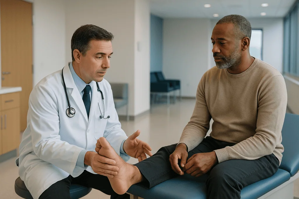
The physical examination is the second layer. The examiner locates the tender point, then moves the ankle to the position that should provoke impingement while pressing there. Reproducing the patient’s exact pain with that maneuver strengthens the suspicion considerably. They will also check ligament stability, because an unstable ankle that keeps re-spraining feeds the cycle of inflamed lining and scar. Range of motion is measured and compared with the other side.
Imaging comes third, and it serves two purposes: to look for spurs or accessory bones, and to rule out things that mimic impingement. Plain X-rays show bony ridges on the front of the tibia and the bony process at the back of the talus. Magnetic resonance imaging, or MRI, uses magnetic fields rather than radiation to show soft tissue, and it can reveal inflamed lining, thickened scar bands, fluid around irritated tendons, and any damage to the smooth cartilage covering the talus. That last item matters because cartilage damage after a sprain can produce pain in a similar location and changes the treatment discussion.
Some clinicians add a diagnostic injection of local anesthetic into the suspected spot. If the pinch disappears for the duration of the numbing, the location is confirmed. Whether that step is used depends on the clinician’s judgment and how clear the picture already is.
No single test settles it. Diagnosis is the examiner’s synthesis of history, hands-on findings and images, which is also why a self-diagnosis from an internet description is unreliable and why the treating team’s assessment should guide the next step.
Ankle impingement treatment: what happens first, before surgery
Nearly every guideline-level approach to impingement begins without a scalpel, and for good reason: the inflamed tissue that causes the pinch often settles when the joint stops being driven into the provoking position over and over.
Activity modification is the first move. This does not mean stopping exercise. It means temporarily avoiding the specific end-range movement that triggers the pinch, such as deep squats for anterior impingement or full pointe work for posterior impingement, while keeping the rest of the body active. For a runner, that may be a change of terrain away from steep hills. For a dancer, it may mean reducing repetitions of the provoking position rather than stopping class.
Managing swelling comes next. Ice after activity, elevation and compression are the same basic tools used for sprains, and Mayo Clinic describes them as reasonable self-care for ankle injuries in the early period. Anti-inflammatory medicines from the nonsteroidal class work by dampening the chemical signals that drive inflammation in the joint lining. Whether they are appropriate, and for how long, is a decision for the prescribing clinician, since they carry stomach, kidney and cardiovascular considerations that depend on the individual.
Footwear and supports can help mechanically. A small heel lift can reduce how far the front of the ankle closes during walking, taking pressure off an anterior pinch. Lace-up braces or taping may help a person whose ankle keeps giving way, interrupting the sprain-and-scar cycle.
Physical therapy is the backbone of this phase and gets its own section below. What ties the whole approach together is time. Clinicians commonly give nonsurgical ankle impingement treatment a sustained trial over several months before considering anything invasive, partly because inflamed tissue takes time to calm and partly because the surgical option is more convincing when the simpler options have genuinely been exhausted rather than sampled.
What physical therapy for ankle impingement actually does
People sometimes hear ‘physical therapy’ and imagine being stretched harder into the position that already hurts. Good rehabilitation for impingement does close to the opposite. It aims to restore normal joint mechanics so that the tissue stops getting caught, and it does this through several distinct strands.
The first strand is joint mobilization. When the talus does not glide smoothly backward as the foot bends up, the front of the joint closes down earlier than it should. A therapist uses hands-on techniques to encourage that backward glide, and teaches the patient self-mobilization with a band. The goal is a cleaner arc of motion, not a bigger one.
Strength is the second strand. The muscles on the outside of the lower leg, the peroneals, are the ankle’s active stabilizers. After sprains they are often weak and slow to fire, which is one reason ankles re-sprain. Rebuilding them reduces the repeated micro-injuries that keep the lining inflamed. Calf strength and hip control are addressed too, because a hip that collapses inward on landing drives the ankle into awkward angles.
Balance and proprioception form the third strand. Proprioception is the body’s sense of joint position, and it is measurably impaired after ligament injury. Single-leg stance work, unstable surfaces and reactive drills retrain the reflexes that protect the joint before it reaches a painful position.
The fourth strand is movement retraining specific to the person’s sport. A dancer’s alignment on pointe, a footballer’s kicking mechanics, a runner’s foot strike: subtle adjustments can reduce how often the joint is forced into the pinch.
How long does this take? There is no single number that applies to every ankle, and the honest answer is that a therapist reassesses progress over weeks and adjusts. What the evidence and clinical consensus support is that rehabilitation is the foundation, whether or not surgery eventually follows, because a joint with poor mechanics can redevelop symptoms even after spurs are removed.
Do injections help ankle impingement?
Injections sit in the middle of the treatment ladder, above rehabilitation and below surgery, and they can serve two roles at once.
The first role is diagnostic. A small amount of local anesthetic placed precisely at the suspected pinch point should numb the pain for a short time if that spot is truly the source. If the pinch persists despite the numbing, the clinician has learned something useful and may look elsewhere. Guidance from imaging, such as ultrasound, helps the needle reach the intended pocket of tissue.
The second role is therapeutic. A corticosteroid is a synthetic version of a hormone the body produces, and when injected into a joint it suppresses the inflammatory activity in the lining. The mechanism is straightforward: less inflammation means less swelling of the synovium, which means a thinner fold of tissue that is less likely to be caught. Relief, when it occurs, typically develops over days rather than instantly, and its duration varies widely from person to person. Some people find a single injection settles the lining enough for rehabilitation to progress; others notice a short-lived effect and no more.
What a steroid injection cannot do is shrink a bone spur or remove an accessory bone. If the pinch is mainly mechanical, coming from bone-on-tissue contact, the injection may calm the reaction without changing the cause. This is one reason clinicians are usually cautious about repeating injections into the same joint. Repeated corticosteroid exposure raises concerns about weakening nearby tendons and softening cartilage, and most treating teams set a limit.
Other injectable substances are sometimes discussed, including preparations derived from the patient’s own blood. The evidence for these in ankle impingement specifically is limited and inconsistent, and they should be presented as unproven rather than as established treatment.
Whether an injection is appropriate, which substance, and how many times, remains a judgment for the treating clinician who has examined the ankle and seen the imaging.
When is ankle arthroscopy considered, and who is usually asked to wait?
Arthroscopy is keyhole surgery: a pencil-thin camera and small instruments are passed into the joint through incisions roughly a centimeter long, and the surgeon works while watching a screen. Mayo Clinic describes it as a procedure used both to diagnose and to treat joint problems, usually performed as an outpatient. In the ankle it allows a surgeon to trim inflamed lining, remove scar bands and shave down bone spurs without opening the joint.
Who is usually considered a candidate? In broad terms, someone whose diagnosis is clear on examination and imaging, whose pain is reliably reproduced at the suspected spot, and who has completed a genuine course of nonsurgical ankle impingement treatment without adequate improvement. Clinicians differ on how long that trial should be, but a sustained period of several months of rehabilitation, activity modification and possibly an injection is a common threshold before surgery enters the conversation.
Pain that limits daily function, not just peak sport, strengthens the case. So does a large bony spur that is clearly the mechanical obstacle, because no amount of rehabilitation will change bone. Where imaging also shows a cartilage lesion on the talus that could be addressed in the same procedure, the surgeon may feel the operation achieves two goals at once.
Who is usually asked to wait? Anyone who has not yet tried structured physical therapy, since surgery on a joint with untreated mechanical problems tends to disappoint. People whose imaging shows widespread arthritis rather than a focal pinch, because trimming a spur does not address a joint that is worn throughout. Those with active infection, poorly controlled diabetes, heavy smoking or circulation problems in the leg may be asked to address those factors first, as they raise the risk of wound and healing complications. Someone whose pain is diffuse and not reproducible at one point may be sent for further investigation rather than surgery.
The decision always rests with the treating team, weighing the individual’s anatomy, goals and health.
What actually happens during ankle arthroscopy for impingement
The operation is shorter and less dramatic than most people picture. According to the NHS, an arthroscopy typically takes between 30 minutes and 2 hours depending on what needs to be done, and most people go home the same day.
Anesthesia comes first. This may be a general anesthetic, where the patient is asleep, or a regional block that numbs the leg while the patient is sedated. The choice is made with the anesthetist beforehand. A tourniquet around the thigh may be used to keep the field clear of blood.
For anterior impingement, the surgeon makes two small incisions at the front of the ankle, one either side of the tendons that lift the foot. Sterile fluid is pumped into the joint to expand it and give the camera a clear view. Through one portal goes the arthroscope, a slender tube with a lens and light; through the other go the working instruments. The surgeon inspects the whole joint, then uses a motorized shaver to remove inflamed lining and scar bands and a small burr to grind down bony spurs on the tibia and talus. The foot is moved through its range on the table to confirm that the pinch has been cleared.
Posterior impingement is approached from the back, with the patient lying face down and two small incisions either side of the Achilles tendon. The surgeon removes the accessory bone or bony process and releases any tissue tethering the tendon that flexes the big toe.
Throughout, the surgeon is also looking for problems that imaging may have missed, such as loose fragments or cartilage damage, and may treat those as planned in advance. The incisions are closed with a stitch or two, a padded dressing is applied, and the patient is woken and monitored before going home with instructions.
What the operation does not do is rebuild ligaments or replace cartilage. Those are different procedures, sometimes combined with arthroscopy when the surgeon judges it appropriate.
Ankle impingement treatment options compared
Seeing the options side by side helps make sense of why clinicians move through them in a particular order. Each has a different target, a different level of intrusion, and a different set of trade-offs.
| Option | What it targets | How invasive | Main trade-offs |
|---|---|---|---|
| Activity modification and footwear changes | The provoking movement | None | Requires patience; may limit sport temporarily |
| Physical therapy | Joint mechanics, strength, balance | None | Takes weeks of consistent effort; results vary |
| Anti-inflammatory medicines (nonsteroidal class) | Inflammation in the lining | Low | Stomach, kidney and heart considerations; short-term use guided by prescriber |
| Corticosteroid injection | Inflamed lining at the pinch point | Low to moderate | Temporary; does not change bone; repeat injections limited |
| Ankle arthroscopy | Spurs, scar, inflamed lining, accessory bone | Surgical, keyhole | Anesthesia; small risk of infection, nerve irritation, clots; recovery weeks to months |
A few observations flow from the table. The first three rows address the movement side of the problem and the inflammatory reaction; only the last row addresses a mechanical bony block directly. That is why a large spur on X-ray shifts the discussion toward surgery sooner, while a purely soft-tissue picture on MRI argues for a longer nonsurgical trial.
The rows are also cumulative rather than either-or. Rehabilitation continues after an injection and resumes after arthroscopy. A person who skips the foundation often ends up returning to it later.
Nothing in this comparison ranks one option as superior for everyone. The right rung of the ladder depends on the individual’s anatomy, how long symptoms have persisted, what they need the ankle to do, and the judgment of the treating team.
Ankle arthroscopy recovery time: what the following days and weeks usually look like
Recovery from keyhole ankle surgery follows a recognizable arc, though the pace differs from person to person and depends heavily on what was done inside the joint.
The first few days are about swelling control. The ankle is kept elevated above heart level as much as possible, the dressing stays clean and dry, and ice is applied over the bandage in intervals. Most people are allowed to put weight through the foot in a supportive boot or shoe from early on when only soft tissue and spurs were removed, though the surgeon’s specific instructions take precedence. Pain relief prescribed by the team is used as directed, and gentle movement of the toes and ankle is often encouraged to keep the joint from stiffening. The NHS notes that after arthroscopy generally, many people can return to light activities within a few days to a few weeks, but that full recovery may take several weeks or months.
The first follow-up visit usually happens within a couple of weeks. Stitches are checked or removed, the wounds are inspected, and the surgeon reviews what was found and done. Physical therapy typically restarts around this point, initially focused on regaining range of motion and reducing swelling, then progressing to strength and balance work.
Weeks three to six tend to be when walking normalizes and the ankle starts to feel like part of the body again. Swelling can linger longer than people expect, particularly at the end of a long day, and that alone is not a sign of trouble.
Return to running and sport is guided by function rather than the calendar: symmetrical range of motion, adequate strength, good single-leg balance and pain-free hopping are the kinds of milestones therapists look for. Athletes in demanding sports may take longer than the general ranges above, especially if cartilage or ligament work was combined with the impingement procedure.
These are typical patterns drawn from general arthroscopy guidance, not guarantees. Your team’s timeline for your ankle overrides any general description.
Risks of ankle arthroscopy and the alternatives, in neutral terms
Every surgery carries risk, and a good consultation lays it out plainly rather than glossing over it. Mayo Clinic and the NHS both characterize serious complications of arthroscopy as uncommon, while listing the ones that do occur.
Infection is the first concern with any incision. Small wounds lower the risk compared with open surgery, but it is not zero, and the treating team gives instructions about wound care and warning signs for exactly this reason. Nerve irritation is a particular consideration in the ankle because several sensory nerves run just under the skin near the portal sites. Numbness or tingling on the top of the foot after anterior arthroscopy, or on the heel after posterior surgery, can occur and often improves over time, though not always completely. Blood clots in the leg veins are a known risk after lower-limb surgery, which is why early movement and, for some patients, preventive measures chosen by the team are part of the plan.
Less common problems include bleeding into the joint, damage to cartilage or tendon during the procedure, stiffness, and persistent pain. Spurs can also re-form over years if the mechanical stress that created them continues. Anesthesia carries its own small set of risks, which the anesthetist discusses individually.
The alternatives are the same options that came before surgery, continued. Some people choose to live with position-specific pain, adapting their activities so the joint is rarely driven into the pinch, and that is a legitimate choice. Others continue rehabilitation and accept an occasional injection. For people with more widespread joint wear, surgeons may discuss entirely different procedures aimed at arthritis rather than impingement.
Open surgery through a larger incision remains an option in certain anatomies or when arthroscopic access is judged inadequate, with a generally longer recovery.
Weighing these against the burden of the current symptoms is the core of the shared decision, and that weighing belongs to the patient and the treating team together.
What people often get wrong about ankle impingement
Because impingement is common in active people and widely discussed in gym and studio changing rooms, it has gathered a fair amount of folklore. Several beliefs deserve correcting.
The first is that a bone spur on an X-ray means surgery is inevitable. Spurs are extremely common in people who load their ankles hard, and many cause no pain at all. The spur is only a problem when tissue is caught against it, and calming that tissue frequently settles symptoms without touching the bone.
The second is that stretching the ankle harder will ‘open up’ the pinch. Aggressive stretching into the painful end range does the opposite, repeatedly compressing the inflamed tissue. Rehabilitation aims to restore how the joint glides, not to force it further.
A third belief is that the pain must be a fresh sprain that never healed. Sometimes it is, but impingement often develops months or years after the original injury, once scar and thickened lining have matured. The ligament may be perfectly intact.
Fourth, people assume that because the ankle feels fine most of the time, the problem is minor and can be ignored indefinitely. Position-specific pain can be lived with, but a joint that keeps being pinched tends to stay inflamed, and repeated sprains from a weak, unstable ankle can add cartilage damage over time. Ignoring it is a choice with consequences worth understanding.
Fifth, there is a myth that arthroscopy is a minor tidy-up with no real recovery. It is less invasive than open surgery, but it is still an operation under anesthesia with weeks of rehabilitation afterward.
Finally, some assume that an injection that wears off has failed. A short-lived response is still informative: it confirms the location of the problem and tells the team that a mechanical cause may be dominating. Treatment decisions are built from exactly that kind of information, and the team, not the folklore, should interpret it.
Questions to ask your care team about ankle impingement treatment
Consultations move quickly, and the most useful questions are the ones that clarify what is actually driving your pain and what each step is meant to achieve. Bringing a short written list is a reasonable habit.
Start with the diagnosis itself. Ask whether the impingement is anterior or posterior, and whether the main problem is soft tissue, bone, or both. Ask what the imaging showed beyond the pinch point, particularly whether the cartilage on the talus and the ligaments look healthy, because those findings shape everything that follows.
On nonsurgical care, ask how long a trial of rehabilitation the team would consider adequate before revisiting the plan, and what specific goals the physical therapist will be working toward. Ask which movements you should avoid for now and which you can safely continue. If an injection is proposed, ask whether its purpose is mainly diagnostic, mainly therapeutic, or both, and how the team will judge the response.
If arthroscopy is raised, ask what exactly the surgeon expects to remove or trim, and whether anything else might be addressed in the same procedure. Ask about the type of anesthesia, whether you can expect to go home the same day, and what weight-bearing instructions you should plan for. Ask what the realistic recovery pattern looks like for someone with your job and activities, and when physical therapy would restart.
On risks, ask which complications the surgeon sees most often in ankle arthroscopy specifically, and how you would recognize the early signs of each. Ask what would happen if symptoms persisted after surgery.
Practical questions matter too: who to call if something feels wrong after hours, when the first follow-up will be, and whether you will need help at home in the first days.
None of these questions has a universal answer. Their value lies in hearing the team’s reasoning for your ankle, which is the only ankle that matters in the decision.
When to call your doctor
Ankle impingement itself is not usually urgent, but some situations around it deserve prompt contact with your care team, and a few warrant emergency attention.
Before any treatment, get in touch if the ankle becomes hot, red and swollen without a clear injury, if you develop a fever alongside joint pain, or if the pain is constant and severe rather than position-specific. Those features do not fit ordinary impingement and can signal infection, inflammatory arthritis or another problem that needs different evaluation. Sudden inability to bear weight after a twist, visible deformity, or numbness in the foot also call for same-day assessment, as they may indicate a fracture or significant ligament injury.
After an injection, contact the team if the joint becomes markedly more painful, swollen and warm over the following one to three days, or if you develop a fever. A brief flare of discomfort in the first day is common; a worsening, hot joint is not.
After arthroscopy, the warning signs are more specific. Call promptly if you notice increasing redness spreading from the incisions, pus or cloudy fluid leaking from the wounds, a temperature that is rising, or pain that is escalating rather than easing after the first few days. Calf pain, tenderness or swelling in the operated leg, particularly if the calf feels tight or warm, can indicate a blood clot and needs urgent assessment. Sudden breathlessness, chest pain or coughing up blood after surgery are emergency symptoms requiring immediate emergency services, because a clot can travel to the lungs.
Numbness or tingling on the top of the foot that appears after surgery should be reported at your follow-up even if it seems minor, so the team can document and monitor it. Any worry that something is not right is a legitimate reason to call; treating teams would rather hear from you early than late.
These lists are prompts, not a substitute for the individual instructions your team gives you.
Frequently asked questions
Can ankle impingement go away on its own?
It can settle, particularly when the cause is inflamed soft tissue rather than a bony block, and when the provoking movement is avoided long enough for the lining to calm. Bone spurs and accessory bones do not disappear, so pain driven mainly by bone-on-tissue contact often recurs when full activity resumes. Structured rehabilitation improves the odds compared with simple rest, because it addresses the joint mechanics that created the pinch.
What does anterior ankle impingement feel like compared with a sprain?
A sprain hurts broadly across the outer ankle after a twist and improves steadily over days to weeks. Anterior impingement produces a sharp, focused pinch at the front of the joint that appears only when the foot bends fully upward and fades when the position is released. Many people with impingement had one or more sprains in the past, sometimes long before the pinching began, so the two are related but feel different.
Is posterior ankle impingement mainly a dancer's problem?
Dancers are the group most often described with it because pointe work drives the foot into extreme plantarflexion thousands of times, but posterior impingement also affects gymnasts, swimmers, footballers and anyone who repeatedly points the foot hard. An accessory bone behind the talus, present from birth in some people, raises susceptibility. The diagnosis is made by examination and imaging, not by occupation.
How long is physical therapy for ankle impingement usually continued before surgery is discussed?
There is no fixed number, and clinicians differ. A sustained trial of several months of structured rehabilitation, activity modification and possibly an injection is a common threshold before surgery is raised. The therapist reassesses regularly and reports progress to the referring clinician. The decision to move toward arthroscopy rests with the treating team, who weigh how much function has been regained against how much the remaining pain limits daily life.
What is the typical ankle arthroscopy recovery time for impingement?
The NHS describes arthroscopy in general as usually allowing a return to light activities within a few days to a few weeks, with full recovery taking several weeks or months. For ankle impingement specifically, weight bearing in a supportive shoe often begins early, therapy restarts around the first follow-up, and return to sport is judged by strength and balance milestones rather than a date. Procedures that also address cartilage or ligaments usually take longer.
Does ankle arthroscopy remove bone spurs permanently?
The surgeon grinds down the spur present at the time of surgery, and the removed bone does not simply grow back in the following months. Over years, however, bone can re-form if the ankle continues to be driven into the same repetitive stress that created the spur originally. That is why rehabilitation and attention to movement patterns continue after surgery rather than ending with it.
Will I need a cast or crutches after ankle arthroscopy?
For impingement alone, a cast is not usually required. Most people are placed in a padded dressing and a supportive boot or shoe, and many are allowed to bear weight early with crutches for comfort in the first days. If ligament repair or cartilage work was combined with the procedure, the surgeon may restrict weight bearing for longer. Your team’s specific instructions override any general pattern.
Can I keep running with ankle impingement?
Often yes, with adjustments. Flat terrain tends to provoke anterior impingement less than uphill running, which forces the foot into deeper upward bend on each stride. A small heel lift and attention to cadence may help. Running through sharp, reproducible pinching is not advisable, as it keeps the lining inflamed. A physical therapist can help you find the modifications that let you train while the joint settles.
Are anti-inflammatory medicines a long-term solution for ankle impingement?
No. Nonsteroidal anti-inflammatory medicines reduce the chemical signals that drive inflammation in the joint lining, which can ease pain and swelling in the short term while rehabilitation progresses. They do not change the mechanics or the bone. Longer use carries stomach, kidney and cardiovascular considerations that vary by individual, so any use should be discussed with, and directed by, the prescribing clinician.
What is the difference between ankle impingement and ankle arthritis?
Impingement is a focal problem: one area of tissue or bone caught at one position, with the rest of the joint surfaces healthy. Arthritis involves wearing of the cartilage across the joint, producing pain that is more diffuse, present through much of the range and often worse after prolonged activity. Imaging distinguishes them. The distinction matters because trimming a spur addresses impingement but does not treat a joint that is worn throughout.
References
This article is for general information only and is not a substitute for professional medical advice. Please consult a qualified doctor about your individual situation.

More from the Blog

Is Night Pain Normal After Rotator Cuff Surgery? What Is Expected and When to Call
Yes, night pain after rotator cuff surgery is expected, especially in the first few weeks, because lying flat removes the support gravity gives the…

Helping Someone With a Dislocated Shoulder: What to Do on the Way to Care and What Not to Try
If someone dislocates a shoulder, keep the arm still in the position it is already in, support it against the body with a sling,…

Why an Elbow Fracture Often Needs a CT Scan Before Surgery: Planning the Fixation
An elbow fracture often needs a CT scan before surgery because plain X-rays flatten the elbow's three interlocking bones into two views, so hidden…
Tenosynovitis on Ultrasound: What the Scan Shows and How It Guides Splinting or Injection
On ultrasound, tenosynovitis appears as fluid or thickened tissue inside the sleeve (sheath) that surrounds a tendon, often with extra blood flow on Doppler…

When Is Tennis Elbow Surgery Considered? What Comes First and What Release Surgery Involves
Tennis elbow surgery is usually considered only after a sustained course of non-surgical care has failed. Mayo Clinic describes roughly 6 to 12 months…

Is It Still Shoulder Impingement? Pain That Does Not Settle With Rehab and When to Go Back
Shoulder impingement not improving after several months of well-performed rehabilitation is a reason to go back, not to push harder. Persistent pain may mean…