

Arthroscopy Complications Explained: Portal Infections, Swelling, Clots and When to Call
Key Takeaways
- Swelling after knee arthroscopy typically peaks around day two to three and then softens over one to two weeks; swelling that turns tense and hot after that point needs review.
- Bruising that migrates down the shin and changes from purple to yellow is gravity acting on a small expected bleed, not a sign of a failed operation.
- Septic arthritis, the dangerous form of post-arthroscopy infection, often produces fever and a hot, immobile joint without any visible pus at the portal wounds.
- The CDC's surgical site infection window runs 30 days, so a fever in week three still counts as a surgical concern.
- A clot in the operated leg usually declares itself as new calf pain or swelling extending below the knee, while sudden breathlessness or chest pain is an emergency-services call.
- Driving after arthroscopy depends on performing an emergency stop without hesitation while off sedating medicines, plus surgeon clearance, rather than on a fixed number of days.
Arthroscopy complications are uncommon but real. Most people have some bruising, swelling and soreness for one to two weeks, which is expected. Warning signs include a portal wound that becomes red, hot or leaks cloudy fluid, fever, rapidly tightening swelling, calf pain or swelling in one leg, breathlessness or chest pain. Those symptoms need same-day medical review, and the surgical team should make every decision about treatment.
Four days after a knee arthroscopy, a patient sits on the edge of the bed with her leg propped on two pillows and stares at a bruise that has crept from the kneecap halfway down the shin. The two tiny wounds look fine. The knee feels like a water balloon. Her partner asks the question everyone eventually asks: is this normal, or is this the thing the surgeon warned us about?
That is the honest heart of arthroscopy complications. The procedure is small, the incisions are shorter than a fingernail, and the recovery is often described as quick. Yet the body still bleeds, swells and occasionally gets infected or clots, and the hard part for patients is not avoiding every risk. It is telling a loud, ordinary recovery from a quiet, dangerous one.
This explainer walks through what actually happens inside the joint, which problems are common and which are rare, how each one tends to announce itself, and when a phone call to the care team should not wait until morning.
What actually happens during an arthroscopy of the knee or shoulder?
An arthroscopy is keyhole surgery on a joint. The surgeon makes two or three small cuts, each usually under a centimeter, and passes in an arthroscope, a thin tube with a camera and light that sends a live image to a screen. Those small cuts are called portals, and they matter later because most arthroscopy complications that patients can see start at a portal.
Sterile fluid is pumped into the joint through one portal to open up the space and wash away blood so the camera can see. Through another portal, the surgeon introduces slim instruments: shavers that trim frayed cartilage, graspers that remove loose fragments, and anchors and sutures that repair torn tissue. In the knee, the most frequent targets are the menisci, the two crescent-shaped shock absorbers between the thigh bone and shin bone. In the shoulder, it is usually the rotator cuff tendons or the labrum, the cartilage rim of the socket. Hips, ankles, wrists and elbows are scoped less often.
According to the NHS, the operation is most often done under general anesthesia, although spinal or regional anesthesia with sedation is also used, and the great majority of people go home the same day. Mayo Clinic describes a typical procedure lasting from under an hour to a little more, depending on what is found and repaired.
At the end, the fluid is drained as far as possible, the portals are closed with a stitch or adhesive strip, and a padded dressing or compression bandage goes on. Some fluid always remains inside the joint and in the soft tissue around it. That leftover fluid, mixed with a little blood, is the origin of the swelling and bruising almost everyone notices in the first week.
How common are arthroscopy complications, really?
Here is the frame that helps most: expected effects happen to nearly everyone, minor complications happen to a few, and serious complications happen to very few. Mayo Clinic, the NHS and Cleveland Clinic all describe arthroscopy as a low-risk procedure in which serious problems are uncommon, while being clear that no joint surgery is risk-free.
The expected effects are swelling, bruising, soreness and stiffness. These are not complications; they are the body responding to irrigation fluid, instrument contact and a few hours of lying still. Minor complications include a portal wound that oozes for longer than expected, a superficial skin infection around a portal, or a knee that stays puffy and stiff for several weeks instead of one or two.
Serious complications, listed by the NHS and Mayo Clinic, include infection inside the joint (septic arthritis), a blood clot in a deep leg vein (deep vein thrombosis, or DVT) that can travel to the lungs (pulmonary embolism), accidental damage to nerves, blood vessels, cartilage or ligaments, and reactions to anesthesia. Published surgical series generally place each of these well below 1 in 100 arthroscopies, but exact rates vary with the joint, the complexity of the repair, the patient’s age and medical history, and how long the operation takes. A simple meniscal trim is not the same risk profile as a multi-anchor reconstruction with a tourniquet in place for over an hour.
Why does this matter to you at four days out? Because rarity cuts both ways. A low baseline rate means the swollen, bruised knee is overwhelmingly likely to be normal. It also means that when a genuine warning sign appears, it is unlikely to be a coincidence and deserves a prompt phone call rather than a wait-and-see weekend.
Who is usually offered arthroscopy, and who is asked to wait?
Arthroscopy is both a diagnostic tool and a treatment. The NHS notes it is used when imaging and examination have not fully explained persistent pain, locking, catching or instability, and when the likely problem can be fixed through the same small portals: a torn meniscus, a loose body of cartilage or bone, a damaged rotator cuff or labrum, inflamed joint lining, or an anterior cruciate ligament in need of reconstruction.
People most often considered for it have mechanical symptoms that interfere with daily life or sport despite a reasonable trial of physiotherapy, activity modification and time. The decision is never made on a scan alone. Many meniscal tears seen on MRI in people over 40 cause no symptoms, and mainstream guidance has become more cautious about operating on degenerative tears in knees that are already arthritic, because the evidence that surgery outperforms structured exercise in that group is weak.
Who is usually asked to wait, or offered something else first?
- Someone whose joint is acutely swollen and hot, where infection must be ruled out before any instrument enters the joint.
- A person with an active skin infection, ulcer or rash near the planned portals.
- Anyone whose diabetes, blood pressure or anticoagulation is not yet stable enough for safe anesthesia.
- A patient with advanced arthritis, where cleaning out the joint is unlikely to change the underlying wear.
- Someone who has not yet completed a rehabilitation program that might resolve the symptoms without surgery.
Smokers, people with poorly controlled blood sugar, and those with a personal or family history of clots carry higher complication risk and may be asked to optimize these first. None of this is a judgment; it is the surgical team shifting the odds in your favor before the first portal is made. The final call always sits with you and that team together.
Is bruising after knee arthroscopy normal?
Yes, and often more of it than people expect. Bruising after knee arthroscopy appears when small vessels inside the joint and in the fat pad around the kneecap bleed a little during surgery. That blood mixes with the irrigation fluid and, over the following days, gravity carries it down through the soft tissue of the shin and sometimes toward the ankle. A bruise that seems to migrate downward over a week is following physics, not spreading disease.
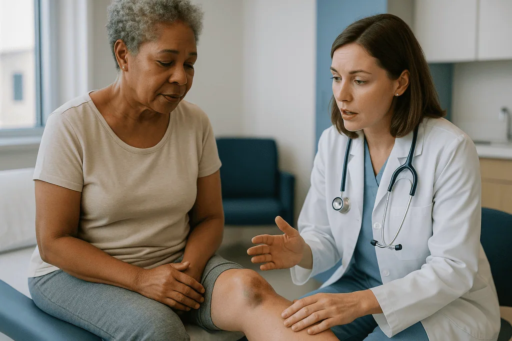
Colors tell the age of the bruise. Fresh blood under the skin looks purple or deep red; as hemoglobin breaks down it turns green and then yellow-brown before fading. MedlinePlus and the NHS both describe bruising and swelling as expected after knee arthroscopy and advise elevation, cold packs over the dressing and gentle movement to help it resolve. Most visible bruising settles within two to three weeks, although in people taking blood thinners or with fair skin it can look dramatic and linger longer.
Two features change the picture. The first is bruising that comes with a joint that is rapidly tightening, extremely painful to bend even a few degrees, and feels hot and tense. That pattern suggests a hemarthrosis, meaning a significant bleed into the joint space, and it needs assessment the same day. The second is bruising accompanied by numbness, tingling or weakness in the foot, which can signal pressure on a nerve or, rarely, compartment syndrome, a dangerous rise in pressure within the muscle compartments of the lower leg that is a surgical emergency.
A useful home test, suggested by many post-operative leaflets, is simply whether things are heading in the right direction. Bruising that fades, swelling that softens and pain that eases day over day is ordinary healing. The reverse trajectory, after the first 48 hours, is what should prompt a call.
Swelling after knee arthroscopy: leftover fluid or a warning sign?
Swelling after knee arthroscopy has three ingredients. First, the sterile saline pumped in during surgery, some of which seeps into the tissue around the knee and takes days to be reabsorbed. Second, the joint lining’s own inflammatory response, which produces extra synovial fluid, the slippery lubricant that normally bathes the joint. Third, a small amount of blood. Together they produce an effusion, the clinical term for excess fluid inside the joint capsule.
Cleveland Clinic and the NHS describe swelling as peaking in the first two to three days and then gradually improving over one to two weeks for simple procedures. After a meniscal repair with sutures, or a ligament reconstruction, the knee may stay puffy for several weeks, and that is within the expected range. Elevation above heart level, cold therapy applied over the dressing in short sessions, and a compression bandage all help by reducing blood flow to the area and encouraging fluid to drain back toward the trunk.
Movement helps too. The calf and thigh muscles act as pumps; gentle ankle circles and quadriceps contractions, as taught by the physiotherapist, push fluid out of the leg and, as a bonus, reduce clot risk.
What should worry you is the quality of the swelling rather than its mere presence. Fluid that is soft, moves when pressed and feels at room temperature is typical. Swelling that becomes tense and shiny, feels markedly hotter than the other knee, is accompanied by fever or chills, or follows a period of improvement with a sudden relapse points toward infection or a bleed and needs prompt review. Surgeons sometimes draw fluid off a very swollen joint with a needle, both to relieve pressure and to send a sample to the laboratory. That is a decision for the surgical team, never a reason for a patient to delay reporting symptoms.
Knee arthroscopy infection signs: portal wound problems versus septic arthritis
Infection after arthroscopy comes in two very different sizes, and knowing which is which saves a lot of anxiety. A superficial portal infection is a skin infection, sometimes called cellulitis, confined to the tissue around one of the small incisions. Septic arthritis is an infection inside the joint itself. The first is uncomfortable and treatable; the second is an emergency that can damage cartilage within days.
The CDC defines a surgical site infection as one that develops at or near the operative site within 30 days of surgery, or within a year when an implant such as a suture anchor has been placed. In practice, portal infections most often show themselves between about day three and day ten, when the body’s initial post-operative inflammation should be fading rather than growing.
Knee arthroscopy infection signs around a portal include spreading redness beyond the edge of the dressing, warmth, increasing rather than decreasing tenderness, and discharge that changes from thin pink fluid to thick, cloudy or yellow material with an odor. Signs that infection has reached the joint are more systemic and more severe: fever or sweats, a knee that becomes too painful to bend or bear weight, a joint that is hot and tense to the touch, and a general sense of being unwell. The two can coexist, so any portal infection should be assessed by a clinician who can judge whether the joint is involved.
Why do portals get infected at all? Bacteria normally living on the skin can be carried into the wound, moisture under a dressing softens the skin barrier, and leaking joint fluid keeps the wound from sealing. Diabetes, smoking, obesity and immune-suppressing medicines all raise the risk, as does a longer operation. Treatment depends on depth: a superficial infection may need wound care and antibiotics chosen by the team, while a joint infection usually requires urgent washout surgery and intravenous antibiotics. Those are decisions for the surgeons, and the patient’s job is simply to report the signs early.
Blood clot after arthroscopy: how DVT and pulmonary embolism show up
A blood clot after arthroscopy is uncommon but is the complication most capable of becoming life-threatening without warning, which is why every discharge leaflet mentions it. Deep vein thrombosis, or DVT, is a clot in one of the large veins deep in the calf or thigh. If a piece breaks away and travels through the heart to the lungs, it becomes a pulmonary embolism, or PE.
Surgery raises clot risk through three mechanisms: blood pools in the veins when a limb is still, the vein wall is irritated by tourniquet pressure and inflammation, and the body’s clotting system is switched on by any tissue injury. Risk rises further with longer operations, a plaster cast or brace that keeps the leg immobile, older age, obesity, smoking, hormonal contraception or hormone therapy, a previous clot, cancer and a family history of clotting disorders. Surgeons weigh these factors before the operation and may recommend compression stockings or a short course of an anticoagulant medicine, a drug that reduces the blood’s tendency to clot. Whether, what and for how long is entirely the prescribing clinician’s decision.
The NHS describes DVT symptoms as throbbing pain, swelling, warmth and red or darkened skin, usually in one calf or thigh, sometimes with swollen veins that are sore to touch. The trap after knee surgery is that the operated leg is already swollen and sore. The distinguishing features are calf pain that is new or disproportionate, swelling that extends below the knee into the calf and ankle rather than staying around the joint, and a leg that feels heavy and tight compared with the other side.
Pulmonary embolism announces itself with sudden breathlessness, sharp chest pain that is worse on breathing in, a racing heartbeat, coughing up blood, or feeling faint. Those symptoms mean calling emergency services immediately, not the surgeon’s office. Most clots form within the first two weeks, but the risk window can extend to around a month, especially with reduced mobility.
Nerve injury, stiffness and the rarer arthroscopy complications
Beyond infection and clots, a cluster of less common problems rounds out the list that Mayo Clinic and the NHS describe for arthroscopy. Each has a mechanism worth understanding, because understanding takes the mystery out of the consent form.
Nerve injury usually involves small skin nerves that run close to the portals. In the knee, a branch of the saphenous nerve crosses the inner side of the joint, and a numb patch of skin below the kneecap or on the inner shin after surgery is relatively common, often temporary, and occasionally permanent. Damage to the larger nerves that move the foot is rare and tends to show up as weakness or persistent tingling rather than a numb patch. In shoulder arthroscopy, nerve stretch from positioning the arm can cause transient tingling in the hand that typically settles within days to weeks.
Blood vessel injury is rarer still. The main artery behind the knee lies within a few centimeters of where meniscal instruments work, which is one reason surgeons are meticulous about instrument angles. Bleeding into the joint (hemarthrosis) is more common than arterial injury and usually resolves, though it may need to be drained.
Stiffness deserves honest mention. A degree of tightness is universal. Arthrofibrosis, the formation of excessive scar tissue inside the joint that limits bending or straightening for months, is uncommon after simple procedures but more frequent after ligament reconstructions, especially when rehabilitation is delayed. Early, guided movement is the main protective factor, which is why physiotherapy begins so soon.
Fluid leakage from a portal for a few days is normal; persistent leakage past a week slows wound sealing and raises infection risk. Accidental damage to cartilage from instruments, breakage of an instrument tip inside the joint, and tourniquet-related skin or muscle irritation are all documented and all rare. Anesthesia carries its own small risks, including nausea, sore throat and, very rarely, serious reactions, which the anesthesia team reviews with each patient beforehand.
Normal recovery or arthroscopy complication? A side-by-side guide
Patients consistently say the hardest part of the first fortnight is deciding whether a symptom belongs in the normal column. The table below summarizes what mainstream guidance from the NHS, Mayo Clinic and MedlinePlus describes as expected after a straightforward knee or shoulder arthroscopy, set against the pattern that suggests a problem. It is a guide to conversation with your care team, not a substitute for it.
| Symptom | Usually expected | Pattern that needs a call |
|---|---|---|
| Swelling | Peaks days 2–3, soft, improves over 1–2 weeks | Tense, hot, worsening after day 3, or sudden relapse after improvement |
| Bruising | Spreads downward, changes color, fades in 2–3 weeks | Accompanied by numbness, foot weakness or a rapidly tightening joint |
| Portal wounds | Thin pink ooze for 2–3 days, then dry | Spreading redness, thick or cloudy discharge, odor, leaking beyond a week |
| Pain | Eases day over day, controlled with prescribed plan | Escalating despite medicine, or too severe to bend the joint at all |
| Temperature | Mild warmth around joint; no fever | Fever, chills or sweats at any point in the first month |
| Calf | Soft, not tender | New calf pain, swelling to ankle, warmth, darkened skin in one leg |
| Breathing | Normal | Sudden breathlessness, chest pain, coughing blood: call emergency services |
| Skin sensation | Small numb patch near a portal | Spreading numbness, pins and needles, or weakness moving the foot or hand |
Two patterns underpin the whole table. The first is direction of travel: after the first 48 to 72 hours, every normal symptom should be moving toward better, even if slowly. The second is the arrival of anything systemic, meaning fever, feeling unwell, breathlessness or faintness, which has no place in an uncomplicated recovery and should always be reported the same day.
What the first days and weeks usually look like, including when you can drive
The timeline below reflects typical ranges described by the NHS, Mayo Clinic and MedlinePlus for simple procedures such as a meniscal trim. Repairs that require stitches in tissue, and reconstructions that require protected weight-bearing or a brace, run slower, and your own surgical team’s schedule overrides anything written here.
Day of surgery. Most people go home within a few hours. The leg or arm is bandaged, often numb from local anesthetic, and the instructions are elevation, cold therapy over the dressing, prescribed pain relief and short, frequent walks or ankle pumps to keep blood moving.
Days 1 to 3. Swelling and soreness peak as the local anesthetic wears off. Crutches are common after knee surgery, mainly for confidence. The dressing is usually left intact unless it soaks through. Physiotherapy exercises start with gentle range of motion and muscle activation.
Week 1. A wound check or dressing change often happens around this point, and stitches, if used, are removed at roughly one to two weeks. The NHS notes that many people with desk-based jobs return to work within about a week after a simple arthroscopy, while physically demanding work may need several weeks.
Weeks 2 to 6. Swelling continues to settle, strength work increases, and walking without aids becomes comfortable. The NHS and Mayo Clinic describe full recovery, meaning a return to sport or heavy activity, as taking from a few weeks to several months depending on what was done.
Driving. No fixed date applies. The standard advice from the NHS is that you must be able to control the vehicle fully, including an emergency stop, without pain or hesitation, must be off any medicine that causes drowsiness, and should check with your surgeon and your insurer. For a left knee with an automatic transmission that can be a matter of days; for a right knee, a manual car or a shoulder in a sling it is commonly one to several weeks. The surgeon’s clearance is the deciding factor.
What exercises are usually recommended after arthroscopic meniscus surgery?
Exercise after meniscal surgery has two jobs: to restore motion and strength, and to prevent complications. Moving the calf and thigh muscles pumps fluid and blood out of the leg, which eases swelling and lowers clot risk, and early controlled bending reduces the chance of excessive scar tissue. The specific program must come from the surgeon and physiotherapist, because a trimmed meniscus and a repaired meniscus follow different rules: after a repair, deep bending and full weight-bearing are often restricted for weeks to protect the stitches.
With that caveat, the early-stage exercises that appear in most mainstream rehabilitation leaflets, including those referenced by MedlinePlus and the NHS, fall into a few families.
- Ankle pumps. Pointing and flexing the foot every hour while awake, which drives the calf muscle pump that protects against DVT.
- Quadriceps sets. Tightening the thigh muscle with the leg straight, holding for a few seconds, which keeps the muscle from switching off, a common reflex response to a swollen knee.
- Heel slides. Lying down and sliding the heel toward the buttock to the point of mild stretch, then back, to regain bending gradually.
- Straight-leg raises. Once the thigh muscle can lock the knee, lifting the straight leg a short distance to build strength without loading the joint.
- Gentle stationary cycling. Often introduced once the knee bends comfortably beyond a right angle, as a low-impact way to restore motion and endurance.
Later stages add balance work, step-ups, controlled squats and, eventually, sport-specific drills. The sensible pace is governed by swelling: if the knee is noticeably puffier the morning after a session, the session was too much. Pain that lingers for more than an hour or two after exercise sends the same message.
What rehabilitation cannot do is repair the joint faster than biology allows or compensate for a protocol that skips the protected phase after a repair. Patients who do best tend to be consistent rather than heroic, doing the prescribed small movements several times a day instead of one long, punishing session.
What people often get wrong about arthroscopy complications
Myth-busting matters here because misconceptions push people in both unhelpful directions: toward panic over normal bruising, and toward dismissing the one symptom that counts.
Myth: small cuts mean a small operation inside. The portals are tiny, but the surgeon may have trimmed cartilage, drilled bone or placed anchors. Internal healing time depends on what was done, not on the length of the scars. The NHS is explicit that full recovery can take weeks to months.
Myth: a big bruise means something went wrong. Bruising that travels down the shin is gravity acting on a small, expected bleed. It is the combination with tense swelling, numbness or fever that matters, not the size or color alone.
Myth: if it were infected I would see pus at the wound. Septic arthritis, the dangerous kind, often shows no wound discharge at all. Its signature is fever, a hot joint and pain out of proportion to the stage of recovery. Waiting for pus can waste critical days.
Myth: a clot would be obvious. Half of deep vein thromboses cause few or subtle symptoms, and the operated leg is already swollen. New calf pain or swelling that extends below the knee is the clue, and breathlessness is the emergency.
Myth: rest is the safest way to avoid complications. Prolonged immobility raises clot and stiffness risk. Early guided movement within the limits set by the team is protective, not reckless.
Myth: cleaning out an arthritic knee will reset it. Mainstream evidence does not support arthroscopic debridement for degenerative knee arthritis as superior to exercise therapy, and surgeons increasingly decline to offer it for that reason.
Myth: complications only happen in the first week. The CDC’s surgical site infection window runs to 30 days, and clot risk persists for about a month. A fever in week three still counts.
The through-line is simple: watch the trajectory and the systemic signs, and let the care team interpret the rest.
Questions to ask your care team about arthroscopy risks
A good pre-operative conversation does more to prevent a bad week than any amount of online reading. Clinicians generally welcome specific questions; they signal a patient who will also notice and report problems early. These are the ones worth bringing, with a note on why each matters.
- What exactly do you expect to do inside the joint, and what will you do if you find something different? A trim and a repair carry different recovery rules and risks.
- What is my personal risk of infection or clot, given my history? Diabetes, smoking, weight, prior clots and medicines all shift the baseline.
- Will I need anything to reduce clot risk, and who decides? Stockings or an anticoagulant may or may not be recommended; understanding the reasoning helps.
- How much swelling and bruising should I expect, and when should it start improving? A concrete timeline turns vague worry into a checkable milestone.
- How should I care for the portal dressings, and when can they get wet? Moisture management is the main thing patients control in infection prevention.
- Which symptoms should make me call the clinic, and which should send me to emergency care? Ask for the number to call out of hours.
- When will physiotherapy start, and what movements are off limits at first? Essential after any repair with sutures or anchors.
- When can I realistically drive, return to work and return to sport? Ask for ranges rather than dates, and for what clearance depends on.
- Should I stop or adjust any of my regular medicines before or after surgery? Never change a prescribed medicine without that answer coming from the prescriber.
- What are the alternatives if I choose not to have surgery now? Structured exercise, injections or watchful waiting may be reasonable for some problems.
Write the answers down. The person who remembers the surgeon’s own description of a worrying swelling, in the surgeon’s own words, is the one who makes the call at the right moment rather than a day late.
When to call your doctor after arthroscopy: red-flag signs
Most recoveries never need an unscheduled call. When one does, speed matters more than certainty, and no clinician minds being contacted about a symptom that turns out to be nothing. Keep the clinic’s daytime and out-of-hours numbers where you can find them without searching.
Contact your surgical team the same day if you notice:
- Fever, chills or sweats at any point in the first month after surgery.
- A portal wound with spreading redness, increasing warmth, thick or cloudy discharge, an unpleasant odor, or fluid still leaking after about a week.
- Swelling that becomes tense, hot and shiny, or that suddenly worsens after a period of improvement.
- Pain that escalates despite the prescribed plan, or a joint too painful to move at all.
- New pain, swelling, warmth or darkened skin in the calf or thigh of either leg, particularly if it extends below the knee.
- Numbness or tingling that spreads, or new weakness moving the foot, ankle, hand or fingers.
- A dressing soaked through with blood that does not stop with firm pressure and elevation.
- Persistent nausea, vomiting or inability to pass urine in the first day or two after anesthesia.
Call emergency services immediately, do not wait for the clinic, if you develop:
- Sudden breathlessness or difficulty breathing.
- Chest pain, especially pain that is sharper when you breathe in.
- Coughing up blood.
- A racing heartbeat with light-headedness, fainting or collapse.
- A lower leg that is extremely painful, pale or cold, with pain that worsens when the toes are stretched, which can indicate compartment syndrome.
These lists are deliberately broad. The NHS and Mayo Clinic both emphasize that infection and clots are treatable when caught early and far harder to manage when they are not. What happens next, whether that is a wound check, a blood test, an ultrasound of the leg, aspiration of the joint or a return to theater, is for the treating team to decide. Your part is to notice the change and to make the call.
Frequently asked questions
Is it normal to experience bruising after a knee arthroscopy?
Yes. Bruising after knee arthroscopy is expected and often spreads down the shin as gravity moves a small amount of blood through the soft tissue. It typically changes color and fades over two to three weeks. Bruising only becomes a concern when it is paired with rapidly tightening swelling, numbness or weakness in the foot, or fever, in which case the surgical team should be contacted the same day.
What are the first knee arthroscopy infection signs I should watch for?
Early signs include redness spreading beyond the portal, increasing warmth and tenderness after day three, and discharge that turns thick, cloudy or smelly. Signs of infection inside the joint are fever, chills, a hot and tense knee, and pain too severe to bend it. Infection inside the joint is urgent, and wound discharge may be absent, so fever plus a hot joint should prompt an immediate call.
How long does swelling after knee arthroscopy last?
For simple procedures such as a meniscal trim, mainstream guidance describes swelling peaking in the first two to three days and improving steadily over one to two weeks. After a meniscal repair or ligament reconstruction, several weeks of mild puffiness is within the expected range. The warning pattern is swelling that becomes tense, hot or suddenly worse after it had begun improving.
How would I know if I had a blood clot after arthroscopy?
A deep vein thrombosis usually causes new throbbing pain, swelling, warmth and red or darkened skin in one calf or thigh, often extending below the knee rather than staying around the joint. A pulmonary embolism causes sudden breathlessness, chest pain worse on breathing in, a racing heart or coughing up blood, and requires an immediate emergency call. Clot risk is highest in the first two weeks but persists for about a month.
When can I start driving again after meniscus surgery?
There is no fixed date. You need to be able to perform an emergency stop without pain or hesitation, be off any medicine that causes drowsiness, and have your surgeon’s agreement; checking with your insurer is also sensible. For a left knee and an automatic car this may take only days, while a right knee or a manual car commonly takes one to several weeks.
What is the procedure for arthroscopy of the knee?
The surgeon makes two or three small cuts called portals, fills the joint with sterile fluid, and inserts a thin camera to view the inside on a screen. Slim instruments pass through another portal to trim or repair the meniscus, remove loose fragments or treat cartilage. The portals are then closed with a stitch or adhesive strip, and most people go home the same day under a padded dressing.
What exercises are recommended after arthroscopic knee surgery for the meniscus?
Early programs usually include ankle pumps, quadriceps sets, heel slides and straight-leg raises, progressing to stationary cycling, balance work and step-ups as swelling allows. The exact plan depends on whether the meniscus was trimmed or repaired, because repairs often restrict deep bending and full weight-bearing for weeks. Always follow the protocol your surgeon and physiotherapist provide rather than a generic list.
How long do portal wounds take to heal after arthroscopy?
Portal wounds usually ooze thin pink fluid for two to three days, then dry and seal within about a week; stitches, if used, are commonly removed at one to two weeks. Leakage persisting beyond a week slows sealing and raises infection risk, so it should be reported. Keep dressings dry and intact as instructed, and ask your team when the wounds may safely get wet.
Can arthroscopy cause permanent nerve damage?
Rarely. The most common nerve effect is a numb patch of skin near a portal, caused by irritation of a small sensory branch, which often improves over months and is occasionally permanent. Injury to the larger nerves that move the foot or hand is uncommon. Spreading numbness, pins and needles, or new weakness after surgery should be reported to the surgical team promptly for assessment.
What are the alternatives if I am worried about arthroscopy complications?
Depending on the diagnosis, alternatives may include structured physiotherapy, activity modification, anti-inflammatory strategies, joint injections or watchful waiting. For degenerative meniscal tears in arthritic knees, mainstream evidence does not show surgery outperforming exercise therapy, so surgeons often recommend rehabilitation first. Discuss your specific findings and goals with your treating team, who can weigh the risks and benefits of each option for you.
References
This article is for general information only and is not a substitute for professional medical advice. Please consult a qualified doctor about your individual situation.

More from the Blog

How Tenosynovitis Starts: Why a Tendon Sheath Swells and How Care Is Planned
Tenosynovitis usually starts when the thin, fluid-lined sheath around a tendon becomes irritated, most often from repeated gripping, twisting or lifting, and less often…
Running and Squatting After Meniscus Surgery: How Return to Sport Is Cleared Stage by Stage
Running after meniscus surgery is cleared in stages rather than on a fixed date. Surgeons and physical therapists typically look for a knee that…

Does Every Meniscus Tear Need Surgery? Degenerative vs Traumatic Tears and the Decision Path
No, most meniscus tears do not need surgery. Degenerative tears, which develop gradually with age-related wear, are usually managed first with physical therapy and…
Ganglion Cyst Aspiration vs Surgical Removal: Recurrence, Recovery and How Doctors Choose
Aspiration drains a ganglion cyst with a needle in minutes and needs little recovery, but the cyst returns in roughly half of cases because…
Shoulder Dislocation in Teen Athletes vs Older Adults: Why Age Changes the Treatment Plan
Age changes the treatment plan for a dislocated shoulder because the injury damages different structures at different ages. In teenagers, the first dislocation often…

When a Repaired Tendon Ruptures Again: Warning Signs and What Re-Repair Involves
A repaired tendon can rupture again, most often in the early months when healing tissue is weakest and loading outpaces strength. Warning signs are…