

Before Craniosynostosis Surgery: The CT Scan, Blood Typing and Anesthesia Review Explained

Key Takeaways
- Craniosynostosis affects about 1 in every 2,500 babies in the United States, and the sagittal suture along the top of the head is the one most often fused.
- A low-dose 3D CT scan is both diagnosis and surgical map, confirming which suture is fused and letting surgeons plan bone cuts before the first incision.
- Blood typing and cross-matching are done so matched blood is ready in the room; open cranial vault remodeling needs transfusion far more often than endoscopic repair.
- Endoscopic strip craniectomy is generally offered to babies up to about 6 months, with a molding helmet afterward, while open remodeling is used for older infants and reshapes the skull on the table.
- Facial and eyelid swelling after surgery peaks in the first day or two and can temporarily close the eyes; it is expected, not a complication.
- A cold or cough in the days before surgery is the most common reason an infant's operation is postponed, because an irritated airway raises anesthesia risk.
Preparing for craniosynostosis surgery usually involves three checks. A low-dose 3D CT scan confirms which skull suture has fused and maps the bones for the surgeon. Blood typing and cross-matching identify matched donor blood in case a transfusion is needed, which is more common in open repairs. An anesthesia review assesses the baby's airway, heart, lungs and recent illnesses. Your surgical team decides the exact plan.
The folder arrives in a plain envelope, thicker than you expected. Inside are a CT appointment, a lab slip that says “type and screen,” and a phone number for the anesthesia clinic. Your baby, meanwhile, is asleep on your shoulder, unbothered, the slightly long, narrow head you first noticed at the pediatrician’s office now warm against your neck.
Most parents describe this stretch of craniosynostosis surgery preparation as the strangest part of the whole journey. The operation itself is scheduled, defined, handled by professionals. The weeks before it belong to you, and they fill up with tests whose purpose nobody fully explained.
This article walks through those three tests in order, then the operation, the recovery and the questions worth asking. It stays with the evidence, names what remains uncertain, and leaves every clinical decision where it belongs: with the team treating your child.
Craniosynostosis surgery preparation: what the pre-op weeks are really for
Craniosynostosis is a condition in which one or more of the flexible seams between a baby’s skull bones, called sutures, close earlier than they should. Those seams normally stay open so the skull can expand as the brain grows, and the brain grows fastest during the first year of life. When a seam fuses too soon, the skull cannot widen across that line, so it lengthens elsewhere, producing the characteristic head shapes doctors recognize on examination (Mayo Clinic).
The CDC estimates the condition affects about 1 in every 2,500 babies in the United States, which makes it uncommon but far from rare in a busy children’s neurosurgery service (CDC). The sagittal suture, running front to back along the top of the head, is the one most often involved.
Craniosynostosis surgery preparation has three jobs, and it helps to keep them separate in your mind. The first is confirmation: an exam is persuasive, but imaging settles which suture is fused and whether more than one is involved. The second is safety planning: the surgery involves bone and a rich blood supply, so the team wants to know your baby’s blood type and have matched units available before the first incision. The third is anesthesia fitness: an infant’s airway, heart and lungs behave differently from an adult’s, and the anesthesiologist needs a clear picture before the day.
None of these steps is unique to your child or a sign that something has gone wrong. They are the standard scaffolding around a planned operation on a very young patient, and each one narrows the range of surprises on the day itself.
Is craniosynostosis a major surgery?
Yes, by any reasonable definition. It takes place under general anesthesia, involves the skull and the tissue layer covering the brain, and in its open form can last several hours (MedlinePlus). Parents deserve that plain answer rather than reassurance dressed up as information.
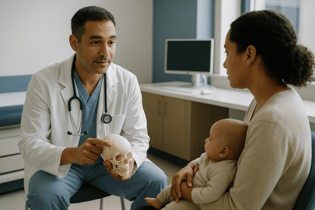
It also helps to say what “major” does not mean here. The brain itself is not operated on. The surgeon works on bone, and the membrane beneath the bone, called the dura, is protected throughout. The operation is planned, not emergency, which means the team has weeks to assemble imaging, blood products and a tailored anesthesia plan. And it is one of the most practiced procedures in pediatric craniofacial surgery, performed by teams that typically include a neurosurgeon and a plastic or craniofacial surgeon working together (Johns Hopkins Medicine).
Why operate at all? The clearest reason is the shape of the skull and face, which becomes harder to correct as bone thickens with age. The second is pressure. A fused suture limits the room the brain has to grow, and in some children, more often when several sutures are involved, this can raise pressure inside the skull. Mayo Clinic notes that, untreated, this can affect development, vision and head shape, which is why surgeons prefer to intervene during infancy (Mayo Clinic).
The risks are real and worth naming in neutral terms: bleeding that may require transfusion, infection, reaction to anesthesia, leakage of the fluid that surrounds the brain, and, uncommonly, the need for a second operation if growth does not proceed as hoped (MedlinePlus). Every one of those is something the pre-operative tests are designed to anticipate.
What are the steps involved in craniosynostosis surgery?
Two broad approaches exist, and the steps differ enough that it is worth walking through each.
In an endoscopic strip craniectomy, the surgeon makes one or two small incisions in the scalp and slides in an endoscope, a thin tube with a camera and light. Through it, a narrow strip of bone along the fused suture is removed, reopening the seam. The incisions are closed, and the operation often takes about an hour. Because the skull is not reshaped on the table, the baby usually wears a custom molding helmet afterward so that natural brain growth does the reshaping over the following months (Mayo Clinic).
In open cranial vault remodeling, the surgeon makes a longer, wavy incision across the top of the scalp, usually hidden within the hairline, and lifts the scalp forward or back. Sections of skull bone are carefully removed, reshaped and repositioned, then fixed in place with small plates and screws that dissolve over time. The scalp is closed over a drain in some cases. This approach takes several hours (MedlinePlus).
Before either version begins, the anesthesia team places an intravenous line, secures the airway with a breathing tube, and often inserts an arterial line, a thin catheter in an artery that gives second-by-second blood pressure readings. A urinary catheter may be placed for longer operations. These steps happen after your baby is asleep and are removed as the child recovers.
Which approach your child is offered depends heavily on age at diagnosis, which suture is involved and whether a syndrome is present. That question deserves its own section.
Who is usually offered surgery, and who is usually asked to wait
Age drives the decision more than almost anything else. Mayo Clinic describes endoscopic repair as generally suited to babies up to about 6 months of age, when the skull bones are thin and soft enough to remove through small incisions and pliable enough to respond to a helmet. Open remodeling is more often used for babies older than 6 months, whose thicker bone needs to be reshaped directly (Mayo Clinic). A child diagnosed late will not typically be offered the endoscopic route.

Timing matters in the other direction too. Very young infants have small blood volumes, and their bodies tolerate blood loss less well. Surgeons therefore balance the advantages of early repair against the physiological reserve of the baby in front of them, and some children are asked to wait a few weeks to grow.
Children whose craniosynostosis is part of a genetic syndrome, such as Apert, Crouzon or Pfeiffer syndrome, are usually managed by a broader craniofacial team. Their care may involve staged operations over years rather than a single repair, and genetic testing is often part of the workup (NHS).
Some babies are not offered surgery at all, at least initially. When a suture fusion is very mild, when the head shape is stable and there is no sign of raised pressure, the team may recommend monitoring rather than operating. This is a legitimate clinical choice, not a delay, and it should come with a clear plan for follow-up measurements and a threshold for revisiting the decision.
A word of caution on shape alone: a flattened head from lying in one position, called positional plagiocephaly, is common and does not involve a fused suture. It is managed differently, usually without surgery (CDC). Distinguishing the two is one reason imaging is requested.
Why a CT scan before craniosynostosis surgery, and what about radiation?
A CT scan uses a rotating X-ray beam and a computer to build cross-sectional pictures of the skull, which software then assembles into a three-dimensional model. For craniosynostosis it does what no examination can: it shows whether a suture is truly fused, how much of its length is involved, and whether a second suture is affected. Mayo Clinic lists CT, sometimes with 3D reconstruction, as the imaging most often used to confirm the diagnosis (Mayo Clinic).
The 3D model is also the surgeon’s map. In open remodeling, the pattern of cuts and the final position of each bone segment are planned against it. In endoscopic repair, it shows precisely where the strip of bone should be removed. Some teams use the reconstruction to print a physical model of the skull to rehearse the operation.
Radiation is the question every parent raises, and it is a fair one. CT delivers more radiation than a plain X-ray, and children are more sensitive to its effects than adults. Pediatric imaging departments respond with low-dose protocols that reduce the exposure substantially while still showing bone detail, since bone is easier to image at low dose than soft tissue. Ask whether a low-dose or “craniosynostosis protocol” scan is being used; most pediatric centers have one.
Sedation is sometimes needed because the scan requires stillness, though modern scanners are fast enough that many babies are simply swaddled, fed and scanned while sleeping. In selected cases, particularly for very young infants with a single clear suture, ultrasound of the suture or examination alone is judged sufficient (NHS). That decision rests with the surgical team, who weigh the value of the information against the exposure.
Skull X-rays are still used occasionally as a first look, but they rarely replace CT when surgery is being planned.
Blood typing and cross-matching: why the lab needs a sample first
A “type and screen” is a laboratory test that identifies a person’s blood group, the A, B, AB or O system, along with Rh status, and checks for antibodies that could react against donor blood. A “cross-match” goes a step further and tests specific donor units against the patient’s sample to confirm compatibility. Both are ordered before craniosynostosis surgery because the skull and scalp have a generous blood supply and blood loss during the operation, particularly the open form, can be significant relative to an infant’s small total volume (MedlinePlus).
The practical meaning is simple. If transfusion becomes necessary during or after surgery, matched blood is already in the room rather than being requested from a blood bank under time pressure. Many open repairs do involve transfusion; endoscopic repairs need it far less often, one of the main reasons that approach was developed (Mayo Clinic).
Parents sometimes ask about donating their own blood for their child, a practice called directed donation. Policies vary by hospital and by blood bank, and a parent’s blood is not automatically the best match; ask your team early if this matters to you, because the process takes time.
Alongside the type and screen, the pre-operative blood draw usually includes a complete blood count, which measures red cells, white cells and platelets, and sometimes clotting studies. A low red-cell count before surgery, common in infants at certain ages, may prompt the team to adjust timing or to discuss supplementing iron under the pediatrician’s direction. That is a decision for the prescribing clinician, not a step to take on your own.
Expect the draw to be a brief, uncomfortable moment for your baby and a hard one for you. Numbing cream, sucrose on a pacifier, swaddling and feeding immediately afterward are all standard comfort measures; ask for them.
The anesthesia review: what the anesthesiologist wants to know
General anesthesia is a medically induced state of unconsciousness in which the child feels nothing and does not remember the operation. Delivering it safely to an infant is a specialty in its own right, and the pre-anesthesia review, whether by phone, video or in a clinic, is where that specialist gathers what they need.
Expect questions about your baby’s birth history, including prematurity and any time spent in a neonatal unit; breathing problems such as noisy breathing, apnea or previous bronchiolitis; heart murmurs or known cardiac conditions; and any earlier anesthesia, including how the child recovered. Family history matters too. A relative who had an unexplained severe reaction to anesthesia, or a family history of certain muscle disorders, changes the drugs the anesthesiologist chooses.
Recent colds are a recurring concern. A child with an active upper respiratory infection has a more irritable airway, which raises the chance of breathing complications under anesthesia. Many teams will postpone if a baby is coughing, wheezing or feverish in the days before surgery. Tell them; a rescheduled operation is a minor inconvenience compared with an avoidable airway problem.
You will receive fasting instructions with specific cutoff times for solid food, formula, breast milk and clear fluids. The times differ by type of feed and are set by the anesthesia team, not the surgeon, so follow the anesthesia clinic’s sheet exactly and call if you are unsure. A full stomach at induction can lead to inhalation of stomach contents, which is the reason fasting exists.
Finally, ask what the plan is for pain control after surgery, whether a parent can be present at induction, and how the team monitors temperature and blood loss in a small body during a long operation. An infant loses heat quickly on an operating table, so warming devices and fluid warmers are routine.
Endoscopic vs open craniosynostosis surgery: a preparation-focused comparison
The table below summarizes how the two approaches differ in the ways that affect preparation and the first days afterward. It is a summary of typical patterns described by mainstream sources, not a prediction for any individual child (Mayo Clinic; MedlinePlus).
| Feature | Endoscopic strip craniectomy | Open cranial vault remodeling |
|---|---|---|
| Typical age window | Up to about 6 months | Usually older than 6 months |
| Incisions | One or two small scalp incisions | Longer incision across the scalp within the hairline |
| What is done to the bone | Strip along the fused suture removed | Bone segments removed, reshaped, repositioned and fixed |
| Operating time | Often around an hour | Several hours |
| Transfusion need | Less common | More common; matched blood always prepared |
| Typical hospital stay | Often about one night | Often several days |
| Helmet afterward | Usually required for months | Usually not required |
| Reshaping achieved | Gradually, through growth in the helmet | Immediately, on the operating table |
Two honest caveats. First, the endoscopic approach depends on months of helmet wear and regular orthotist visits, so families weighing the shorter operation against the longer commitment afterward should discuss both halves. Second, neither approach is “better” in the abstract; the right one is the one that fits your child’s age, suture and anatomy, and it is the surgeon’s call.
The team may also mention other named operations, such as a spring-assisted repair or a fronto-orbital advancement for sutures near the forehead. These are variations on the same principles, and the preparation described here applies to them as well.
The pre-op checklist: feeding, illness and the night before
Once the three big tests are done, the rest of preparation is domestic, and that is where families often feel most useful. A few practical patterns come up repeatedly in pediatric surgical guidance and in what parents say afterward.
Keep your baby well. In the last two weeks, that means being cautious about crowded indoor spaces and about visitors with colds. A respiratory infection is the single most common reason an infant’s operation is postponed, and there is no test that can undo a cough the morning of surgery.
Follow the fasting sheet exactly. Write the cutoff times on the refrigerator, set alarms, and plan the last feed so that your baby is as comfortable as possible. Ask whether a clear fluid is permitted close to the time; many anesthesia teams allow this, but the specifics are theirs to set.
Bring what soothes. A familiar blanket, pacifier or small toy can travel into the pre-operative area, and often into the recovery room. Ask whether one parent can hold the baby while the anesthesia begins; many hospitals allow it.
Plan the household. Open repairs often mean several nights in hospital, and someone will want to stay. Sort out siblings, pets, work and food before the day, because the day itself will not leave room.
Prepare yourself for the first sight afterward. Facial and eyelid swelling after skull surgery is expected and can be dramatic for a few days, sometimes closing the eyes temporarily (MedlinePlus). Parents who know this in advance describe the moment as hard but manageable; those who do not describe it as frightening. The swelling is not a complication, and it settles.
Write down your questions. The morning of surgery is not the time to remember them.
What should I expect after craniosynostosis surgery? The first week
Recovery begins in a recovery room or intensive care unit, depending on the approach and the hospital’s routine. Your baby will have an intravenous line and monitoring leads, and possibly a soft head dressing and a small drain. Nurses check the wound, the fontanelle and the child’s alertness frequently in the first hours.
Swelling is the dominant feature of the first few days. It typically peaks a day or two after the operation and then subsides over the following week, with eyelid puffiness sometimes severe enough that the eyes close for a time (MedlinePlus). Gentle head elevation, which the nursing team manages, helps. The bruising that follows can track down toward the eyes and cheeks and is not a sign of harm.
Feeding usually restarts within hours once the child is awake and the anesthesia team is satisfied. Many babies are fussy and off their normal feeding rhythm for a few days; most return to their usual pattern before discharge.
Length of stay differs by approach. Mayo Clinic describes endoscopic repair as usually requiring about one night in hospital, while open remodeling more often means several days (Mayo Clinic). Discharge depends on the child eating, having pain controlled with the medicines the team prescribes, a stable wound and no fever, not on a fixed number of days.
Before you leave, you should receive written instructions on wound care, bathing, sleep positioning and which signs mean you should call. If a helmet is planned, the fitting appointment will usually be booked for after the swelling has settled.
Expect your baby to look different, and then, over weeks, to look like your baby again with a new head shape. That transition is slower than the operation and, for many families, more emotional.
How painful is craniosynostosis surgery recovery?
The truthful answer is that infants cannot tell us, so pain is judged by behavior, and the picture that emerges from clinical experience is one of discomfort that is real but manageable and that fades over days rather than weeks.
Scalp incisions are less painful than many parents expect, because the scalp heals quickly and the skull bone itself has few pain fibers. What babies seem to mind most is the tight, swollen feeling across the face and eyes, the strangeness of being handled while sore, and, in the first day or two, the after-effects of anesthesia. Fussiness, disrupted sleep and reluctance to feed are the usual signs, and they generally ease as swelling recedes.
Pain is treated with medicines chosen and dosed by the anesthesia and surgical teams, typically starting with stronger options in hospital and stepping down to simpler ones for home. This article will not describe doses or schedules; those belong on your discharge sheet and in a conversation with the prescribing clinician. Ask before discharge exactly how to recognize pain in your child, how to know if the plan is working, and whom to call if it is not.
Comfort measures matter as much as medicine. Skin-to-skin contact, feeding, a quiet dim room, and holding the baby upright against your chest rather than lying flat all help, and nurses will show you positions that protect the incision.
Children who have had endoscopic repair typically have a shorter, milder recovery than those who have had open remodeling, in line with the smaller incision and shorter operation (Mayo Clinic). Either way, most parents report that by the end of the first week their child is behaving largely like themselves, with a healing scar and a face that has stopped looking swollen.
The following weeks: helmets, follow-up and how the skull shape changes
After open remodeling, follow-up is mostly about the wound and the shape. The incision is usually checked within a couple of weeks, dissolvable sutures need no removal, and the scar softens and hides in the hair as it grows. The head shape you see at discharge is close to the final shape, refined slightly by growth.
After endoscopic repair, the work has only begun. The custom molding helmet is fitted once swelling settles and is worn most of the day and night for several months, with regular visits to an orthotist, a specialist who makes and adjusts the helmet, to accommodate growth (Mayo Clinic). The helmet does not squeeze the skull; it leaves room where growth is wanted and gently limits it where it is not, so the brain’s own expansion does the reshaping. Skin checks for pressure marks and daily cleaning become part of the routine.
Both groups return for surgical follow-up over the following months and years. Head circumference is measured against growth charts, the shape is photographed, and the team watches for signs that the sutures are restricting growth again or that pressure is rising. Repeat surgery is uncommon, but it is a known possibility, particularly in children with several fused sutures or a syndrome (NHS).
Developmental follow-up is often built in as well. Most children with a single fused suture develop normally, and monitoring is a precaution rather than a prediction. Children with syndromic craniosynostosis may have hearing, vision, breathing and dental needs that are followed by a multidisciplinary team (Johns Hopkins Medicine).
What surprises many families is how ordinary life becomes again. Within weeks, the medical routine shrinks to appointments and, for helmet wearers, a daily cleaning ritual. The photographs from the first days start to feel like they belong to a different child.
What people often get wrong about craniosynostosis surgery preparation
“The surgeon operates on the brain.” The operation is on the skull bone and the scalp. The dura, the tough membrane covering the brain, is protected throughout, and the brain itself is not cut or handled. The purpose is to give the brain room, not to alter it.
“A flat spot means my baby needs this surgery.” Most flat spots are positional plagiocephaly, from time spent lying in one position, and involve no fused suture. They are managed with repositioning and sometimes a helmet, not surgery. Only a fused suture, confirmed on examination and usually imaging, is craniosynostosis (CDC).
“The CT scan is optional and just for pictures.” The 3D reconstruction is the surgical plan. It tells the surgeon which suture, how much of it, and whether others are involved. Some very young infants can be assessed without it, but when it is requested, it is doing real work.
“Blood typing means my baby will definitely need a transfusion.” It means the team is prepared. Many endoscopic repairs need no transfusion at all; open repairs more often do. Having matched blood ready is a safety step, not a forecast.
“Something I did during pregnancy caused this.” In most cases the cause is unknown. Research points to a mix of genetic and environmental factors, and a minority of cases are linked to specific gene changes or syndromes. It is not caused by how you carried, delivered or positioned your baby (CDC).
“Earlier is always better, so I should push for surgery now.” Earlier repair opens the endoscopic option, but very young infants have less physiological reserve. Timing is a balance the team strikes for each child, and a recommendation to wait a few weeks is not neglect.
“Once it is fixed, it is finished.” Follow-up continues for years, and a small number of children need further surgery. Monitoring is part of the treatment, not an afterthought.
Questions to ask your care team
Good consultations are conversations, and you are allowed to steer them. Bring these written down, and add your own.
- Which suture or sutures are fused, and how sure are you? Is there any sign of raised pressure inside the skull?
- Which operation are you recommending for my child, and why this one at this age rather than the other approach or waiting?
- Will a CT scan be needed? If so, is a low-dose pediatric protocol used, and will my baby need sedation for it?
- How likely is a transfusion with the approach you are planning, and what is your policy on directed donation from a parent?
- Who will give the anesthesia, and can I speak with them before the day? What should I tell you about colds, breathing or family history?
- How long do you expect the operation to take, and how will I be updated while I wait?
- What will my baby look like afterward, and how long does the swelling usually last in your experience?
- How will pain be assessed and managed in hospital and at home, and who do I call if I think the plan is not working?
- If a helmet is part of the plan, for how many months, who fits and adjusts it, and how often will we visit?
- What is the follow-up schedule over the first two years, and what would make you consider a second operation?
- Does my child need genetic testing or evaluation by other specialists?
- Which signs at home mean I should call immediately, and which number do I use out of hours?
You may not get every answer in one appointment, and some answers will be honest uncertainties. A team that says “we do not know yet, but here is how we will find out” is giving you good information.
When to call your doctor
Most recoveries are uneventful, and the discharge sheet you receive will be tailored to your child. Even so, certain signs should prompt a call to the surgical team or a visit to emergency care without waiting for the next appointment. Mainstream guidance on skull surgery in infants points to the following red flags (MedlinePlus; NHS).
- Fever, particularly in the first two weeks after surgery, or a baby who feels hot and unusually unwell.
- Redness spreading from the incision, warmth, a foul smell, pus, or an incision that is opening.
- Clear or watery fluid leaking from the wound or from the nose, which could indicate a leak of the fluid that surrounds the brain.
- A soft spot or incision area that bulges or feels tense when the baby is calm and upright.
- Repeated vomiting, especially forceful vomiting or vomiting without feeding.
- Unusual sleepiness, difficulty waking, a limp or floppy baby, or a high-pitched or inconsolable cry that is not relieved by comfort or the prescribed pain plan.
- Any seizure, jerking movements, or episodes of staring and unresponsiveness.
- Rapid or labored breathing, pauses in breathing, or bluish lips.
- A sudden change in head shape, or rapid new swelling after the initial swelling had settled.
- For helmet wearers, a skin sore or blister under the helmet that does not resolve when the helmet is adjusted.
Trust your instinct. Parents are usually the first to notice that something is off, and surgical teams would far rather take a call about a symptom that turns out to be nothing than miss one that matters. Keep the after-hours number somewhere you can find it half-asleep, and use it.
Frequently asked questions
Is craniosynostosis a major surgery?
Yes. It is performed under general anesthesia on the skull of an infant, and the open version can last several hours and may require a blood transfusion. The brain itself is not operated on; surgeons work on bone and scalp while the membrane over the brain is protected. Because it is planned rather than emergency surgery, the team has weeks to arrange imaging, blood products and a tailored anesthesia plan.
What are the steps involved in craniosynostosis surgery?
After anesthesia and line placement, an endoscopic repair removes a narrow strip of bone along the fused suture through one or two small incisions, and a helmet shapes the head afterward. An open repair uses a longer scalp incision, removes and reshapes sections of skull, and fixes them with dissolvable plates. Both end with wound closure and monitored recovery.
Why does my baby need a CT scan for craniosynostosis?
The scan confirms which suture has fused, how much of it is involved and whether more than one is affected, then produces a 3D model the surgeon uses to plan the operation. Pediatric centers typically use low-dose protocols to reduce radiation. In some very young infants with a single clear suture, examination or ultrasound is judged sufficient, a decision made by the surgical team.
Will my baby need a blood transfusion during craniosynostosis surgery?
Not necessarily, but the team prepares as if it might. Blood typing and cross-matching are done before surgery so matched units are available immediately. Open cranial vault remodeling involves more bone work and more blood loss relative to an infant’s small volume, so transfusion is more common; endoscopic repair needs it far less often. Ask your surgeon about their expectations for your child’s specific operation.
How painful is craniosynostosis surgery recovery?
Infants cannot describe pain, but behavior suggests real discomfort that is manageable and fades over days. Scalp incisions are less painful than expected; the tight swelling across the face seems to bother babies most. Pain is treated with medicines chosen by the surgical and anesthesia teams, and fussiness and disrupted feeding usually ease within the first week as swelling settles.
What should I expect after craniosynostosis surgery?
Expect marked facial and eyelid swelling that peaks in the first day or two and may temporarily close the eyes, along with bruising, fussiness and disrupted feeding for a few days. Endoscopic repairs often mean about one night in hospital; open repairs usually several days. You should leave with written wound-care instructions, a pain plan and clear red-flag signs to watch for.
What are the main craniosynostosis surgery risks?
The recognized risks are bleeding that may require transfusion, infection of the wound or bone, reactions to anesthesia, leakage of the fluid surrounding the brain, and, uncommonly, the need for a second operation if skull growth does not proceed as hoped. The pre-operative CT scan, blood typing and anesthesia review exist specifically to anticipate and reduce these risks. Your team can explain how they apply to your child.
Endoscopic vs open craniosynostosis surgery: which is better for my baby?
Neither is better in the abstract. Endoscopic repair suits babies up to about 6 months, uses small incisions and a short operation, but requires months of helmet wear. Open remodeling is used for older infants, reshapes the skull immediately and needs no helmet, but involves a longer operation, more blood loss and a longer stay. The right choice depends on your child’s age, suture and anatomy.
Why do surgeons want to operate so early in infancy?
Infant skull bone is thin and soft, which makes it easier to remove or reshape and allows the endoscopic approach. The brain also grows fastest in the first year, so reopening a fused suture early gives it room and lets natural growth help shape the head. Waiting too long makes bone thicker and reshaping harder, while operating too early is balanced against a very young infant’s smaller reserves.
Can a cold delay my baby's craniosynostosis surgery?
Yes, and it is the most common reason infant operations are postponed. An active respiratory infection makes the airway more irritable under anesthesia, raising the chance of breathing complications. Tell the anesthesia team about any cough, wheeze, runny nose or fever in the days before surgery. A short delay is a minor inconvenience compared with an avoidable airway problem during the operation.
References
This article is for general information only and is not a substitute for professional medical advice. Please consult a qualified doctor about your individual situation.

More from the Blog

Which Specialist Evaluates Neurodegenerative Diseases, and When Is a Referral Made?
A neurologist usually evaluates suspected neurodegenerative disease, often one who focuses on memory, movement or nerve-and-muscle disorders; geriatricians and old-age psychiatrists share this work…

Carotid Doppler, CT Angiography or MRI: Which Scans Map Cerebrovascular Disease?
Carotid Doppler (duplex ultrasound) is usually the first scan for suspected carotid narrowing because it uses no radiation or contrast and measures blood flow…

When Does Numbness or Tingling in the Feet Call for a Peripheral Neuropathy Evaluation?
Numbness or tingling in the feet usually warrants a peripheral neuropathy evaluation when it lasts more than a few weeks, spreads upward, affects both…

Is It Expected or Urgent? Blood Pressure Swings and Fainting in Autonomic Neuropathy Care
In autonomic neuropathy, brief lightheadedness on standing that settles within seconds is a known feature and belongs in your next routine review. Call your…

Genetic Testing for Huntington’s Disease: What the HTT Result Means for Planning Care
Genetic testing for Huntington's disease counts the CAG repeats in the HTT gene from a blood sample. A result of 40 or more repeats…

Preparing for Autonomic Disorder Testing: Caffeine, Medicines and the Symptom Diary
Preparing for autonomic testing usually means skipping caffeine, alcohol and nicotine for the window your laboratory specifies, wearing loose clothing without skin lotions, bringing…