Craniosynostosis
Learn about craniosynostosis in babies: what it is, common symptoms and types, how doctors confirm it with imaging, surgical treatment options and outlook.

Quick answer
Craniosynostosis is a birth condition in which one or more of the joints (sutures) between a baby's skull bones fuse too early. This restricts skull growth, often causing an unusual head shape and sometimes pressure on the growing brain. It is diagnosed by physical examination and imaging, and is usually treated with surgery in infancy.
What is craniosynostosis?
Craniosynostosis is a condition in which one or more of the flexible joints between the bones of a baby’s skull close too early. These joints are called sutures. In a healthy newborn, the sutures stay open and slightly flexible so the skull can expand as the brain grows rapidly during the first years of life. They normally fuse gradually over childhood and into early adulthood. When a suture fuses before birth or in early infancy, the skull cannot grow in the direction that suture would normally allow. The brain keeps growing, so the skull compensates by expanding in other directions. This often produces an unusual head shape and, in some cases, may put pressure on the developing brain.
Craniosynostosis in babies is a congenital condition, meaning it is present at birth, although the change in head shape may not be obvious until a few weeks or months later. It affects boys slightly more often than girls, and most children who have it are otherwise healthy. In a smaller number of children, it occurs as part of a wider genetic syndrome that affects other parts of the body, such as the face, hands, or feet.
Craniosynostosis is usually managed by a team that includes a pediatric neurosurgeon (a surgeon who operates on the brain, skull, and nervous system in children) and a craniofacial or plastic surgeon (a surgeon who specializes in the bones and soft tissues of the head and face). At Acibadem, this condition is assessed within the Neurosurgery department in cooperation with pediatric and craniofacial specialists.
Craniosynostosis symptoms
The most noticeable sign of craniosynostosis is a head shape that looks different from what parents or doctors expect, and that does not improve as the baby grows. Other signs depend on which suture is affected and whether the condition is putting pressure on the brain.
- An unusual or asymmetrical head shape, such as a long and narrow head, a wide and short head, or a head that appears flattened or pointed on one side or at the front.
- A hard, raised ridge along the line of the fused suture that can be felt through the scalp.
- A soft spot (fontanelle) that feels absent, very small, or unusually firm, or one that closes earlier than expected.
- Slow or no growth of the head relative to the rest of the body over several months.
- Uneven facial features, such as one eye socket or ear sitting higher or further forward than the other, or a forehead that looks flat on one side and bulging on the other.
- Signs of raised pressure inside the skull, which are less common and may include persistent irritability, poor feeding, vomiting, unusual sleepiness, a high-pitched cry, or a bulging soft spot.
- Developmental delays in some children, particularly when more than one suture is involved or when a syndrome is present.
Symptoms differ by type. When only one suture is fused, the main concern is often the head shape itself, and many babies feed, sleep, and develop normally. When several sutures are fused, the skull has less room to compensate, and the risk of increased pressure on the brain is higher. Because the head shape can change slowly, the condition is sometimes first recognized during a routine well-baby visit rather than by parents at home.
It is important to know that not every unusual head shape is craniosynostosis. A far more common cause of a flattened head is positional plagiocephaly, which happens when a baby spends a lot of time lying in the same position. In positional flattening the sutures remain open, the head shape often improves with time and repositioning, and no surgery is needed. A doctor can help tell the difference.
Causes and risk factors
In most cases, doctors cannot identify a specific cause. This is called nonsyndromic craniosynostosis, and it usually affects a single suture. Researchers believe it results from a combination of genetic and environmental factors that influence how the skull bones develop before birth, but the exact mechanism is often unknown.
In a smaller group of children, craniosynostosis is part of a genetic syndrome. Examples include Apert syndrome, Crouzon syndrome, Pfeiffer syndrome, and Saethre-Chotzen syndrome. These conditions are caused by changes in specific genes that guide bone growth, and they often affect other structures such as the face, hands, and feet. Syndromic cases are more likely to involve several sutures.
Factors that have been associated with a higher chance of craniosynostosis include:
- A family history of craniosynostosis or a related genetic syndrome.
- Certain maternal health conditions during pregnancy, such as thyroid disease, which some studies have linked to a higher risk.
- Exposure to certain medications during pregnancy, such as some fertility drugs and some antiseizure medications, which have been studied as possible contributing factors.
- Multiple pregnancy (twins or more) and limited space in the womb, which may restrict head growth.
- Male sex, since the condition is somewhat more common in boys.
- Metabolic conditions in the baby, such as rickets or an overactive thyroid, which can occasionally lead to early suture fusion.
Having one or more risk factors does not mean a baby will develop the condition, and many affected babies have no identifiable risk factors at all.
Diagnosis
Diagnosis usually begins with a physical examination. A pediatrician or specialist will look carefully at the shape of the head from several angles, feel along the sutures for ridges, check the size and firmness of the soft spots, and measure the head circumference. They will also ask about the pregnancy, birth, family history, and how the baby’s head shape has changed over time.
If the examination suggests craniosynostosis, imaging is used to confirm it and to plan any treatment. Common tests include:
- Skull X-rays, which can sometimes show a fused suture, although they are less detailed than other options.
- Computed tomography (CT) scan, an imaging test that uses X-rays to build detailed cross-sectional pictures of the skull. A CT scan with three-dimensional reconstruction is generally considered the most reliable way to confirm which sutures have fused and to show the overall shape of the skull. Doctors use low-dose techniques where possible to limit radiation exposure in infants.
- Ultrasound of the sutures, which is sometimes used in young infants because it does not involve radiation, although it does not show as much detail.
- Magnetic resonance imaging (MRI), which uses magnets and radio waves rather than radiation and may be recommended if there are concerns about the brain itself.
When a genetic syndrome is suspected, the care team may recommend genetic testing and a consultation with a clinical geneticist (a doctor who specializes in inherited conditions). An eye examination may also be arranged, since raised pressure inside the skull can affect the nerve at the back of the eye. Other specialists, such as an ear, nose, and throat doctor or a developmental pediatrician, may be involved depending on the findings.
Craniosynostosis treatment options
Treatment depends on the type of craniosynostosis, the number of sutures involved, the age of the child, and whether there are signs of pressure on the brain. The goals are to allow the brain enough room to grow, to relieve or prevent raised pressure inside the skull, and to improve the shape of the head and face.
Observation. In very mild cases where a single suture is involved, the head shape is only slightly affected, and there are no signs of raised pressure, a doctor may recommend careful monitoring rather than immediate surgery. This involves regular check-ups to measure head growth and watch for any changes. Positional flattening that is mistaken for craniosynostosis is managed with repositioning and, in some cases, a molding helmet, but helmets alone do not treat a fused suture.
Medication. There is no medication that can reopen a fused suture. Medicines may be used to manage related problems, for example pain relief after surgery, or treatment of an underlying metabolic condition if one is identified.
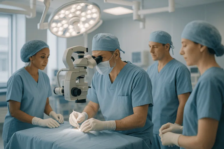
Craniosynostosis surgery. Surgery is the main treatment for most children with confirmed craniosynostosis. The two broad approaches are:
- Endoscopic (minimally invasive) surgery. This is usually offered to younger infants, often within the first few months of life, before the skull bones become thick and rigid. The surgeon makes one or two small incisions in the scalp and uses a thin tube with a camera, called an endoscope, to remove the fused suture. Because the skull is not reshaped during the operation, the child usually wears a custom-made molding helmet for several months afterward to guide the skull into a more typical shape as the brain grows. This approach generally involves a shorter operation, less blood loss, and a shorter hospital stay, but it depends on the child being young enough and on consistent helmet use.
- Open cranial vault remodeling. This is typically performed in older infants, often between about six and twelve months of age, or in children with more complex or syndromic craniosynostosis. The surgeon makes a longer incision across the scalp, removes the affected sections of skull, reshapes them, and secures them in a new position, often using small plates and screws that dissolve over time. This operation allows direct correction of the head shape but is more extensive and usually requires a longer recovery.
Both approaches are performed under general anesthesia by a team that typically includes a pediatric neurosurgeon and a craniofacial surgeon. A blood transfusion is sometimes needed, particularly with open surgery. Your child’s surgical team will explain which approach they recommend and why, along with the specific risks, which can include bleeding, infection, problems with wound healing, and, less commonly, the need for a second operation if the head shape does not develop as expected or a suture refuses again.
Rehabilitation and follow-up. After surgery, children are followed for several years to monitor head growth, skull shape, and development. Children with syndromic craniosynostosis may need additional operations as they grow, for example to address the position of the mid-face or jaw, and may benefit from input from speech therapists, hearing specialists, eye doctors, dentists, and developmental teams. Physical or occupational therapy may be recommended if there are motor delays.
Living with craniosynostosis and outlook
For most children with nonsyndromic craniosynostosis affecting a single suture, the long-term outlook after appropriate treatment is generally good. Many go on to develop normally, attend regular school, and take part in everyday activities without restriction. Head shape often improves considerably after surgery, although a small degree of asymmetry can remain and is not usually a medical concern.
The outlook is more variable when several sutures are involved or when craniosynostosis is part of a genetic syndrome. These children may face additional challenges, including breathing or feeding difficulties related to the shape of the face, hearing or vision problems, dental issues, and learning or developmental differences. Ongoing, coordinated care from a multidisciplinary team helps identify and address these issues early. Outcomes vary from child to child, and your care team can give you a clearer picture based on your child’s specific situation.
Parents often find the period around diagnosis and surgery stressful. It can help to keep a record of your child’s head measurements and milestones, to prepare a list of questions before appointments, and to ask the care team to explain imaging results in plain terms. If a molding helmet is prescribed after endoscopic surgery, following the recommended wearing schedule is an important part of treatment. Support groups for families of children with craniofacial conditions can also be a useful source of practical advice and reassurance.
Frequently asked questions
Can craniosynostosis in babies correct itself without surgery?
A suture that has truly fused will not reopen on its own, and the head shape caused by craniosynostosis generally does not improve without treatment. However, positional flattening, which is much more common and does not involve fused sutures, often improves with repositioning and time. This is why an accurate diagnosis is essential before any decisions are made. In some very mild cases of confirmed craniosynostosis, a doctor may recommend monitoring rather than immediate surgery.
What are the main types of craniosynostosis?
Types of craniosynostosis are usually named after the suture involved. Sagittal synostosis, the most common type, affects the suture running from front to back along the top of the head and produces a long, narrow skull. Metopic synostosis affects the suture in the middle of the forehead and can cause a triangular forehead. Coronal synostosis affects the sutures running from ear to ear across the top of the head and may involve one side or both. Lambdoid synostosis, the rarest, affects the suture at the back of the head. Doctors also distinguish between nonsyndromic cases, which occur alone, and syndromic cases, which are part of a wider genetic condition.
At what age is craniosynostosis surgery usually performed?
Timing depends on the type of surgery. Endoscopic procedures are generally performed in the first few months of life, while the skull bones are still thin and the brain is growing quickly enough to help reshape the head with a helmet. Open remodeling surgery is more often performed later in the first year. Your child’s surgical team will recommend timing based on the sutures involved, the child’s age at diagnosis, and overall health.
Does craniosynostosis affect brain development?
Many children with single-suture craniosynostosis develop normally, especially when the condition is recognized and treated in infancy. Some studies have suggested a slightly higher chance of mild learning or behavioral differences in this group, but findings vary and most children do well. The risk of developmental problems is higher when several sutures are fused, when raised pressure inside the skull goes untreated, or when a genetic syndrome is present. Regular developmental follow-up is recommended for all affected children.
What are the early craniosynostosis symptoms parents should watch for?
The earliest sign is usually a head shape that appears unusual and does not improve over the first weeks or months. Parents may also notice a firm ridge along a suture line, a soft spot that seems very small or has already closed, or a face that looks uneven. Signs such as persistent vomiting, extreme irritability, poor feeding, or unusual sleepiness are less common but more urgent and should be assessed promptly.
Is craniosynostosis hereditary?
Most cases of nonsyndromic craniosynostosis are not clearly inherited, although they may occasionally run in families. Syndromic forms are often caused by identifiable gene changes and can be passed from parent to child, although they can also occur for the first time in a family. If a syndrome is suspected or confirmed, a genetic counselor can explain the pattern of inheritance and what it may mean for future pregnancies.
Will my child need more than one operation?
Children with single-suture craniosynostosis usually need only one operation, although a small number require a second procedure if the head shape does not develop as hoped. Children with syndromic or multi-suture craniosynostosis are more likely to need additional surgeries over the years, for example to address the mid-face, jaw, or airway as they grow. Long-term follow-up allows the team to identify any need for further treatment early.
When to see a doctor
If you notice that your baby’s head shape looks unusual, is becoming more asymmetrical over time, or is not growing as expected, arrange an appointment with your pediatrician or family doctor. Head shape is routinely checked at well-baby visits, so mention any concerns at your next scheduled visit if there are no other symptoms. Early assessment allows more treatment options, including less invasive approaches.
Seek urgent medical attention if your baby shows any of the following signs, which may indicate raised pressure inside the skull or another serious problem:
- Repeated or forceful vomiting, especially without diarrhea or other signs of a stomach illness.
- A bulging or very tense soft spot when the baby is calm and upright.
- Unusual sleepiness, difficulty waking, or a marked drop in alertness.
- Persistent, inconsolable crying or a high-pitched cry.
- Poor feeding or refusal to feed over more than a few hours.
- Eyes that appear to look downward, do not move together, or seem to bulge.
- Seizures, or episodes of stiffening, jerking, or unresponsiveness.
- Breathing difficulties, particularly in children with syndromic craniosynostosis.
- After surgery: fever, increasing swelling, redness or discharge at the wound, or any of the signs listed above.
These symptoms can have many causes, and most babies who have them do not have craniosynostosis. However, they should always be evaluated promptly by a medical professional.
Medically reviewed by the Acıbadem International Medical Board September 8, 2026
See our medical review board →
Update history
- PublishedSeptember 8, 2026
- Medical review approvedSeptember 8, 2026
- Last content updateSeptember 8, 2026