

Bone Density: The Silent Number Behind Fracture Risk
Key Takeaways
- Peak bone mass arrives around age 30; after that, the realistic goal shifts from building bone to keeping it.
- A T-score of -2.5 or below defines osteoporosis, yet most fragility fractures occur in people with milder osteopenia: the number is a guide, not a verdict.
- Women can lose up to 20% of their bone density in the 5–7 years after menopause, the steepest drop of adult life.
- A DXA scan takes about 15 minutes and uses roughly one-tenth the radiation of a chest X-ray.
- Swimming and cycling barely load the skeleton, bone responds to weight-bearing impact and resistance training, ideally two or more days a week.
- About 1 in 4 adults 65 and older falls each year, so fixing rugs, lighting, and footwear lowers fracture risk faster than any change in bone density can.
Bone density measures the mineral packed into your bones, usually reported as a T-score from a DXA scan. A T-score of -1.0 or higher is considered normal; -2.5 or lower indicates osteoporosis. Lower density raises fracture risk, but weight-bearing exercise, adequate calcium and vitamin D, and fall prevention can slow bone loss and, in some cases, modestly rebuild strength.
It often starts with a tape measure, not a broken bone. At an annual checkup, someone in their sixties learns they’re an inch shorter than the height printed on their driver’s license. No pain, no injury, no warning, just a quiet subtraction that may signal the spine has been slowly compressing for years.
Bone loss works like that. There are no symptoms while it happens, no ache to send you to a clinic. The first sign is frequently the last one you’d want: a wrist that snaps catching a fall, or a hip that gives way on an icy step. Roughly half of women and a quarter of men over 50 will break a bone because of weakened bone, according to the National Institutes of Health.
The good news is that this story has a number attached to it, one you can learn, track, and influence. Here’s what that number actually means, and what the evidence says you can do about it.
What Is Bone Density, Really?
Picture the inside of a healthy bone and you won’t see solid ivory. You’ll see a honeycomb: a lattice of mineral struts wrapped in a dense outer shell. Bone density describes how much calcium and other minerals are packed into that structure, measured in grams per square centimeter of bone.
Bone is living tissue, and it never stops rebuilding itself. Specialized cells called osteoclasts dissolve old, worn bone while osteoblasts lay down fresh mineral in its place. Through childhood and your twenties, the builders outpace the demolition crew, and bones grow denser. Somewhere around age 30, most people hit their peak bone massthe strongest their skeleton will ever be.
After that, the balance tips. Removal starts to edge out replacement, and density drifts downward, slowly for most adults and much faster for women in the years around menopause, when falling estrogen accelerates breakdown. When the honeycomb’s walls thin and its holes widen, the bone can look normal from the outside while losing much of its ability to absorb force.
That’s why bone density is called a silent number. Unlike blood pressure, you can’t feel it rise or fall. Unlike cholesterol, it won’t show up on a routine blood panel. It has to be measured directly, and for millions of adults, it never is until something breaks.
What Does a DXA Scan Actually Measure?
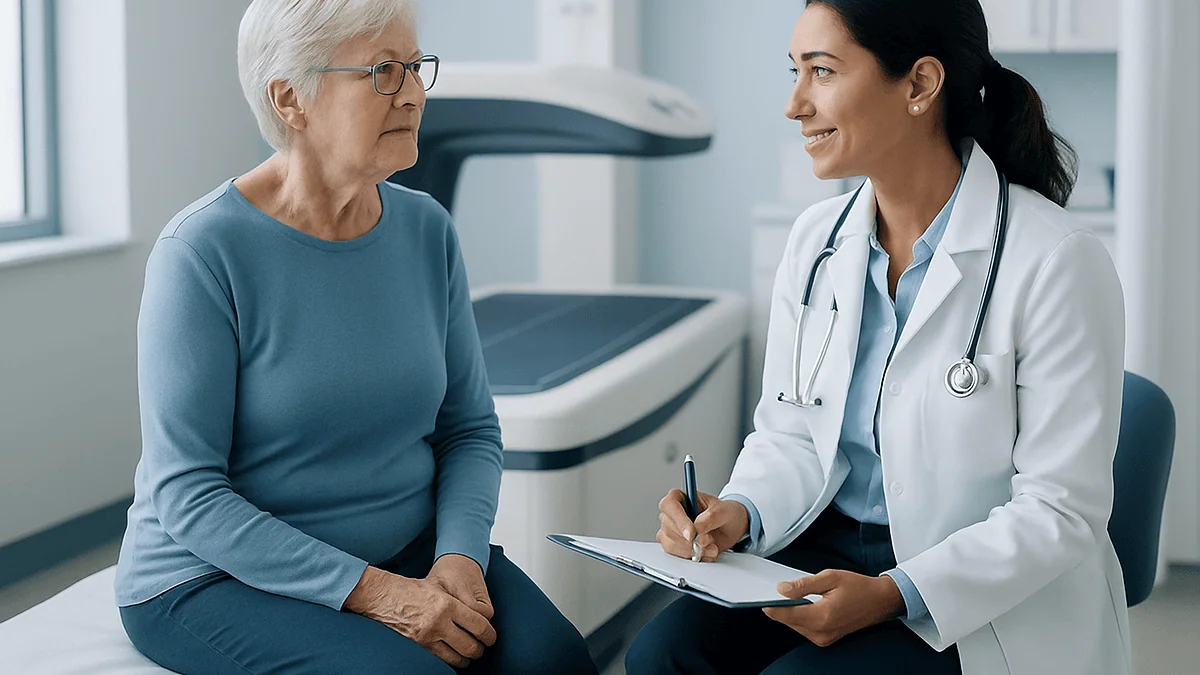
The standard test is a DXA scan (dual-energy X-ray absorptiometry), sometimes written DEXA. You lie fully clothed on a padded table while a scanning arm passes over your hip and lower spine: the two sites where fractures matter most. The whole appointment takes about 15 minutes, nothing touches you, and the radiation exposure is roughly one-tenth that of a chest X-ray, per MedlinePlus.
The result arrives as a T-score: a comparison between your bone density and that of a healthy young adult at peak bone mass. Each whole point below zero represents one standard deviation of loss.
| T-score | What it means |
|---|---|
| -1.0 and above | Normal bone density |
| Between -1.0 and -2.5 | Low bone mass (osteopenia) |
| -2.5 and below | Osteoporosis |
| -2.5 and below, plus a fragility fracture | Severe (established) osteoporosis |
Your report may also include a Z-score, which compares you to people of your own age and sex rather than to young adults. Clinicians lean on the Z-score for children, premenopausal women, and men under 50, because in those groups an unusually low reading can point toward an underlying medical cause rather than ordinary aging.
One practical tip: if you repeat the test years later, try to use the same machine or facility. Different scanners can produce slightly different readings, and consistency makes the trend, the part that matters, easier to trust.
What Is Normal Bone Density by Age?
Here’s the honest, slightly surprising answer: there is no separate “normal” chart for each birthday. The T-score always compares you to a healthy 30-year-old, no matter your age. A 75-year-old and a 45-year-old are graded against the same yardstick, because fracture risk tracks with absolute bone strength, not with how you rank among your peers.
What changes with age is what’s typical, and typical is not the same as ideal. The general arc looks like this:
- Up to about 30: density climbs to its lifetime peak. Roughly 90% of peak bone mass is banked by the late teens.
- 30s to mid-40s: a long plateau, with losses so gradual they’re hard to detect.
- Menopause and the 5–7 years after: women can lose up to 20% of their bone density as estrogen declines: the steepest drop of adult life.
- After 65–70: both sexes lose roughly 0.5–1% per year, and men’s losses begin to catch up.
So when people search “normal bone density by age,” what they usually want to know is whether their loss is on schedule. That’s what the Z-score answers. A Z-score near zero means you’re aging like your peers; a Z-score of -2.0 or lower suggests something beyond ordinary aging, a nutrient absorption problem, a hormonal condition, certain long-term medicines, and deserves a closer look with your clinician.
What Happens If Bone Density Is Low?
For a long stretch, nothing you’d notice, and that’s precisely the problem. Low bone density produces no pain, no stiffness, no fatigue. The skeleton simply becomes less able to withstand everyday forces, and the margin for error shrinks year by year.
The consequences show up as fragility fractures: breaks caused by forces that healthy bone would shrug off, like a fall from standing height or, in severe cases, a hard cough or an awkward twist. Three sites bear the brunt:
- The wrist, often the first break, because we instinctively throw out a hand to catch a fall.
- The spine, where vertebrae can compress silently. Many vertebral fractures are never diagnosed: they announce themselves only as gradual height loss or a rounding upper back.
- The hip, the most serious. Hip fractures almost always require surgery, and many people never fully regain their previous mobility and independence.
There’s a meaningful distinction between the two “low” categories. Osteopenia (T-score between -1.0 and -2.5) means bone mass is below normal but not yet at the osteoporosis threshold: a caution flag, not a diagnosis of disease. Osteoporosis (-2.5 or below) means the honeycomb has thinned enough that fracture risk is substantially elevated. Neither label is a prediction; both are information. And information, in this case, arrives early enough to act on, which is exactly what the fracture doesn’t offer.
Why the Number Isn't the Whole Story
Here’s a statistic that reframes everything: most fragility fractures happen in people who do not have osteoporosis on paper. Their T-scores sit in the osteopenia range, worse than normal, better than -2.5. How? Simple arithmetic. Far more people have moderately low density than severely low density, so the moderate group produces more total fractures even though each individual’s risk is lower.
Density also isn’t the only property that makes bone strong. Researchers talk about bone qualitythe architecture of the internal lattice, the rate of turnover, the accumulation of microscopic damage, none of which a DXA scan captures. Two people with identical T-scores can carry different real-world risks.
That’s why clinicians increasingly pair the T-score with a broader risk calculation, often using a tool called FRAX, which estimates your 10-year probability of a major fracture. It folds in age, sex, body weight, smoking status, alcohol use, parental hip fracture, prior fractures, and certain medical conditions alongside the density number.
And then there’s the variable no scan measures at all: whether you fall. A fragile bone that never meets the floor doesn’t break. A moderately weak bone that meets the floor twice a year eventually will. In my read of the evidence, this is the most under-discussed truth in bone health: the T-score tells you how breakable the bone is, but your balance, vision, and living environment decide how often it gets tested.
Who Should Get a Bone Density Test, and When?
Screening guidelines in the United States are refreshingly concrete. A DXA scan is generally recommended for:
- All women 65 and older, regardless of risk factors.
- Postmenopausal women under 65 who carry elevated risk, low body weight, a parent who broke a hip, smoking, heavy alcohol use, or conditions such as rheumatoid arthritis or celiac disease.
- Anyone over 50 who breaks a bone from a minor fall. A fragility fracture is itself a red flag that warrants testing, yet studies consistently find that most people who suffer one never get scanned afterward.
- Men, typically starting around 70, or earlier with risk factors. Recommendations for men vary among expert groups, so it’s worth a direct conversation with your clinician.
A few situations justify earlier testing at any age: unexplained height loss of more than 1.5 inches, long-term use of medicines known to thin bone (ask your prescriber whether any of yours do), early menopause or surgical removal of the ovaries, and hormonal or digestive conditions that interfere with calcium absorption.
How often to repeat the scan depends on the first result. Someone with robust density may not need another test for a decade or more; someone near the osteoporosis threshold might be rechecked in two years. Because bone changes slowly, testing more often than every two years rarely reveals anything a scanner can reliably distinguish from measurement noise.
Can You Improve Bone Density?
Yes, within honest limits, and the limits matter. This is a place where wellness marketing tends to overpromise, so let’s stick to what trials actually show.
For adults past peak bone mass, exercise and nutrition changes typically produce gains of about 1–3% in bone density over a year or two at the sites being loaded. That sounds modest, and it is. But the comparison isn’t gain versus zero; it’s gain versus the 0.5–1% you’d otherwise lose annually. Swinging the trajectory from minus one to plus one is a meaningful shift, compounded over a decade.
More encouraging still: fracture risk improves more than the density number suggests. Exercise strengthens the muscles that protect bone, sharpens the balance that prevents falls, and appears to improve bone quality in ways DXA can’t see. In studies of older adults, structured exercise programs reduce falls by roughly a quarter, and a prevented fall is a prevented fracture, whatever your T-score says.
For people already diagnosed with osteoporosis, prescription treatments exist that can slow bone breakdown or stimulate new bone formation, and they meaningfully reduce fracture risk in clinical trials. Which option fits, or whether one is needed at all, is a decision for you and your clinician, weighing your full fracture risk, not the T-score alone.
The realistic headline: you probably can’t rebuild the skeleton of your 30-year-old self. You can absolutely change how fast you’re losing it, and how likely it is to break.
Which Exercises Actually Build Bone?
Bone follows a blunt engineering rule: it strengthens where it’s stressed. Cells embedded in the bone matrix sense mechanical strain and signal the building crew to reinforce that spot. No strain, no signal. That single mechanism explains almost everything about which exercise works.
Two categories do the heavy lifting, per Harvard Health and the NHS:
- Weight-bearing impact activityanything done on your feet against gravity with a bit of jolt. Brisk walking, jogging, stair climbing, hiking, dancing, pickleball, tennis. Higher impact generally means a stronger bone signal, scaled to what your joints and heart can safely handle.
- Resistance traininglifting weights, using resistance bands or machines, or bodyweight moves like squats and push-ups, ideally two or more days a week. Muscles pulling hard on their bony anchors is one of the most direct strains bone ever feels.
Notice what’s missing. Swimming and cycling are superb for the heart but nearly weightless for the skeleton, buoyancy and the saddle absorb the load bone needs. Enjoy them, just don’t count them as bone work.
Add a third element as the years advance: balance training. Tai chi, yoga, or simple heel-to-toe walking drills won’t add density, but they demonstrably cut fall risk in older adults, arguably the fastest way to lower fracture risk in the short term.
Progression matters more than perfection. Bone adapts to novelty, so a walk that’s grown easy is a walk your skeleton has stopped noticing. Add hills, add a weighted backpack, add a faster interval, gradually, and with a clinician’s blessing if you already have low density.
Calcium and Vitamin D: What the Evidence Actually Shows
Think of calcium as the brick and vitamin D as the forklift. Bone stores about 99% of the body’s calcium, and without enough vitamin D, the gut absorbs only a fraction of the calcium you eat. Short either one for long enough and the body solves the problem ruthlessly, by withdrawing calcium from your skeleton to keep blood levels steady.
The strongest evidence supports a food-first approach. Reliable calcium sources include:
- Milk, yogurt, and cheese, still the most concentrated everyday sources.
- Canned salmon and sardines eaten with their soft bones.
- Kale, bok choy, and broccoli. (Spinach is calcium-rich on paper, but its oxalates block most absorption: a detail worth knowing.)
- Calcium-set tofu and fortified cereals, juices, and plant milks.
Vitamin D is harder to eat. Fatty fish, egg yolks, and fortified milk contribute, and skin makes it from sunlight, less efficiently with age, darker skin tones, northern winters, and indoor lives. The NIH Office of Dietary Supplements publishes recommended intakes by age for both nutrients, and they’re worth checking against your actual diet.
What about supplements? The evidence is more modest than the marketing. Trials show supplements help people who are genuinely deficient or can’t meet needs through food, but in well-nourished healthy adults, calcium and vitamin D pills alone have not convincingly prevented fractures. They’re a gap-filler, not a strategy, and more is not better, since very high calcium intakes carry their own risks. If you’re unsure where you stand, a clinician can assess your intake and, if needed, check your vitamin D level.
What Drink Is Good for Bone Density?
The unglamorous truth: the best drink for your bones is probably already in your refrigerator. A glass of milk delivers calcium, protein, and (in the U.S.) added vitamin D in one package, and it remains the most-studied bone-supporting beverage there is. Lactose-intolerant? Lactose-free milk carries identical nutrition.
Fortified plant milks, soy, almond, oat, can match dairy’s calcium when fortified, with soy also contributing comparable protein. One practical note: the added calcium settles at the bottom of the carton, so shake it well or a good share of the benefit stays behind. Calcium-fortified orange juice is another reasonable option.
A few drinks deserve honest footnotes rather than fear:
- Alcohol: heavy, sustained drinking clearly harms bone: it suppresses bone-building cells and raises fall risk. Moderation is the operative word.
- Cola: some observational studies have linked heavy cola intake with lower bone density in women, possibly through phosphoric acid or simply because soda displaces milk. The evidence is associative, not proof, but a daily multi-can habit isn’t doing your skeleton favors.
- Coffee and tea: largely exonerated. Moderate caffeine has, at most, a small effect on calcium balance, easily offset by adequate intake. Splash milk in your coffee and you may come out ahead.
- Bone broth: despite the name, analyses find it contains surprisingly little calcium. Enjoy it as soup, not as therapy.
No beverage is a treatment. But swapping one daily soda for fortified milk is the kind of small, boring trade that actually shows up in nutrition math.
The Bone-Heart Connection Most People Miss
Bones and the cardiovascular system seem like distant neighbors, but they age on strikingly parallel tracks, and researchers keep finding the paths crossing. Observational studies have repeatedly noted that people with lower bone density tend to show more calcium deposits in their arteries, a marker of cardiovascular risk. It’s a curious inversion: mineral leaving the place it belongs and appearing where it doesn’t.
To be clear about what the evidence shows: these are associations, not proof that one condition causes the other. Scientists suspect shared biology, chronic inflammation, hormonal shifts, and overlapping cell-signaling pathways that regulate mineral placement, but the mechanisms are still being mapped, and low bone density is not currently used to diagnose or predict heart disease.
What’s beyond dispute is that the two systems share a risk ledger. Smoking damages blood vessels and starves bone-building cells. Physical inactivity weakens the heart and silences the strain signals bone needs. Heavy alcohol use, poor protein intake, and diets thin on fruits and vegetables burden both.
Which leads to a genuinely useful piece of efficiency: the American Heart Association’s target of 150 minutes of moderate activity per week, plus two days of muscle-strengthening work, is almost a word-for-word match for bone-health exercise guidance. Choose weight-bearing versions, brisk walking over swimming, for instance, and every session pays two accounts at once. For readers of this section of the magazine, that may be the most practical takeaway of all: you don’t need a separate program for your skeleton. You need to point the one you have at the ground.
Fracture Prevention Is Also Fall Prevention
A fragility fracture usually requires two ingredients: a vulnerable bone and a moment of impact. Medicine and media spend most of their attention on the first ingredient. The second is often easier to change, and faster.
The CDC reports that about one in four adults 65 and older falls each year, and falls cause the vast majority of hip fractures. Yet fall risk responds to intervention within weeks, not the years bone remodeling requires. The highest-yield moves:
- Audit the home. Loose rugs, trailing cords, dim stairways, and clutter cause a remarkable share of falls. Grab bars in the bathroom and a nightlight on the path to it are inexpensive insurance.
- Get vision and hearing checked yearly. Both systems feed your balance more than most people realize.
- Review your medicines with a clinician or pharmacist. Some prescriptions, alone or in combination, cause dizziness or drowsiness that quietly raises fall risk. Never stop anything on your own; do ask the question.
- Train balance deliberately. Tai chi programs have reduced falls in older adults by significant margins in controlled studies.
- Wear real shoes. Backless slippers and smooth soles are overrepresented in fall statistics.
Two habits round out the list. Quitting smoking benefits bone measurably, smokers reach lower peak mass and lose density faster. And maintaining adequate protein and overall nutrition preserves the muscle that both cushions falls and, often, prevents them entirely.
When to See a Doctor About Your Bones
Because bone loss is silent, the signals that warrant a medical conversation are mostly indirect, and easy to dismiss as ordinary aging. Don’t dismiss these:
- You’ve broken a bone from a minor fall or bump after age 50. This is the single strongest signal on the list. Any fragility fracture should trigger a bone density evaluation, and research shows it usually doesn’t.
- You’ve lost more than 1.5 inches of height compared with your young-adult measurement, or you notice your posture rounding forward. Silent spinal compression fractures are a common cause.
- New, persistent back pain without an obvious injury, particularly in the mid or lower back.
- You went through menopause before 45, or had your ovaries removed, and have never been screened.
- A parent broke a hip. Family history independently raises your risk.
- You take long-term medicines for inflammation, seizures, acid reflux, or hormone-sensitive conditions, some can thin bone over time. Ask your prescriber whether yours carry that risk.
- You have a condition that affects absorption or hormones, such as celiac disease, inflammatory bowel disease, rheumatoid arthritis, or thyroid disorders.
And even without any of the above: if you’re a woman 65 or older, or a man around 70, and have never had a DXA scan, that alone is reason enough to raise it at your next visit. The test is quick, painless, low-radiation, and typically covered by insurance for screening at these ages. Few 15-minute appointments deliver as much usable information.
Bone Density Myths Worth Retiring
Bone health attracts folklore the way any silent condition does: the gap between what’s happening and what’s felt gets filled with stories. A few that deserve a respectful burial:
“Osteoporosis is a women’s disease.” Women are at higher risk, but roughly one in four men over 50 will suffer a fracture related to bone loss, and men who break a hip tend to fare worse afterward than women do. Men are also far less likely to be screened or treated: a gap the data doesn’t justify.
“Milk actually causes bone loss.” This claim circulates persistently online, usually citing observational studies with conflicting findings. The weight of evidence, reflected in guidance from the NIH and NHS, supports dairy as a legitimate calcium and protein source. If you avoid it for other reasons, fine, fortified alternatives work, but the fear itself isn’t supported.
“It’s too late to do anything after 70.” Wrong on two counts. Exercise trials in adults in their seventies and eighties show measurable gains in strength, balance, and fall reduction. And fall prevention, the fastest lever, works at any age.
“A supplement routine means my bones are covered.” Pills fill nutrient gaps; they don’t replace mechanical loading. A skeleton that’s never stressed has no reason to stay strong, no matter how well it’s supplied.
“I feel fine, so my bones must be fine.” The entire premise of this article, in five words: bone loss doesn’t feel like anything. That’s why the number exists, and why it’s worth knowing yours before a fracture announces it for you.
Frequently asked questions
What is normal bone density by age?
There is no separate normal chart for each age: a DXA T-score always compares you to a healthy young adult, and -1.0 or above counts as normal at any age. What changes with age is typical loss: density peaks around 30, declines slowly through midlife, drops fastest in the 5–7 years after menopause, and falls roughly 0.5–1% yearly in later decades. The Z-score compares you to your own age group.
Can you improve bone density?
Yes, modestly. In studies, weight-bearing exercise and resistance training in adults produce gains of roughly 1–3% at the bones being loaded, and, just as valuably, they halt the yearly loss that would otherwise occur. Adequate calcium, vitamin D, and protein support the process. For diagnosed osteoporosis, prescription treatments can slow breakdown or stimulate new bone; whether one fits your situation is a conversation for your clinician.
What drink is good for bone density?
Milk remains the best-studied option, combining calcium, protein, and added vitamin D. Lactose-free milk is nutritionally identical, and fortified soy or other plant milks can match dairy’s calcium, shake the carton, since added calcium settles. Calcium-fortified orange juice also helps. Moderate coffee and tea are fine. Heavy alcohol harms bone, and despite its name, bone broth contains very little calcium.
What happens if bone density is low?
Usually nothing you can feel, low bone density is symptomless, which is exactly why it’s dangerous. Weakened bone breaks under forces healthy bone would tolerate, most often at the wrist, spine, and hip. Spinal fractures can occur silently, showing up only as height loss or a rounding back. A low result on a DXA scan is a prompt to act, through exercise, nutrition, fall prevention, and a treatment discussion, before a fracture happens.
Is osteopenia the same as osteoporosis?
No. Osteopenia means bone density is below normal (T-score between -1.0 and -2.5) but hasn’t reached the osteoporosis threshold of -2.5 or lower. It’s a caution flag rather than a disease diagnosis. That said, most fragility fractures actually occur in people with osteopenia simply because so many more people fall into that category, so it’s worth taking seriously as a moment to change trajectory.
How often should a bone density test be repeated?
It depends on your first result and overall risk. Someone with strong density may reasonably wait many years, sometimes a decade or more, before retesting, while someone near the osteoporosis threshold might be rechecked in about two years. Testing more often than every two years rarely helps, because bone changes too slowly for scanners to reliably distinguish real change from measurement variation. Your clinician will set the interval.
Does walking increase bone density?
Walking helps preserve bone, but on its own it produces only modest density gains, because bone responds to strains beyond what it’s used to. Brisk walking, hills, stairs, or a weighted backpack strengthen the signal, and adding resistance training two or more days a week does considerably more. Walking’s underrated benefit is indirect: it maintains the leg strength and balance that prevent the falls that cause most fractures.
Do men need bone density tests?
Yes, roughly one in four men over 50 will break a bone due to bone loss, and men who fracture a hip tend to have worse outcomes than women. Screening is generally suggested for men around age 70, or earlier with risk factors such as smoking, heavy alcohol use, low body weight, prior fractures, or long-term use of bone-thinning medicines. Guidelines for men vary, so ask your clinician directly.
Is a DXA bone density scan safe and painful?
It’s painless and among the lowest-radiation imaging tests in medicine. You lie clothed on a padded table for about 15 minutes while a scanner arm passes over your hip and spine without touching you. The radiation dose is roughly one-tenth that of a chest X-ray, less than a day of natural background exposure. Tell the technologist if you’re pregnant or had recent imaging with contrast material, which can affect timing.
Does coffee weaken your bones?
Not meaningfully at moderate intakes. Caffeine causes a small, temporary increase in calcium excretion, but studies show the effect is easily offset by adequate calcium in the diet, adding milk to your coffee more than compensates. Concerns apply mainly to very high caffeine consumption combined with low calcium intake. Heavy cola drinking has shown associations with lower bone density in some studies, but ordinary coffee and tea have been largely exonerated.
References
This article is for general information only and is not a substitute for professional medical advice. Please consult a qualified doctor about your individual situation.

More from the Blog

Before a Peripheral Angiogram: Kidney Tests, Contrast Allergy History and Medicine Review
Before a peripheral angiogram, care teams typically check kidney function with a blood test, ask in detail about any previous reaction to iodinated contrast…
Aortic Diseases and How They Are Treated: Medication, Endovascular Repair or Open Surgery
Aortic disease is usually treated in one of three ways, chosen by size, location, growth and symptoms. Small, stable aneurysms are managed with blood…
Is a Stent Procedure Painful? Local Anesthesia, the Wrist or Groin Puncture and Afterward
A coronary stent procedure is usually uncomfortable rather than painful. The only sharp moment is typically the local anesthetic injection at the wrist or…
Pain Control After Robotic Cardiac Surgery: Breathing Exercises, Wound Care and First Walks
Pain after robotic cardiac surgery is usually felt around the small chest-wall incisions and the drain site rather than in the breastbone, and for…
How a Coronary Stent Is Placed: Balloon, Mesh Tube and the Wrist or Groin Route
A coronary stent is placed during a procedure called percutaneous coronary intervention. A cardiologist threads a thin tube through an artery in the wrist…

What Does Protein Do for Your Body? Beyond Muscle
Protein does far more than build muscle. Your body breaks dietary protein into amino acids, then uses them to make enzymes that run digestion…