

Cartilage Restoration Recovery: Why Weight-Bearing Is Limited and How Rehab Protects Repair

Key Takeaways
- Articular cartilage has no blood supply and no nerves, so it heals poorly on its own and cannot signal pain when a fresh repair is being overloaded.
- Weight-bearing is restricted after cartilage restoration because immature repair tissue is vulnerable to shear, the sideways drag of walking, far more than to gentle compression.
- Motion without load is prescribed from the start because joint fluid, pumped by movement, is the only way nutrients reach the repair.
- Microfracture produces fibrocartilage, a scar-like tissue tougher but less cushioning than the original, which is why grafting and cell-based techniques were developed.
- No vitamin or supplement has been shown to rebuild cartilage; vitamin C is needed for collagen and vitamin D for bone, but correcting a deficiency is different from restoring tissue.
- Repair tissue continues to mature for many months after walking feels normal, and the return to running or sport is often the riskiest point in the whole recovery.
After cartilage restoration surgery, weight-bearing is limited because the new repair tissue is soft and easily sheared for weeks; loading it too early can crush or dislodge it before it matures. Recovery usually involves a protected weight-bearing phase on crutches, early controlled motion to nourish the cartilage, then staged strengthening over months. The surgical team sets the exact timeline based on the procedure, the joint and the size of the defect.
Six days after knee surgery, the stairs in your own house look different. Twelve steps you never counted before. Crutches under both arms, a brace locked straight, and a sheet from the physical therapist that says, in bold, “toe-touch only.” Your leg feels strong enough. The incisions are small. And yet the instruction is to treat this knee as if it were made of wet clay.
That instruction is the heart of cartilage restoration recovery, and it is also the part patients find hardest to believe. Most orthopedic operations reward early movement and early loading. This one asks for movement without load, for weeks, and then a slow climb back that stretches across most of a year.
The reason is biology, not caution for its own sake. What follows explains what is happening inside the joint during those weeks, why the rules are written the way they are, and where the evidence is honest about limits.
What is cartilage restoration, and what does recovery have to protect?
Articular cartilage is the smooth, glassy layer that caps the ends of bones inside a joint. It is only a few millimeters thick, yet it lets the knee, hip or ankle glide with less friction than ice sliding on ice. According to Cleveland Clinic, this tissue has no blood vessels and no nerves, which explains two facts at once: it does not hurt when it first wears, and it cannot repair itself the way skin or bone can.
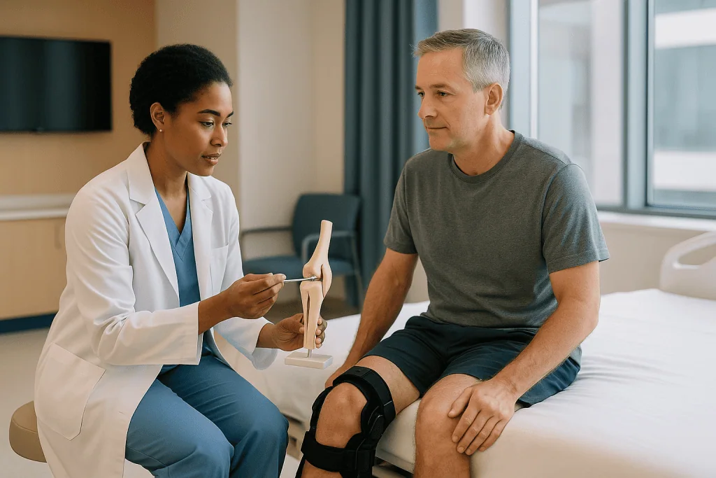
Cartilage restoration is a family of surgical techniques designed to fill a focal defect, a discrete hole or crater in that surface, with tissue that can carry load. It is not the same as joint replacement, which resurfaces the whole compartment with metal and plastic. Restoration targets one damaged patch and leaves the rest of the joint alone.
Recovery, then, has a specific job. Whatever was placed or stimulated in that crater, whether marrow cells, a bone-and-cartilage plug, or cultured cells under a membrane, begins as something far weaker than mature cartilage. Early on it is closer to a firm gel than to the tough, pressurized tissue around it. Mature articular cartilage resists compression because water is trapped inside a dense mesh of collagen and large sugar-protein molecules called proteoglycans. New repair tissue has not yet built that mesh.
Every rule in the rehab plan flows from this gap between what the tissue can tolerate now and what it will eventually tolerate. Protect it while it is fragile, feed it with gentle motion, then load it progressively as it stiffens. The sequence matters more than any single exercise.
What actually happens in cartilage restoration surgery?
Surgeons choose among several techniques depending on the size, depth and location of the defect, as well as the patient’s age and goals. The main approaches differ in where the repair tissue comes from and how much bone is involved.
| Technique | What the surgeon does | What fills the defect | Why loading is restricted |
|---|---|---|---|
| Microfracture | Creates small holes in the bone beneath the defect | A marrow clot that matures into fibrocartilage, a scar-like cartilage | The clot is soft and can be crushed or washed out |
| Osteochondral autograft (OATS) | Moves one or more bone-and-cartilage plugs from a low-load area of the same joint | Living hyaline cartilage on a bone base | Plugs must knit into surrounding bone before bearing full force |
| Osteochondral allograft | Implants a matched donor bone-and-cartilage segment | Donor hyaline cartilage on donor bone | Bone integration takes months; early load risks collapse |
| Autologous chondrocyte implantation (ACI or MACI) | Harvests cells, grows them in a lab, then implants them under a membrane or scaffold in a second operation | Cartilage-like tissue built by the patient’s own cells | Cells are unprotected until they lay down a matrix |
MedlinePlus describes microfracture as a way to prompt the body to grow new cartilage, and it is usually done arthroscopically, through small incisions with a camera. The grafting and cell-based procedures often need a larger opening because a precise fit is essential; a plug that sits even slightly proud of the surface will take abnormal force.
Surgeons frequently correct other problems in the same operation. A leg that is bowed or knock-kneed sends more force through one side of the joint, so a realignment osteotomy, a controlled cut and reshaping of bone, may be added. A loose kneecap track or a torn meniscus might be addressed too. Each add-on changes the recovery rules, which is one reason two people with “cartilage surgery” can have very different instructions.
Why is weight-bearing limited after cartilage restoration?
Picture a freshly poured concrete slab. It looks solid within hours. Drive a truck across it that first day and it cracks, not because the mix was wrong but because curing takes time. Repair cartilage cures far more slowly than concrete, and the truck is your own body weight multiplied by the physics of walking.
Walking on level ground pushes force through the knee that is several times body weight, and stairs or a deep squat push it higher still. That force arrives in two forms. Compression squashes the tissue straight down. Shear drags across it sideways as the joint surfaces roll and slide. Immature repair tissue handles gentle compression reasonably well, since compression actually signals cells to make matrix. Shear is the enemy. It can peel a cell-covered membrane from its bed, fragment a marrow clot, or rock a bone plug loose before it has fused.
There is also the question of where the defect sits. A lesion on the weight-bearing part of the femoral condyle, the rounded end of the thigh bone, is loaded every time the foot hits the ground. A lesion under the kneecap is loaded most when the knee bends under weight, as in stairs or rising from a chair. Surgeons tailor the restrictions to the location: a kneecap repair may allow more walking with the leg straight but forbid loaded bending, whereas a condyle repair does the reverse.
None of this is about pain. Because cartilage has no nerves, the repair cannot warn you when it is being overloaded. A knee that feels fine at week three is not evidence that the tissue is ready. The restriction exists precisely because the usual feedback loop is silent.
How long is non weight bearing after cartilage surgery, and why motion still matters?
Timelines vary with the technique and the joint, and only the operating surgeon can give the number that applies to you. That said, the shape of the schedule is consistent. MedlinePlus notes that after microfracture people typically need crutches for a period measured in weeks, with a return to full activity measured in months rather than weeks. Protocols for grafts and cell-based repairs generally run longer, because bone integration and matrix formation are slower processes than clot formation.
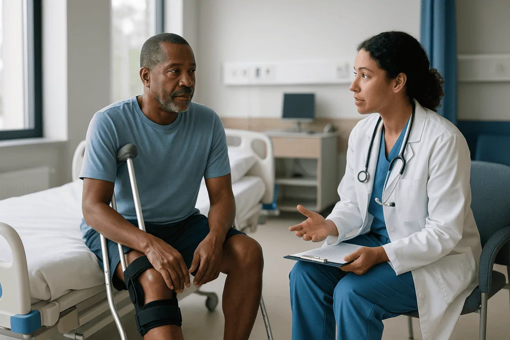
Most plans do not mean “no weight at all” for the full period. Common stages include toe-touch weight-bearing, where the foot rests on the floor for balance only; partial weight-bearing, often a set percentage of body weight that a therapist teaches you to feel using a bathroom scale; and progression to full weight as the tissue matures. A brace locked in extension is often used during walking so that the repair is loaded only in the position the surgeon considers safe.
What surprises many patients is that the same team insisting on crutches also insists on bending the knee from day one. This is not a contradiction. Cartilage has no blood supply, so it draws nutrients from joint fluid the way a sponge draws water: cyclic gentle pressure pumps fluid in and out. Motion without load provides exactly that pumping. Continuous passive motion machines, devices that slowly flex and straighten the leg while you lie still, are sometimes prescribed for several hours a day for this reason, and Cleveland Clinic describes motion as central to cartilage nutrition.
So the formula is unusual but logical. Move the joint through a safe arc many times a day. Do not stand on it. Let the tissue eat without asking it to work.
Who is cartilage restoration usually for, and who is usually asked to wait?
The procedures described here are aimed at focal defects: a distinct hole in otherwise healthy cartilage, typically caused by a twisting injury, a direct blow, or a condition called osteochondritis dissecans, in which a piece of bone and its cartilage loses blood supply and can separate. Mayo Clinic notes that osteochondritis dissecans most often affects children and young adults and that in younger people whose growth plates are still open, the fragment may heal without surgery.
Candidates commonly share several features. The defect is limited in size and surrounded by intact cartilage that can share the load. The joint is stable, or instability such as a torn ligament is being corrected at the same time. Leg alignment is normal or is being corrected. Body weight is within a range that keeps joint forces manageable, and the person is able and willing to follow a long, restrictive rehab. That last point is not a moral test; it is a practical one, since a perfect graft cannot survive being walked on in week two.
People are usually asked to wait, or steered toward other options, in several situations. Widespread thinning across the joint surface, the pattern of established osteoarthritis, is not a focal problem, and filling one crater in a joint that is wearing everywhere rarely helps. Active infection, inflammatory arthritis that is not controlled, and smoking are common reasons to pause. Some surgeons also delay when a person cannot arrange the help needed for weeks on crutches, because the recovery environment is part of the treatment.
Alternatives include structured physical therapy, activity modification, bracing, injections aimed at symptoms rather than repair, and, for advanced wear, partial or total joint replacement. Which path fits is a judgment the treating team makes with the patient after imaging and examination.
What do the first days and weeks of cartilage restoration recovery look like?
The first week is mostly about swelling control, wound care and learning to move safely on crutches. Expect ice or a cooling sleeve, elevation above heart level for stretches of the day, and a set of exercises that look almost too gentle: tightening the thigh muscle while the leg lies flat, sliding the heel toward the buttock within a set range, moving the ankle up and down to keep blood flowing in the calf.
Pain and swelling management are handled with a plan from the surgical team. Anti-inflammatory medicines are sometimes limited after certain cartilage or bone procedures because of concerns about their effect on healing, so it is worth asking rather than assuming. Any decision to start, stop or adjust a medicine belongs to the prescribing clinician.
Days spent largely off your feet raise the risk of a blood clot in the deep leg veins, and the calf pumps, hourly position changes and hydration in your instructions exist for that reason. Some people are given a clot-preventing medicine; that choice depends on individual risk and is made by the team.
Weeks two through six or so are the plateau that tests patience. The knee often feels better than the rules suggest it should. Physical therapy visits focus on restoring full straightening first, since a knee that cannot fully extend walks badly forever, then gradual bending, hip and core strength that do not load the repair, and stationary cycling without resistance once the arc of motion allows a full pedal turn.
Sleep is frequently disrupted by the brace and by finding a comfortable position. Low mood in this phase is common and worth mentioning to the team; it is a normal response to enforced stillness, not a sign that recovery is going wrong.
How does a cartilage repair rehab protocol protect the repair?
A rehab protocol is a staged plan, written by the surgeon and therapist, that matches what you are allowed to do to what the tissue can tolerate at each point. The phases have different names in different clinics, but the logic underneath is shared.
The protection phase covers roughly the period on crutches. Goals are simple: full extension, steadily increasing flexion, a quiet joint without a large effusion, and a thigh muscle that fires on command. The quadriceps often shuts down after knee surgery, a reflex inhibition triggered by swelling, and re-awakening it early prevents a wasting that takes months to reverse.
The transition phase begins as weight-bearing is advanced. Walking is retrained deliberately, because weeks on crutches teach a stiff-legged, hip-hiking gait that overloads other joints. Closed-chain strengthening, exercises with the foot on the ground such as shallow squats and leg presses, is introduced within a limited range chosen to keep the repair out of its highest-contact angles. Balance work starts here too.
The remodeling phase, often the second half of the first year, adds load and speed. Repair tissue continues to stiffen for many months; MedlinePlus frames full recovery from microfracture in terms of months, and grafts and cell-based repairs are generally treated as maturing over a similar or longer span. Running, jumping and cutting are reintroduced one at a time, each earning its place by the knee staying quiet afterward.
The protocol protects the repair in two ways. It caps the load at each stage, and it builds the muscles and movement patterns that will share load once full activity returns. A strong hip and a well-aligned squat are not extras; they are the long-term insurance policy for the patch.
Can knee cartilage regenerate naturally?
The honest answer has two halves. Articular cartilage in an adult does not regrow on its own in any meaningful way once it is lost. Cleveland Clinic and the NIH’s arthritis institute both describe its lack of blood supply and its very limited capacity for self-repair. A shallow scuff may smooth over time; a full-thickness crater does not fill itself with the same material.
The other half is more encouraging. In children and adolescents whose growth plates remain open, Mayo Clinic notes that osteochondritis dissecans lesions can heal with rest and activity restriction, because the underlying bone is still actively remodeling. And in adults, what the body does produce after injury, or after microfracture, is fibrocartilage. This is a tougher, more collagen-heavy tissue than the original hyaline cartilage, with less of the water-holding proteoglycan that gives true cartilage its cushioning. Fibrocartilage can carry load and relieve symptoms, but it wears differently and may not last as long under heavy use.
This distinction is why the restoration techniques exist at all. Grafting moves living hyaline cartilage into the defect rather than waiting for scar cartilage to form. Cell-based methods try to coax the patient’s own cells into building something closer to the native tissue. Neither has been shown to produce a perfect copy, and the evidence base is still evolving; the aim is durable, functional tissue, not a return to the joint you were born with.
What the body does do well, at any age, is adapt the muscles and movement around a joint. Stronger hips and thighs, better alignment and a healthy weight all reduce the force any given patch of cartilage has to absorb. That is not regeneration, but it is the part within your control.
What helps cartilage heal faster after restoration surgery?
People often ask this hoping for a supplement or a device. The evidence points somewhere less glamorous: the factors that most reliably influence healing are the ones that shape the environment the tissue matures in.
Following the load rules is first. Cartilage repair does not speed up when you push it; it fails. The single most important thing a patient controls is not exceeding the prescribed weight-bearing and range at each stage.
Motion is second. Because nutrients reach cartilage through joint fluid pumped by movement, the daily repetitions of passive or gentle active bending are not busywork. Skipping them starves the repair.
Not smoking matters. Nicotine narrows small blood vessels and is associated with poorer bone healing across orthopedic surgery, and the grafting techniques depend on bone knitting to bone. Surgeons commonly ask patients to stop before and after surgery; help with quitting is available through primary care.
Swelling control is underrated. A joint distended with fluid inhibits the thigh muscle, stretches the capsule and raises pressure across the repair. Ice, elevation and pacing are simple tools with a real mechanism.
Adequate protein, overall calorie balance and controlled blood sugar support tissue building in general, and anyone with diabetes will usually be asked to keep it well managed around surgery. Sleep supports the hormonal signals of tissue repair; it also happens to be the first thing pain and a brace disrupt, which is worth raising with the team.
What has not been shown to speed cartilage repair, in mainstream evidence, includes oral collagen products, most joint supplements, and consumer light or magnetic devices. Some may be harmless; none replace the protocol. When a claim is uncertain, the honest position is that the evidence does not currently support it.
What vitamin restores cartilage?
None does. No vitamin, mineral or supplement has been shown in mainstream evidence to rebuild lost articular cartilage in adults, and any product implying otherwise is promising something the biology does not allow. That said, several nutrients are genuinely involved in the tissues around a repair, and being deficient in them is not helpful.
Vitamin C is required for the body to make collagen, the protein scaffold of cartilage, bone, tendon and skin. The NIH Office of Dietary Supplements describes this role and notes that deficiency impairs connective tissue formation. Most people eating fruit and vegetables get enough; more than enough does not build extra cartilage.
Vitamin D supports calcium absorption and bone health, according to the NIH Office of Dietary Supplements. Since grafting procedures rely on bone healing beneath the cartilage, surgeons sometimes check vitamin D levels around surgery. Whether to supplement, and how, is a decision for the clinician who sees your blood test, not a self-prescribed step.
Glucosamine and chondroitin, the two most marketed “cartilage” supplements, have been studied mainly in osteoarthritis rather than in surgical repair. Large trials have shown inconsistent results for pain and no convincing evidence that either rebuilds cartilage. Cleveland Clinic and the NIH both characterize the evidence as mixed at best. Some people report modest symptom relief; that is different from restoration.
Omega-3 fats, turmeric and collagen peptides sit in a similar category: plausible mechanisms in the laboratory, weak or inconsistent human evidence, and no demonstrated effect on the tissue formed after restoration surgery.
The practical takeaway is modest. Eat a varied diet with enough protein, correct any deficiency your team identifies, and tell the surgeon about every supplement you take, because some interact with anesthesia or affect bleeding.
Can you fully recover from cartilage damage?
“Fully” is the word to examine. If it means a joint that is structurally identical to an uninjured one, the current evidence does not support that outcome for adults; the repair tissue differs from native cartilage in composition and behavior, and long-term studies show it can wear over time. If it means a joint that is comfortable, stable and capable of the activities that matter to you, that is a realistic goal for many people, though not a guaranteed one, and the strength of the evidence varies by technique, defect size and patient factors.
Several patterns appear consistently across published reviews without needing a headline percentage. Smaller defects tend to do better than large ones. Younger patients tend to do better than older ones. Repairs in a well-aligned, stable joint with healthy surrounding cartilage tend to outlast repairs in a joint that is already wearing broadly. A single, clearly defined traumatic defect is a more favorable starting point than diffuse damage.
Return to sport is where expectations most often collide with biology. Reviews of cartilage repair describe many athletes returning to their sport, but frequently after a longer timeline than for ligament surgery and sometimes at a modified level. A person whose sport involves repeated pivoting and jumping loads the repair very differently from a cyclist or swimmer, and surgeons weigh this when discussing goals.
It is also possible for a repair to fail: the graft does not integrate, the tissue breaks down, or symptoms persist. When that happens, options include revision procedures or, eventually, joint replacement. This is not a reason to avoid restoration when it is indicated; it is a reason to enter it with clear eyes.
The best predictor within your reach remains adherence to the protocol. The repair you protect is the repair most likely to last.
What people often get wrong about cartilage restoration recovery
The myths here are rarely foolish. Most come from applying the rules of other operations to this one.
“It feels fine, so it must be healed.” Cartilage has no nerves. The absence of pain says nothing about whether the repair tissue is ready for load. Timelines are set by biology, not by comfort.
“Non weight bearing means keep it still.” The opposite. Motion without load is the main way the repair is nourished. Stillness starves it and stiffens the joint.
“Crutches are a sign of weakness, and I can wean off early.” Crutches are a load-limiting device, like a cast. Weaning early because the leg feels strong transfers force onto tissue that has not built the structure to take it.
“More exercise means faster healing.” Within the allowed limits, consistency helps. Beyond them, extra repetitions or heavier resistance risk the repair. The protocol is a ceiling as much as a floor.
“A supplement will regrow the cartilage.” No vitamin or supplement has been shown to do this. Correcting a real deficiency supports healing in general; megadoses do not build cartilage.
“Once I am walking normally, recovery is over.” Walking returns long before the tissue finishes maturing. Repair tissue continues to stiffen for many months, and the highest-risk moment for damage is often the return to running or sport, when the knee feels normal and the guardrails feel unnecessary.
“Cartilage restoration and joint replacement are two versions of the same thing.” They answer different problems. Restoration fills a focal hole in an otherwise healthy joint; replacement resurfaces a joint with widespread wear. Neither is a substitute for the other.
Each of these misunderstandings tends to produce the same behavior: doing more, sooner. The tissue being protected has one vulnerability above all others, and that is exactly it.
Questions to ask your care team before and after cartilage restoration
A good consultation leaves you knowing not only what will be done but what you will be asked to do. These questions tend to surface the details that shape daily life during recovery.
- Which technique are you recommending for my defect, and what made you choose it over the alternatives?
- How large is the defect, where exactly is it, and how does that location change my weight-bearing and bending rules?
- Will anything else be corrected in the same operation, such as alignment, the kneecap track or a meniscus, and how does that change the timeline?
- How many weeks of protected weight-bearing do you expect, and what will tell you it is safe to progress?
- Will I use a brace, a continuous passive motion machine, or both, and for how many hours a day?
- Which medicines, including over-the-counter anti-inflammatories and supplements, should I avoid around surgery, and why?
- What is my personal risk of a blood clot, and what will be done to lower it?
- Who runs my physical therapy, will they use your written protocol, and how often will you and the therapist communicate?
- What signs should make me call you between visits?
- How will you assess the repair over time, and is follow-up imaging planned?
- What activities are realistic goals for me, and when might each be considered?
- If the repair does not take, what are the next options?
- What help will I need at home in the first weeks, and for how long?
Write the answers down or ask permission to record them. Recovery from cartilage restoration is long enough that details fade, and the written protocol will be your reference on the days motivation flags. Any change to the plan, whether progression, medication or activity, should come from the treating team rather than from a well-meaning comparison with someone else’s surgery.
When to call your doctor during cartilage restoration recovery
Most of recovery is uneventful in the medical sense: slow, occasionally frustrating, but predictable. A small number of signs need a same-day call to the surgical team or, in some cases, emergency care.
Contact the team promptly if you notice any of the following:
- A calf that becomes painful, swollen, warm or discolored, especially on one side. The NHS lists these as signs of a deep vein thrombosis, a clot in a deep leg vein, which needs urgent assessment.
- Sudden breathlessness, chest pain that worsens with breathing, or coughing up blood. These can indicate a clot that has traveled to the lungs and are an emergency.
- Fever, or an incision that becomes increasingly red, hot, swollen or leaks cloudy fluid. Infection inside a joint is uncommon but serious, and early treatment matters.
- A rapid, tense swelling of the joint, or a sudden new sensation of catching, locking or giving way. These may signal bleeding into the joint or a problem with the graft.
- Pain that escalates sharply rather than easing, or pain that is out of proportion to the stage of recovery.
- Numbness, tingling or weakness in the foot that is new or spreading, or a foot that looks pale or feels cold.
- A fall, or any moment where you accidentally put full weight through the leg before you were cleared to. Even if it feels fine afterward, the team may want to check.
Slower concerns also deserve a message rather than silence: a knee that is not regaining straightening on schedule, persistent difficulty sleeping, or a mood that has sunk low enough to affect your ability to keep up with exercises. None of these is a failure, and all are easier to manage early.
When in doubt, the team would rather hear from you than not. Their answer, not this article, decides what happens next.
Frequently asked questions
What helps cartilage heal faster after surgery?
Following the weight-bearing and range-of-motion limits exactly, moving the joint gently and often, controlling swelling, not smoking, and eating enough protein are the factors with the clearest mechanisms. Cartilage repair does not speed up under extra load; it fails. No supplement or device has been shown in mainstream evidence to accelerate the maturation of repair tissue after cartilage restoration.
What vitamin restores cartilage?
None. No vitamin has been shown to rebuild lost articular cartilage in adults. Vitamin C is required for collagen production and vitamin D supports bone healing beneath a graft, according to the NIH Office of Dietary Supplements, so a genuine deficiency is worth correcting under medical guidance. Taking more than the body needs does not create new cartilage.
Can knee cartilage regenerate naturally?
In adults, full-thickness cartilage loss does not regrow on its own; the body produces fibrocartilage, a scar-like tissue, at best. Children and adolescents with open growth plates are the exception, and Mayo Clinic notes that some osteochondritis dissecans lesions in young people heal with rest. Muscles and movement patterns around the joint can adapt at any age.
Can you fully recover from cartilage damage?
A comfortable, stable, functional joint is a realistic goal for many people after restoration, but a structurally identical joint is not what current evidence supports. Repair tissue differs from native cartilage and can wear over time. Smaller defects, younger age, good alignment and strict adherence to the rehab protocol are consistently associated with better outcomes.
How long is non weight bearing after cartilage surgery?
It depends on the technique, the joint and the defect location, and only the surgeon can give your number. MedlinePlus describes a crutch period after microfracture measured in weeks and full recovery measured in months. Graft and cell-based repairs are generally protected for a similar or longer period because bone integration and matrix building are slow.
What is a typical microfracture recovery timeline?
Broadly, several weeks of protected weight-bearing with daily controlled motion, a gradual return to walking as the marrow clot matures, then progressive strengthening over the following months. MedlinePlus frames full recovery from microfracture in months. Return to impact sport comes last and is decided by the team based on strength, swelling and symptoms.
Why do I have to bend my knee if I cannot stand on it?
Because cartilage has no blood supply. It absorbs nutrients from joint fluid, and that fluid is pumped in and out by movement. Bending the knee without weight feeds the repair; standing on it crushes or shears the repair. The two instructions target different forces and are meant to be followed together, not traded against each other.
What does a cartilage repair rehab protocol include?
Staged goals matched to tissue maturity: first full straightening, gradual bending and swelling control while on crutches; then retrained walking and limited-range strengthening as weight-bearing increases; finally load, speed and sport-specific movement in the remodeling phase. The protocol caps activity at each stage while building the strength that will protect the repair long term.
Are glucosamine and chondroitin worth taking after cartilage restoration?
The evidence does not show that either rebuilds cartilage, and studies in osteoarthritis have produced mixed results for pain. Some people report modest symptom relief. They have not been shown to improve the tissue formed after restoration surgery. Tell your surgical team about any supplement before taking it, since some interact with other medicines.
Is cartilage restoration the same as knee replacement?
No. Restoration fills a focal defect in an otherwise healthy joint using marrow stimulation, a bone-and-cartilage graft or cultured cells, and preserves the natural surfaces. Replacement resurfaces a joint with widespread wear using metal and plastic. They address different problems, and the treating team determines which, if either, suits a particular joint.
References
This article is for general information only and is not a substitute for professional medical advice. Please consult a qualified doctor about your individual situation.

More from the Blog

ACL Reconstruction Over Forty: When Age Matters Less Than Activity and Knee Stability
Age alone is rarely what decides ACL reconstruction over 40. Surgeons weigh how often the knee gives way, how active you want to be,…
Returning to Work After Arthroscopy: Desk Jobs, Standing Jobs and Manual Work Compared
Most people can return to work after arthroscopy within a few days to a few weeks for seated desk work, several weeks for jobs…
How Tendon Repair Surgery Works: Stitching Torn Ends, Grafts and Protecting the Repair
Tendon repair surgery reconnects the two ends of a torn tendon, usually by stitching them together with a strong core suture and a finer…
The First Days Home After Meniscus Surgery: Crutches, Bracing and How a Companion Can Help
In the first days after meniscus surgery, most people manage at home with crutches, a cold pack, elevation and the pain plan their surgical…

When Does Bursitis Need a Doctor? Rest and Ice First, Then the Signs That Call for a Visit
Most bursitis settles with rest, ice, padding and avoiding the movement that caused it, usually within a few weeks. See a doctor if pain…

When to See a Doctor for a Sprain or Strain: The Signs That Call for a Specialist Assessment
See a doctor for a sprain or strain if you cannot bear weight on the joint, the limb looks misshapen, you felt or heard…