Cartilage Restoration
Cartilage restoration repairs damaged joint cartilage, most often in the knee, using techniques such as microfracture, grafting, or cell-based repair to reduce pain and improve mobility.
Quick answer
Cartilage restoration is a group of surgical techniques that repair or replace focal areas of damaged articular cartilage, most often in the knee. Options include marrow stimulation (microfracture), transferring plugs of the patient's own cartilage and bone, donor tissue transplantation, and cell-based repair. It suits localised defects rather than widespread arthritis, and recovery follows a staged rehabilitation programme lasting several months.
What Is Cartilage Restoration?
Cartilage restoration is a group of surgical techniques used to repair or replace damaged articular cartilage: the smooth, resilient tissue that covers the ends of bones inside a joint and lets them glide against each other with almost no friction. It is designed for focal defects, meaning localised patches of damage, rather than the widespread cartilage loss of advanced arthritis. It is most often performed in the knee, though selected techniques are also used in the ankle, hip, shoulder and other joints when the pattern of injury and the patient’s overall condition suit them.
Articular cartilage is unusual tissue. It has no blood vessels of its own, which is why it heals poorly after injury compared with skin, muscle or bone. Beyond letting bones glide smoothly, it distributes load during walking, running, jumping and everyday movement. When a defect develops, the joint surface becomes rough or uneven. That roughness produces pain, swelling, catching, grinding and inflammation, and in some patients the damaged area enlarges over time, particularly when the joint is exposed to repeated impact, abnormal alignment, meniscus loss or ligament instability. Cartilage restoration exists to interrupt that process before it becomes harder to treat.
What does knee joint cartilage do?
Knee joint cartilage covers the end of the thigh bone, the top of the shin bone and the back of the kneecap, allowing these surfaces to move against each other under considerable force many thousands of times a day. Together with the menisci, the two crescent-shaped shock absorbers between the bones, it spreads load across the joint so that no single point takes excessive pressure. When a patch of this surface is lost or damaged, the load that patch used to carry concentrates on its edges and on the exposed bone beneath. This is why even a relatively small defect in a weight-bearing zone can cause pain out of proportion to its size, and why untreated defects tend to affect the surrounding healthy surface over time.
Does cartilage grow back on its own?
Articular cartilage does not reliably grow back on its own. Because it lacks a blood supply, the usual biological repair response that heals a cut or a broken bone never properly reaches an injured cartilage surface. Very small, superficial areas of damage may partially fill with fibrocartilage: a scar-like tissue that is stiffer and less durable than the original hyaline cartilage, but this repair tissue does not match the mechanical quality of what was lost. Deeper defects, and defects that also involve the underlying bone, have essentially no capacity to restore themselves. This limited healing ability is the central reason cartilage restoration techniques were developed: they either stimulate a repair response the tissue cannot mount alone, or they bring healthy cartilage into the defect from elsewhere.
Can cartilage be restored?
Yes, focal cartilage damage can be repaired or replaced surgically, although the honest answer comes with conditions. Restoration works best when the damage is a defined defect surrounded by healthy tissue, when the joint is stable and well aligned, and when the patient can commit to a structured rehabilitation programme. The approach may involve stimulating the body’s own healing response, transferring healthy cartilage and bone from a less critical area of the same joint, using donor tissue, or implanting cartilage-forming cells into the defect. The technique is tailored to the individual lesion rather than selected from a standard list.
The main categories of cartilage restoration are:
- Microfracture or marrow stimulation: small openings are made in the bone beneath the cartilage defect to release marrow elements, which form a clot that matures into repair tissue.
- Osteochondral autograft transfer: small plugs of the patient’s own cartilage and underlying bone are moved from a low-load area of the joint into the damaged area.
- Osteochondral allograft transplantation: donor cartilage and bone tissue are shaped to replace larger or deeper defects when appropriate.
- Cell-based cartilage repair: cartilage cells are collected, processed in a specialised laboratory, and later implanted into the defect as part of a staged treatment plan.
- Cartilage repair combined with other procedures: meniscus repair or transplantation, ligament reconstruction, or realignment osteotomy may be added when those factors are contributing to cartilage overload.
The purpose is never simply to fill a defect visible on an MRI scan. A well-planned cartilage restoration procedure aims to improve the mechanics of the whole joint so that the repaired area sits in the best possible environment to function over the years that follow.
When Articular Cartilage Damage Starts to Limit Your Life
Articular cartilage damage is frustrating precisely because it tends to affect people who are otherwise active and healthy. It often begins quietly: discomfort after sport, a twisting injury that never quite settles, or swelling that comes and goes. Over months it can become pain on stairs, stiffness after sitting, reduced confidence during exercise, or the growing sense that the joint is no longer dependable.
For many patients, the hardest part is uncertainty. You may have been told that cartilage does not heal well on its own, which, as explained above, is broadly true. You may be worried about developing arthritis, needing joint replacement earlier than expected, or giving up activities that matter to your identity and your health. And you probably have practical questions: which technique fits my particular injury, how long is recovery, what imaging is needed, and how do I know a proposed surgical plan is actually appropriate for me?
This page walks through those questions in order. The most important idea to carry with you is that cartilage restoration is a precise orthopaedic decision, not a single procedure. The right plan depends on the size and depth of the defect, the condition of the underlying bone, limb alignment, ligament stability, meniscus health, your age, your activity goals and your expectations for recovery. Two patients with defects that look similar on a scan can need very different treatment.
Who May Need Cartilage Restoration?
Cartilage restoration is considered for patients who have a localised cartilage injury and ongoing symptoms despite appropriate non-surgical treatment. These patients are often too young or too active for joint replacement, or they have a defect that is limited enough to be treated with a joint-preserving approach instead. The typical candidate wants to keep their natural joint working for as long as possible.
Symptoms vary with the joint involved and the size and position of the defect. In the knee, patients commonly report pain during activity, swelling after exercise, stiffness, a catching or locking sensation, difficulty with stairs, discomfort when squatting, or a reduced ability to run and pivot. Some remember a specific event: a sports trauma, a fall, a twist on a planted foot. Others develop symptoms gradually, especially where there is a history of meniscus surgery, ligament injury, malalignment or repetitive overload.
Diagnosis begins with a detailed orthopaedic evaluation. The physician reviews your symptoms, previous injuries, operations, activity level and treatment history, then examines the joint for swelling, range of motion, tenderness, stability, limb alignment, gait and movement patterns. Prior imaging, operative reports and physiotherapy notes all add useful context to this assessment, so it is worth gathering them before any specialist appointment.
Imaging plays a central role. Weight-bearing X-rays show joint space, limb alignment, early arthritis and bone structure. MRI provides detailed information about the cartilage itself, bone marrow changes beneath it, the menisci, the ligaments and the surrounding soft tissues. In some cases additional imaging is used to study alignment, bone defects or complex joint anatomy. Diagnostic arthroscopy, looking inside the joint with a camera, is occasionally needed when symptoms and imaging do not fully match, but most cartilage restoration plans can be developed with modern imaging and careful clinical review.
Cartilage restoration is not appropriate for every patient with joint pain, and a responsible surgeon will say so plainly. It is generally reserved for damage that is focal rather than widespread. Advanced osteoarthritis, diffuse cartilage loss throughout the joint, severe inflammatory arthritis, uncontrolled infection or major untreated instability all reduce the likelihood of a durable result. In those situations, other treatments, including joint replacement or, in the knee specifically, knee arthroplastymay be safer and more effective. Matching the treatment to the true stage of the disease matters more than any individual technique.
Conditions and Injuries Cartilage Restoration Can Address
Cartilage restoration is most commonly used for focal chondral or osteochondral defects. A chondral defect affects only the cartilage surface; an osteochondral defect involves both the cartilage and the bone beneath it. Restoration may be considered after traumatic injuries, sports-related cartilage damage, osteochondritis dissecans, cartilage defects following kneecap instability, or localised damage associated with previous meniscus or ligament injuries.
Can bone cartilage be restored?
Defects that involve both cartilage and the underlying bone can be treated, but they need techniques that rebuild both layers. Marrow stimulation alone is rarely enough when the bone is damaged, because the repair has no sound foundation. Osteochondral autograft transfer and osteochondral allograft transplantation both move a composite plug of cartilage and bone into the defect, restoring the structural base as well as the gliding surface. Which option fits depends on the size and depth of the bone involvement, the location of the lesion and the tissue available.
Location matters as much as depth. In the knee, defects occur on the femoral condyles, the patella, the trochlea or the tibial plateau, and each site has different mechanical demands. Cartilage injury behind the kneecap is influenced by how the patella tracks in its groove, while a defect on a femoral condyle is affected by limb alignment and meniscus function. A cartilage procedure performed without addressing these contributing factors often fails to deliver the improvement the patient hoped for.
For this reason, cartilage restoration frequently sits inside a broader joint preservation strategy. A patient with a cartilage defect and a torn anterior cruciate ligament may need ligament reconstruction to restore stability. A patient whose bowed or knock-kneed alignment overloads one side of the knee may need an osteotomy to shift pressure away from the damaged area. A patient with meniscus deficiency may need meniscus repair or transplantation to restore load distribution. When several of these problems coexist, the operation becomes a form of complex joint reconstruction, staged or combined according to what the joint can tolerate. These decisions are highly individual.
In selected cases, restoring the cartilage surface can help patients delay or avoid replacement of the joint. Whether that is realistic depends on the biological and mechanical condition of the joint at the time of surgery. The aim is to intervene at the right stage, when the injury is significant enough to justify an operation, but not so advanced that a repair is unlikely to survive the loads placed on it.
How Is Cartilage Restoration Performed?
How do surgeons restore knee cartilage?
Surgeons restore knee cartilage by matching one of the four main techniques, marrow stimulation, autograft transfer, allograft transplantation or cell-based repair, to the specific defect, and by correcting any mechanical problem that would overload the repair. Small, contained defects in patients with stable, well-aligned joints may suit simpler arthroscopic techniques. Larger defects, deeper defects and defects with bone involvement generally need grafting or staged cell-based procedures. There is no universally best method; there is only the method that best fits a particular lesion in a particular joint.
Preparation and treatment planning
Preparation begins with confirming the diagnosis and selecting the technique. The orthopaedic team evaluates the size, depth and location of the defect, the quality of the surrounding cartilage, the condition of the underlying bone, and any associated problems such as meniscus tears, ligament instability or malalignment. This planning stage is where the operation is really decided.
Before surgery, patients typically have blood tests, an anaesthesia assessment, cardiac evaluation where indicated, and a structured review of their current medicines and supplements. Any adjustment to medication before or after the operation is decided by the treating doctor and the anaesthesia team as part of that review: it is planned for each patient individually. Nicotine impairs tissue healing, so surgeons generally advise patients who smoke to stop well before cartilage surgery. Patients with diabetes or other chronic conditions may need their general health optimised first, since the quality of biological healing directly affects how well a cartilage repair matures.
Physical preparation matters too. Strengthening the quadriceps, improving hip and core control, reducing swelling and restoring range of motion before surgery all make the early recovery easier. Many teams recommend a period of preoperative physiotherapy, particularly where stiffness or muscle weakness is present. Where a patient’s imaging and records are reviewed remotely before an in-person examination, the final surgical plan is still only confirmed after the joint has been assessed directly.
During the procedure
Cartilage restoration is performed under regional or general anaesthesia. The operation may be entirely arthroscopic, using small incisions and a camera inside the joint, or it may require a small open incision, depending on the technique and the position of the defect. Many procedures combine both: arthroscopy to inspect the whole joint and treat associated problems, followed by a targeted incision for the precise cartilage work.
In microfracture, the surgeon removes unstable cartilage edges and prepares the defect so it has clean, stable borders. Small holes or channels are then created in the exposed bone. Marrow elements flow into the defect and form a clot that matures into repair tissue over the following months. Microfracture is usually reserved for smaller defects in selected patients, and the early recovery must be handled carefully, because loading the joint too soon can disrupt the healing clot before it has organised.
In osteochondral autograft transfer, one or more cylindrical plugs of healthy cartilage and bone are harvested from a low-load area of the patient’s own joint and press-fitted into the defect. This delivers mature, living cartilage directly into the damaged zone. It is typically used for certain small to medium defects, limited by the amount of donor tissue the joint can spare.
In osteochondral allograft transplantation, a matched donor graft containing both cartilage and underlying bone is shaped to fit the patient’s defect precisely. This option is considered for larger defects, deeper defects, or cases where the bone beneath the cartilage is also damaged. Because donor tissue must be sourced and matched, the timing and availability of this procedure can vary.
Modern cartilage surgery relies on detailed imaging, arthroscopic visualisation, specialised instruments and careful intraoperative measurement. Imaging defines the lesion and any associated joint problems before the operation; arthroscopy allows direct assessment of cartilage quality, meniscus tissue, ligaments and joint surfaces; and patient-specific planning may be used for alignment correction or graft sizing. The technology supports precision, but the surgeon’s decision-making remains the most important element of care.
Operative time varies widely. A small arthroscopic procedure is relatively brief, while grafting, cell implantation, or combined ligament, meniscus or alignment surgery takes considerably longer. Your surgeon will explain the expected plan, whether treatment is staged across two operations, and whether a hospital stay is recommended. Some patients go home the same day; others stay in hospital for monitoring, pain management and early rehabilitation instruction.
What does knee cartilage regeneration involve?
Knee cartilage regeneration usually refers to cell-based cartilage repair, in which the patient’s own cartilage cells are used to grow new repair tissue inside the defect. Treatment happens in stages:
- Stage one: a small sample of healthy cartilage is taken arthroscopically from a low-load area of the joint.
- Stage two: the cells are processed and multiplied in a specialised laboratory environment.
- Stage three: in a later procedure, the prepared cells or cell-containing material are implanted into the cartilage defect, where they mature into repair tissue over many months.
This approach is considered for selected larger defects when the surrounding joint environment is favourable, stable ligaments, acceptable alignment and reasonably healthy neighbouring cartilage. It asks more of the patient in terms of time and staged surgery, and the choice between cell-based repair and grafting is one of the key discussions to have with the surgical team.
Recovery and rehabilitation
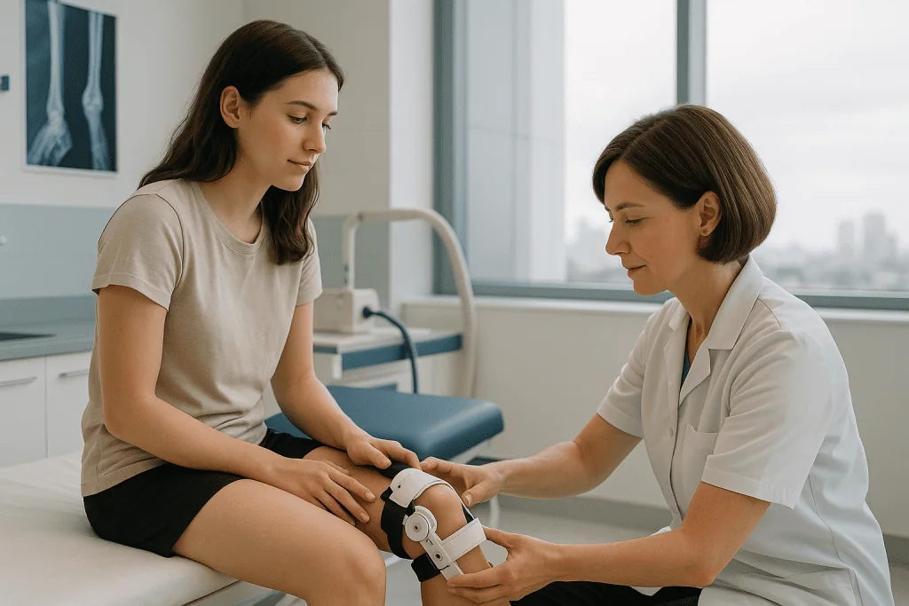
Recovery after cartilage restoration is deliberately gradual. Repair tissue matures slowly, and the treated area must be protected while it develops strength. Most patients use crutches for a period, especially when the defect sits in a weight-bearing zone of the knee, and a brace may be prescribed depending on the procedure and location. Physiotherapy usually begins early, focusing on swelling control, safe range of motion, muscle activation and protection of the repair.
Rehabilitation is highly procedure-specific. A patient who has had microfracture on a weight-bearing surface may face stricter early weight-bearing limits than someone treated in a non-weight-bearing area. A patient recovering from patellar cartilage repair follows a protocol that carefully controls knee bending and stair activity. Combined procedures such as osteotomy or ligament reconstruction change the timeline again, because two healing processes must be respected at once.
Desk work and light daily routines often resume within weeks. Running, pivoting sports, heavy labour and other high-impact activity usually take several months and are reintroduced only on the basis of strength, symptoms, imaging where appropriate, and the surgeon’s assessment. The purpose of rehabilitation is not to rush the joint back to activity but to build a stable, well-controlled movement pattern that protects the repair for the long term.
Recovery Timeline After Cartilage Restoration
Recovery varies with the technique, the defect location and whether additional procedures were performed at the same time, but most patients move through a staged pathway similar to the one below. Treat it as a shape, not a schedule: your own protocol will be set by your surgical and physiotherapy team.
| Time Period | What Patients Can Expect |
|---|---|
| Day 1 | Pain control, swelling management, wound care instructions and crutch training begin, along with guidance on safe movement. Some patients start gentle motion exercises very early, sometimes using a machine that moves the knee passively. |
| First week | The focus is protecting the repair, reducing swelling, activating the thigh muscles and following weight-bearing restrictions precisely. Physiotherapy is introduced or continued. |
| First month | Range of motion and muscle control gradually improve. Depending on the procedure, patients remain partially weight-bearing or progress carefully under supervision. |
| Three to six months | Strength, balance and functional training advance. Low-impact conditioning, cycling, swimming, controlled gym work, increases if symptoms and healing progress allow. |
| Longer term | Return to higher-impact activity is considered only after adequate healing, strength, movement quality and physician clearance. Cartilage maturation continues well beyond the early recovery period. |
Why Acting Early Matters
Cartilage damage tends to progress when its underlying cause goes untreated. A small focal defect can enlarge if the joint keeps absorbing abnormal force. Loose cartilage fragments irritate the joint lining and drive recurrent swelling. Pain changes the way you walk, walking changes weaken the muscles that protect the joint, and reduced activity erodes overall joint health. Each step makes the next one more likely.
Early evaluation does not automatically mean surgery. In many patients, structured non-surgical care, activity modification, targeted physiotherapy, anti-inflammatory strategies, injections where appropriate, weight management and strengthening, reduces symptoms to a manageable level. But when pain, swelling or mechanical symptoms persist despite that care, prolonged delay tends to narrow the range of joint-preserving options that remain on the table.
Timing matters most for younger or active patients with focal defects, patients with meniscus or ligament problems, and those with alignment issues. When the joint environment is corrected before widespread arthritis develops, a cartilage repair has a far better chance of functioning as intended. Waiting until cartilage loss becomes diffuse shifts the conversation from repair towards replacement, including, where the knee is concerned, procedures such as robotic-assisted knee replacement that address the whole joint surface rather than a single defect.
The costs of delay are concrete: increasing defect size, worsening bone changes beneath the cartilage, progressive meniscus damage, muscle deconditioning, reduced range of motion and lower tolerance for rehabilitation. A careful specialist assessment establishes whether watchful management is reasonable in your case or whether intervention deserves earlier consideration.
Can you restore knee cartilage naturally?
No non-surgical method has been shown to regrow lost articular cartilage. Supplements, diets, exercises and injections cannot rebuild a structural defect in the joint surface, and claims to the contrary should be treated with caution. What natural measures genuinely can do is protect the cartilage you still have and reduce symptoms: maintaining a healthy body weight lowers the load on every step, strong thigh and hip muscles absorb force before it reaches the joint surface, and low-impact conditioning keeps the joint nourished and mobile, cartilage receives its nutrition from joint fluid, and movement helps circulate it. These measures form the foundation of non-surgical care and remain valuable before and after any operation. They manage the problem; they do not reverse it.
Benefits of Cartilage Restoration
When the patient is well selected and the rehabilitation is followed carefully, cartilage restoration can offer several meaningful benefits for focal cartilage damage.
| Benefit | What It Means for You |
|---|---|
| Reduced joint pain | Treating the damaged surface may decrease pain during walking, stairs, exercise and the other activities that previously triggered symptoms. |
| Improved mobility | A smoother, more functional joint surface can improve movement confidence and reduce swelling-related stiffness. |
| Joint preservation | For selected patients, restoration is part of a strategy to keep the natural joint and postpone more invasive procedures. |
| Return to activity | With rehabilitation, many patients aim to return to work, fitness, recreational sport or active daily routines, depending on the procedure and the joint’s condition. |
| Correction of contributing problems | When combined with meniscus, ligament or alignment treatment, cartilage repair can address the mechanical causes that overloaded the joint in the first place. |
What Influences a Good Outcome?
The result of cartilage restoration depends on far more than the surgical technique. Patient selection, biological healing capacity, joint mechanics, rehabilitation quality and realistic expectations all shape the outcome. The strongest candidates generally have a focal defect, stable ligaments, correctable or acceptable alignment, healthy surrounding cartilage and a genuine willingness to follow a structured recovery plan for many months.
Defect size and location come first. Small defects may respond well to marrow stimulation or graft transfer in selected cases, while larger defects generally need grafting or cell-based repair. Damage on the kneecap or trochlea poses different mechanical challenges from damage on a femoral condyle, and deep defects involving bone require techniques that rebuild both layers.
Age and activity level matter, but they are not decisive on their own. Younger patients tend to have better biological healing potential, yet older patients with focal defects and otherwise healthy joints may still be candidates for certain procedures. High-level athletes bring demanding return-to-sport goals; other patients prioritise pain reduction and reliable daily function. These goals should be discussed openly before treatment, because they change what counts as a good result.
Joint alignment is among the most important mechanical factors. If the leg directs excessive force through the damaged side of the knee, a cartilage repair placed there will simply be overloaded. In those cases an osteotomy may be recommended to shift pressure away from the repair. Untreated ligament instability creates abnormal joint movement and stresses the repaired area in a similar way, and meniscus deficiency compromises outcomes because the meniscus absorbs and distributes load the repair would otherwise take alone.
Rehabilitation adherence is critical. Cartilage restoration demands patience, especially during the early protected phase. Advancing weight-bearing or impact activity too quickly raises the risk of persistent pain or failure of the repair; excessive immobilisation, on the other hand, produces stiffness and muscle loss. A guided programme balances protection with progressive movement, and following it is not optional extra: it is half the treatment.
General health influences healing too. Smoking, poorly controlled diabetes, inflammatory conditions, obesity, low fitness and certain medicines can all affect tissue repair and recovery. Addressing the modifiable risks before surgery improves the environment in which the repair must mature; your physician will explain how these factors apply to your individual situation.
Can cartilage grow back after surgery?
In a limited and carefully qualified sense, yes: surgery can produce new tissue in the defect, either by stimulating a marrow-based repair, by transplanting living cartilage, or by implanting cultured cells that mature into repair tissue. What grows or is placed there is functional, but it is not always identical to the original hyaline surface, marrow stimulation in particular tends to produce fibrocartilage, which is less durable under high loads. This is why defining success realistically matters. Cartilage restoration is intended to reduce pain and improve function, not to create an indestructible joint. Some patients return to demanding sport; others achieve meaningful improvement in daily life while permanently moderating high-impact exercise. The right measure of success depends on your joint, your procedure and your goals.
How Cartilage Restoration Is Approached at Acibadem
At Acibadem, cartilage restoration is treated as a planning problem before it is a surgical one. Cases are evaluated within the Orthopedics & Joint Center, where orthopaedic surgeons focus on sports medicine, joint preservation, arthroscopy, ligament reconstruction, meniscus surgery and related subspecialties. When a case is complex, multidisciplinary discussion helps ensure that associated factors, alignment, instability, bone involvement, previous surgery, are weighed before a plan is recommended. That kind of review is particularly relevant for patients who have already been told elsewhere that their options are limited.
The diagnostic pathway uses high-resolution MRI, weight-bearing X-rays, alignment studies where needed and arthroscopic evaluation in selected cases to define the injury in detail. Surgical technology includes advanced arthroscopy systems, specialised cartilage instruments, graft preparation tools and imaging-supported planning for alignment or combined reconstruction procedures. The value of these tools lies in how they help the surgeon visualise the joint, size the defect, prepare the repair area and protect the surrounding healthy tissue, not in the equipment for its own sake.
Personalised planning is central, and it cuts both ways. Some patients are best served by non-surgical management and rehabilitation; others benefit from microfracture, grafting, cell-based repair or a combined procedure. The recommendation is based on medical suitability rather than a preference for any one technique, and patients receive a clear explanation of the expected recovery timeline, the limitations after surgery, the follow-up requirements and the central role of physiotherapy before any decision is made.
For patients travelling from abroad, Acibadem International coordinates the practical side of care: organising medical records and imaging review, appointment planning, interpretation in more than 20 languages, hospital admission support, communication with the clinical teams, and structured follow-up recommendations that a local physiotherapist or physician can carry forward after the patient returns home. Because cartilage repair requires active patient participation long after the operation itself, that continuity of communication is part of the treatment, not an add-on. A trustworthy cartilage programme is also one that is willing to say when surgery is not the right option, when additional procedures are needed, and when recovery will take longer than the patient hopes.
Weighing Your Options
If cartilage pain is limiting your activity, or you have been diagnosed with a focal defect, the essential first step is a specialist evaluation that defines exactly what is damaged and why. The appropriate treatment may turn out to be non-surgical care, arthroscopic repair, grafting, cell-based restoration, a combined joint preservation procedure, or, where the damage has become diffuse, a replacement operation instead. Each of these is the right answer for a different joint at a different stage.
Useful material for any specialist review includes recent MRI images, weight-bearing X-rays, previous operative reports, physiotherapy records and an honest summary of your symptoms and activity goals. Second opinions are common and reasonable in this field, precisely because the choice between repair, reconstruction and replacement can change the next decade of a joint’s life.
Cartilage restoration is a careful process, not a quick fix. With an accurate diagnosis, a surgical plan that respects the mechanics of the whole joint, and disciplined rehabilitation, many patients reduce their pain, improve their function and put themselves in a far stronger position to make informed decisions about preserving their joint for the long term.
Preparation
- Before cartilage restoration, the orthopedic team reviews imaging such as MRI and assesses joint alignment, ligament stability, and activity goals. Blood tests and anesthesia evaluation may be required. Patients are usually advised to stop smoking and avoid certain blood-thinning medications before surgery.
Aftercare
- After surgery, weight-bearing is usually limited with crutches, followed by a structured rehabilitation program to protect the repaired cartilage. Pain control, wound care, and follow-up imaging or examinations are planned by the care team. Return to sports or high-impact activity is gradual and may take several months.
Frequently Asked Questions
What affects the cost of cartilage restoration?
Cost depends on the cartilage defect, joint involved, technique selected, imaging needs, graft or scaffold materials, hospital stay, anaesthesia, surgeon fees, and rehabilitation. A personalised quote requires medical record review by an orthopaedic specialist.
How can I get a personalised quote from Acibadem?
You can request a free consultation and share your medical history, MRI or X-ray reports, previous surgery notes, and current symptoms. The clinical team can then advise whether cartilage restoration may be suitable and what the proposed package may include.
Does the quoted package usually include rehabilitation?
Some packages include early physiotherapy guidance and discharge instructions, while longer rehabilitation may be arranged separately. Because cartilage restoration recovery is highly protocol-based, the rehabilitation plan should be clarified before travel.
Why do different cartilage restoration methods have different costs?
Methods vary in complexity. Microfracture may require fewer specialised materials, while grafting, scaffold-assisted repair, or cell-based repair may involve graft preparation, laboratory services, implants, or more detailed surgical planning.
Can the final cost change after I arrive?
It can change if new imaging, examination findings, or intra-operative findings show that additional treatment is needed, such as meniscus repair, ligament reconstruction, or alignment correction. The team should explain any change in plan before proceeding whenever possible.
Is cartilage restoration right for every patient with joint pain?
No. Cartilage restoration is usually considered for selected cartilage defects rather than widespread arthritis. Suitability is decided by a specialist after assessing symptoms, imaging, joint alignment, stability, age, activity goals, and overall joint condition.
Medically reviewed by the Acıbadem International Medical Board August 31, 2026
See our medical review board →
Update history
- PublishedJune 8, 2026
- Medical review approvedAugust 31, 2026
- Last content updateAugust 31, 2026
References1
- Cartilage Disorders medlineplus.gov