

Diagnosing Gigantism: Growth Hormone and IGF-1 Tests, Glucose Suppression Testing and MRI

Key Takeaways
- IGF-1 is the first blood test for suspected gigantism because the liver releases it steadily, whereas growth hormone is released in pulses that make a single random level unreliable.
- The oral glucose suppression test confirms the diagnosis: in healthy children growth hormone falls to a very low level after a glucose drink, while in gigantism it fails to suppress or even rises.
- A pituitary MRI with contrast is ordered after hormone tests point to excess, not before, because tiny incidental pituitary spots are common and do not by themselves mean disease.
- Mayo Clinic classifies pituitary tumors under one centimeter as microadenomas and those one centimeter or larger as macroadenomas; children with gigantism more often have the larger type.
- A child investigated for too much growth hormone is also checked for too little of the other pituitary hormones, because a growing tumor can compress normal gland tissue and cause hypopituitarism.
- Gigantism and acromegaly share the same cause and the same tests; the only difference is whether the growth plates were still open when the excess began.
Gigantism is diagnosed by combining a child's growth record with blood tests and imaging. Doctors usually measure IGF-1, a steadier marker of growth hormone activity, then confirm with an oral glucose suppression test, in which growth hormone normally falls after a sugary drink but stays raised in gigantism. A pituitary MRI then looks for the noncancerous tumor that is usually responsible. Specialists interpret all results together.
The shoe box tells the story before any chart does. A parent lines up the sneakers bought over eighteen months and realizes they have climbed three sizes, that the school photo shows a child a full head above the back row, and that the jaw looks somehow broader than it did last summer. Most of the time the explanation is ordinary: tall parents, an early growth spurt, a late one. Occasionally it is not.
Pediatricians who suspect excess growth hormone follow a well-worn path of gigantism diagnosis tests, and the order matters. A screening blood test comes first, a confirmatory suppression test second, and a scan of the pituitary gland only once the hormone picture points that way. Reversing the order creates confusion, because scans sometimes find harmless specks and single hormone readings swing wildly across a day.
This explainer walks through each step, what the numbers mean, what the family can expect in the clinic, and which questions are worth asking before anyone agrees to anything.
What is gigantism, and how do gigantism diagnosis tests fit together?
Gigantism is excessive linear growth in a child or adolescent caused by too much growth hormone, released while the growth plates are still open. A growth plate is the band of cartilage near the ends of long bones where new bone forms; once it hardens at the end of puberty, height is fixed. The same hormone excess in an adult cannot add inches and instead thickens bone and soft tissue, a condition called acromegaly. MedlinePlus notes that the most common cause of gigantism is a noncancerous tumor of the pituitary gland, a small hormone-producing gland at the base of the brain.
Growth hormone itself does little directly to bone. It travels to the liver and other tissues and triggers production of insulin-like growth factor 1, or IGF-1, which is the molecule that actually drives cartilage cells to multiply. That two-step relay is the key to understanding the tests. Growth hormone is the trigger, IGF-1 is the messenger, and the pituitary tumor is the broken switch that will not turn off.
The diagnostic logic runs in reverse order along that chain. A clinician first asks whether the messenger is raised, since IGF-1 sits at a fairly steady level across the day. If it is, the next question is whether the trigger can be switched off by the body’s normal brake, which is what the glucose suppression test checks. Only then does imaging look for the physical source. Each step narrows the possibilities, and no single result is treated as proof on its own, which is why specialists in pediatric endocrinology, the study of hormones in children, review the whole picture before putting a name to it.
Where is the pituitary gland located in the brain, and why does that matter for testing?
The pituitary sits in a bony cradle called the sella turcica, roughly behind the bridge of the nose and beneath the center of the brain. Mayo Clinic describes it as pea-sized, yet it governs growth, thyroid activity, cortisol release, puberty, breast milk production and water balance. A thin stalk connects it to the hypothalamus, the brain region that tells the pituitary when to speed up or slow down.

Geography explains two things families often wonder about. First, why a scan of the head is needed at all for a condition that shows up in shoe size: the problem starts millimeters from the brain, not in the legs. Second, why eye tests sometimes join the workup. Directly above the gland lies the optic chiasm, the crossing point of the nerves that carry vision from each eye. A pituitary tumor that grows upward can press on it, which typically narrows the outer edges of vision first, often without the person noticing until a formal test maps the gap.
Location also shapes the imaging choice. Standard head scans designed for stroke or injury may skim past the sella entirely. A dedicated pituitary MRI uses thin slices focused on that small region, usually with and without a contrast agent, a dye given through a vein that makes blood-rich tissue stand out. Normal pituitary tissue and tumor tissue take up contrast at different rates, and that difference is what lets a radiologist, a doctor who interprets scans, outline a tumor only a few millimeters wide. The gland’s tucked-away position is why the request on the form reads ‘pituitary protocol’ rather than simply ‘brain’.
Who is usually tested for gigantism, and who is usually asked to wait and re-measure?
Tall children are common. Gigantism is, in MedlinePlus’s words, very rare. The job of the first appointment is to tell the two apart without putting every tall ten-year-old through a two-hour blood test, and the main tool for that is the growth chart.
Pediatricians plot height against age and compare the trajectory with the child’s expected adult height, estimated from both parents. A child who has always tracked along a high percentile and whose parents are tall is growing as predicted. A child whose curve bends sharply upward, crossing percentile lines over successive visits, is growing faster than their own history says they should. That change in velocity, not height alone, is what prompts testing. Features that travel with growth hormone excess add weight: enlarging hands and feet, coarsening of facial features, a widening gap between the front teeth, excessive sweating, headaches, or delayed puberty, which MedlinePlus lists among the findings clinicians look for.
Who is usually asked to wait? A child whose growth is proportionate to family height, whose growth velocity is steady, and whose examination is otherwise unremarkable will often be re-measured in several months rather than tested immediately. Puberty itself causes a normal, temporary surge in growth hormone and IGF-1, so a teenager mid-spurt may have readings that look alarming out of context and settle on repeat. Some clinicians also prefer to wait when a child has an acute illness or has been fasting poorly, because both can distort hormone readings.
None of this is a brush-off. Serial measurement is itself a diagnostic test, and a careful height record taken on a properly calibrated wall stadiometer tells the specialist more than a single lab value ever could. The decision to test, and when, always rests with the clinician who has examined the child.
What blood tests are used to diagnose pituitary tumors that make growth hormone?
Two blood tests do most of the work, and they behave very differently. IGF-1 is the first choice. Because the liver releases it steadily, a single morning sample reflects overall growth hormone exposure rather than the moment the needle went in. MedlinePlus describes IGF-1 testing as the usual starting point when growth hormone excess is suspected, precisely because of that stability. The result must be read against a reference range for the child’s age and pubertal stage, since a healthy fourteen-year-old in mid-puberty normally runs higher than a healthy eight-year-old. A value above the age-matched range raises suspicion; it does not confirm anything.

Growth hormone itself is a more awkward measurement. The pituitary releases it in pulses, mostly during deep sleep and after exercise, with troughs between that can be nearly undetectable. MedlinePlus notes that a random growth hormone level is therefore hard to interpret: a high reading might catch a normal pulse, and a low one might sit in a normal trough. Children, who have more vigorous pulses than adults, make this problem worse. For that reason a lone growth hormone number is rarely used to diagnose or exclude gigantism.
Other hormones travel on the same requisition. Prolactin, the hormone of milk production, is often raised because some tumors make both hormones or because a large tumor interferes with the signal that normally holds prolactin down. Thyroid-stimulating hormone, cortisol or its pituitary driver, and the sex hormones may be checked to see whether the rest of the gland is working, a question explored later in this article. A fasting glucose is frequently added, since growth hormone excess pushes blood sugar up.
The pattern, not any one line on the report, is what the endocrinologist reads.
How does the growth hormone suppression test work, step by step?
The oral glucose suppression test is the confirmatory step, and it rests on a simple piece of physiology. In a healthy person, a rise in blood sugar signals the pituitary to shut off growth hormone within an hour or two. A tumor making growth hormone ignores that signal. So the test deliberately raises blood sugar and watches what the hormone does.
MedlinePlus describes the sequence. The child arrives having fasted as the laboratory instructs, usually overnight. A nurse places a small cannula, a thin flexible tube, into a vein so that repeated samples do not mean repeated needles. A baseline blood sample is drawn. The child then drinks a measured glucose solution, which many describe as tasting like flat, very sweet soda. Blood samples follow at set intervals, typically over about two hours, and each is sent for growth hormone measurement.
Interpretation hinges on the lowest value recorded, called the nadir. In a healthy child, growth hormone falls to a very low level, below a threshold set by the laboratory’s specific assay. In gigantism it fails to drop adequately; some children even show a paradoxical rise. Mayo Clinic frames the result the same way for acromegaly: suppression excludes the diagnosis, failure to suppress supports it. The exact cutoff differs between laboratories because assays are not interchangeable, so a result is always read against the lab’s own reference, not a number found online.
Side effects are usually limited to mild nausea from the drink and the tedium of sitting still. Children with known diabetes, or whose glucose is already markedly high, may be unsuitable for the test, and the team decides case by case. Puberty, poorly controlled diabetes, liver disease and some medicines can blunt suppression, which is another reason the result is interpreted alongside IGF-1 rather than alone.
How do you prepare a child or teenager for the tests?
Much of the anxiety around hormone testing comes from not knowing the shape of the day. Children handle fasting, cannulas and scanners far better when the steps are explained plainly and in advance.
For the blood tests, the practical preparation is fasting. MedlinePlus advises that growth hormone and suppression testing usually require no food for a set period beforehand; the laboratory will specify the hours, and plain water is usually allowed. Scheduling the appointment first thing in the morning shortens the hungry stretch. Bring a snack for afterward, a tablet or book for the two hours of waiting, and a sweater, because clinic rooms run cool and sitting still with a cannula is colder than it sounds. Tell the team about every medicine and supplement the child takes, since some affect growth hormone or blood sugar; the clinician decides whether anything should be paused, and parents should not stop a prescribed medicine on their own.
For the cannula, numbing cream applied before the appointment is often offered to children; asking about it ahead of time avoids a scramble. A comfort object, a parent’s hand, and honest language help. ‘You will feel a sharp pinch for a few seconds, then it stops’ holds up better than ‘it won’t hurt’.
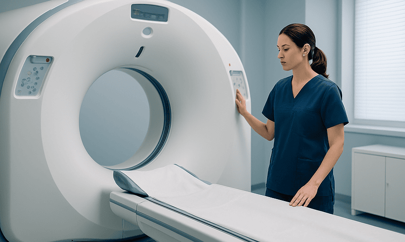
For the MRI, the challenge is stillness and noise rather than pain. The scanner knocks and buzzes loudly; headphones or ear protection are routine. Younger children may be offered sedation or a play-based preparation session with a child-life specialist, a professional who helps children cope with medical procedures. Remove anything metal, mention any implants or orthodontic work, and ask whether a parent may stay in the room. Contrast dye is given through the same kind of cannula and can feel briefly cool as it goes in. None of this is dangerous; all of it goes more smoothly when expected.
What does a pituitary MRI for gigantism show, and what can it miss?
Once blood tests point to growth hormone excess, imaging answers the next question: where is it coming from? Magnetic resonance imaging, or MRI, uses magnetic fields and radio waves rather than radiation to build detailed cross-sections of soft tissue. Mayo Clinic identifies MRI as the preferred scan for locating a pituitary tumor because it shows the gland and the structures around it in far greater detail than a CT scan, which uses X-rays.
A pituitary protocol MRI takes thin slices through the sella, before and after contrast. The radiologist reports the size of any tumor, whether it stays within the sella or pushes upward toward the optic chiasm, and whether it extends sideways into the cavernous sinuses, the venous channels either side of the gland that carry important nerves and arteries. Mayo Clinic’s pituitary tumor guidance divides tumors by size: microadenomas measure under one centimeter, macroadenomas one centimeter or more. Children with gigantism more often have the larger type, which is part of why their hormone levels tend to be high.
The scan has limits worth stating honestly. Small tumors can hide against normal tissue, and a hormone-confirmed case with a clean scan is not a contradiction; it usually prompts repeat imaging with finer technique rather than abandoning the diagnosis. The opposite problem is more common and more misleading. Pituitary glands frequently contain tiny incidental spots that produce no hormone and never will. Found on a scan ordered for headaches, such a spot does not make the diagnosis, and that is exactly why imaging follows hormone testing rather than leading it.
Finally, MRI shows anatomy, not function. It cannot say what a tumor is secreting. The blood tests say that; the scan says where.
Gigantism diagnosis tests at a glance: what each one measures
Families often leave a first appointment holding three or four requisition forms and no sense of how they relate. The table below sets the main gigantism diagnosis tests side by side, with the question each one is designed to answer.
| Test | What it measures | Question it answers | Main limitation |
|---|---|---|---|
| Growth chart and velocity | Height over time versus family expectation | Is this child growing faster than their own history predicts? | Needs accurate serial measurements months apart |
| IGF-1 blood test | Steady marker of overall growth hormone exposure | Is growth hormone activity raised? | Must be read against age and pubertal stage; puberty raises it normally |
| Random growth hormone | Hormone level at one moment | Rarely used alone | Pulsatile release makes single values unreliable |
| Oral glucose suppression test | Lowest growth hormone after a glucose drink | Can the body switch growth hormone off? | Fasting required; cutoffs vary by laboratory assay |
| Pituitary MRI with contrast | Size and extent of any tumor | Where is the excess coming from? | Shows anatomy, not function; incidental spots are common |
| Other pituitary hormones | Prolactin, thyroid, cortisol and sex hormone axes | Is the rest of the gland working? | Several may need repeat or stimulation testing |
| Visual field test | Peripheral vision map for each eye | Is the tumor pressing on the optic chiasm? | Only meaningful if the tumor extends upward |
Read top to bottom, the table is also the usual order of events. The chart raises the question, IGF-1 screens, the suppression test confirms, MRI localizes, and the remaining tests describe what the tumor has done to its neighbors. Mayo Clinic and MedlinePlus describe essentially this sequence. Skipping steps saves little time and tends to generate results nobody can interpret.
What is hypopituitarism, and which other tests check the rest of the gland?
Hypopituitarism is the condition in which the pituitary makes too little of one or more of its hormones. MedlinePlus explains that pituitary tumors are among the leading causes, because a growing mass squeezes the normal gland beside it. So a child being investigated for too much of one hormone is, at the same time, checked for too little of the others. The irony is real and the testing is routine.
The team usually assesses each pituitary axis in turn. The thyroid axis is checked with thyroid-stimulating hormone and free thyroxine. The adrenal axis, which governs cortisol, the stress hormone, is checked with a morning cortisol and sometimes a stimulation test, in which a synthetic signal is given and the response measured. The gonadal axis, which drives puberty, is assessed through luteinizing hormone, follicle-stimulating hormone and estrogen or testosterone, interpreted against the child’s pubertal stage. Prolactin, as noted earlier, is frequently raised rather than low. The posterior pituitary, which releases the hormone that controls urine concentration, is assessed by asking about excessive thirst and urination and, if needed, with blood and urine tests.
Vision assessment joins the list when the MRI shows the tumor reaching toward the optic chiasm. A formal visual field test maps the outer edges of sight in each eye, and an eye specialist may examine the optic nerves directly.
Some children are referred for genetic counseling. MedlinePlus lists several inherited conditions in which gigantism can occur, including McCune-Albright syndrome, multiple endocrine neoplasia type 1, neurofibromatosis and Carney complex. A family history of pituitary tumors, very early onset, or an unusually large tumor can prompt testing for specific gene changes. A positive result may matter for relatives as well as for the child, and the counselor’s role is to explain what a result does and does not mean before any sample is taken.
What do the days and weeks after testing usually look like?
Hormone results and scan reports rarely arrive on the same day, and the waiting is often the hardest part of the process. Laboratories vary in turnaround, so the honest answer to ‘how long’ is the one the clinic gives for its own lab; it is reasonable to ask for that estimate before leaving.
If IGF-1 comes back normal for age and the child’s growth velocity is steady, the pathway often ends there, with a plan to re-measure in several months. If IGF-1 is raised, the suppression test is booked, sometimes within weeks, and the MRI is usually arranged once the hormone picture is clear. Children who have had sedation for the scan are observed until fully awake and typically go home the same day; those who had contrast dye can drink normally afterward.
When the results confirm growth hormone excess and locate a tumor, the next appointment is usually a multidisciplinary one. The pediatric endocrinologist, a neurosurgeon who operates on the pituitary, a radiologist and often an eye specialist review the case together. The NHS and Mayo Clinic both describe surgery through the nose and the sphenoid sinus as the usual first-line approach for pituitary tumors that are accessible, with medicines and radiotherapy as options when surgery is not possible or does not fully control hormone levels. Medicines used in this setting work by mimicking the body’s natural growth hormone brake, by blocking growth hormone’s effect at its receptor, or by acting on dopamine pathways that restrain some tumors. Which, if any, is appropriate is a decision for the treating team based on tumor size, hormone levels and the child’s age; no article can make it.
Whatever path is chosen, repeat IGF-1 testing and periodic imaging become part of life for a while, because the same tests that made the diagnosis are the ones that show whether the condition is being controlled.
What people often get wrong about gigantism and the pituitary
Searches around this topic turn up a few persistent misunderstandings. Correcting them is part of a good diagnosis.
‘My child is very tall, so this must be gigantism.’ Height alone almost never is. MedlinePlus describes gigantism as very rare, and the great majority of tall children are tall because their parents are. A change in growth velocity, together with the physical features described earlier, is what warrants testing.
‘One growth hormone blood test will give the answer.’ It will not. Growth hormone is released in pulses, and MedlinePlus is explicit that random levels are difficult to interpret. IGF-1 plus a suppression test is the standard combination, and both are read against age-specific ranges.
‘You can activate or balance the pituitary gland naturally.’ Sleep and exercise do raise normal growth hormone pulses, which is why healthy children grow mostly at night. That has nothing to do with a tumor that secretes hormone regardless of signals, and no diet, herb or supplement has been shown in mainstream evidence to shrink a pituitary adenoma or lower IGF-1 in gigantism. Products marketed to ‘boost’ or ‘reset’ the pituitary should be mentioned to the clinician, because some interfere with test results.
‘A pituitary tumor means brain cancer.’ MedlinePlus and Mayo Clinic both describe the tumors behind gigantism as almost always noncancerous. They cause harm by size and by hormone output, not by spreading.
‘The MRI is the real test; the blood work is a formality.’ The reverse is closer to the truth. Incidental pituitary spots are common on scans, and a spot without raised IGF-1 and failed suppression is not gigantism.
‘If the scan is clear, the diagnosis is wrong.’ Small tumors can escape detection. A clear scan with convincing hormone results usually leads to repeat, finer imaging rather than reassurance.
Gigantism vs acromegaly: same hormone, different stage of life
The two conditions share a cause, a set of tests and much of their treatment, yet they look nothing alike, and the difference is entirely about timing. Growth hormone excess before the growth plates close produces gigantism: the long bones keep lengthening, and the child becomes unusually tall. After the plates fuse, the same excess produces acromegaly: height is fixed, so the hormone thickens bones in the hands, feet, jaw and brow and enlarges soft tissues and internal organs. Mayo Clinic notes that acromegaly in adults is most often diagnosed in middle age, usually years after features first begin to change because the process is slow.
The diagnostic tests are identical in principle. IGF-1 screens, the glucose suppression test confirms, and MRI localizes in both. Interpretation differs in two ways. First, children’s IGF-1 reference ranges shift with age and puberty, so pediatric results need pediatric ranges. Second, children’s growth hormone pulses are larger, which makes random levels even less useful than in adults and gives the suppression test added weight.
Why does the distinction matter beyond vocabulary? Because an adolescent with gigantism may still have open growth plates, and controlling the hormone before they close changes the course of their height in a way that is not possible later. Pediatric teams therefore tend to move more quickly through the pathway than adult teams do. A person whose excess begins in late adolescence can carry features of both: tall stature from the childhood phase and coarsening features from the adult phase.
The NHS describes acromegaly as rare and gigantism as rarer still, which explains why many general pediatricians will see at most one case in a career and why referral to a specialist center experienced in pediatric pituitary disease is standard rather than a sign of alarm.
Questions to ask your care team
A list on paper beats a list in your head when the appointment runs short. These questions are phrased to draw out specifics rather than reassurance.
- Which growth percentile is my child on now, which were they on at the last two visits, and how does that compare with the height predicted from both parents?
- Is the IGF-1 result being compared with a reference range for my child’s age and pubertal stage, and what range did the laboratory use?
- Do you plan a glucose suppression test, and if so, how long should my child fast and what should we bring?
- Are there medicines or supplements my child takes that could distort the results, and which of them, if any, do you want paused? We will not stop anything without your say.
- Will the MRI be a dedicated pituitary protocol with contrast, and will sedation or a preparation session be offered?
- Which other pituitary hormones are you checking, and what would it mean if one of them is low?
- Does the tumor on the scan sit near the optic nerves, and should my child have a visual field test?
- Is genetic testing or counseling appropriate for our family, and what would a result change?
- How long does your laboratory usually take to return these results, and who will call us?
- If the diagnosis is confirmed, which specialists will be involved, and will they meet as a team?
- What would each treatment option mean for my child’s final height, schooling and daily life, and what are the risks and alternatives of each?
- What symptoms should prompt us to contact you before the next scheduled appointment?
Write the answers down during the visit. Hormone pathways are hard to hold in memory on a stressful day, and the notes will be useful at the follow-up when results are explained.
When to call your doctor
Most of the gigantism pathway unfolds over scheduled appointments, and a tall, otherwise well child is not an emergency. A few situations, however, should prompt a call to the care team the same day or a visit to urgent or emergency care, whether or not testing has been completed.
Seek care urgently if a child develops a sudden, severe headache unlike any before, especially with vomiting, a stiff neck, double vision, drooping of an eyelid or a sudden loss of vision in part or all of one or both eyes. These can signal bleeding into or rapid swelling of a pituitary tumor, which Mayo Clinic describes as a rare but serious complication called pituitary apoplexy, and it needs immediate assessment.
Contact the team promptly, within a day, for any of the following: a steady worsening of headaches over days or weeks; new difficulty seeing things at the edges of vision, such as bumping into door frames or missing objects to one side; unusual thirst with frequent urination, including waking repeatedly at night to drink; marked tiredness, dizziness on standing or fainting, which can point to low cortisol; persistent nausea with weight loss; or any sudden change in behavior or alertness.
After a suppression test, mild nausea or lightheadedness that settles within hours is expected; prolonged vomiting, confusion or shakiness is not and should be reported. After an MRI with contrast, a spreading rash, facial swelling or difficulty breathing is rare but needs emergency care.
Finally, call if growth seems to be accelerating noticeably between visits, if shoe or ring sizes are changing month to month, or if a parent’s instinct simply says something has shifted. Clinicians would far rather hear about a change that turns out to be nothing than miss one that mattered. Every decision about testing, timing and treatment belongs to the treating team, and the quickest way to reach them is the number on the clinic letter.
Frequently asked questions
What blood tests are used to diagnose pituitary tumors that cause gigantism?
IGF-1 is the main screening test, followed by an oral glucose suppression test that measures growth hormone repeatedly after a sugary drink. Clinicians usually add prolactin, thyroid hormones, cortisol or its pituitary driver, sex hormones and a fasting glucose to see how the rest of the gland is functioning. No single blood value makes the diagnosis; the pattern across these tests, read against age-specific ranges, does.
Is the IGF-1 blood test for gigantism enough on its own?
No. A raised IGF-1 raises suspicion but is not proof, because normal puberty, and some other conditions, can push it above the childhood range. Guideline practice is to confirm with a glucose suppression test and then localize the source with a pituitary MRI. Conversely, a normal age-matched IGF-1 in a child with steady growth velocity usually makes gigantism unlikely and often ends the workup.
How long does the growth hormone suppression test take?
The test itself typically takes about two hours after the glucose drink, with blood samples drawn at set intervals through a single cannula, according to MedlinePlus. Add time for fasting beforehand, usually overnight as the laboratory instructs, and for check-in. Children can bring something to read or watch, and a snack for afterward. Results depend on the laboratory’s turnaround and are interpreted by the endocrinologist alongside IGF-1.
Where is the pituitary gland located in the brain?
The pituitary sits at the base of the brain in a small bony hollow called the sella turcica, roughly behind the bridge of the nose. It is about the size of a pea and hangs from the hypothalamus by a thin stalk. The optic chiasm, where the vision nerves cross, lies directly above it, which is why a tumor growing upward can affect peripheral vision.
What is hypopituitarism, and why is it checked in gigantism?
Hypopituitarism means the pituitary produces too little of one or more of its hormones. MedlinePlus lists pituitary tumors among its leading causes, because a growing tumor compresses the normal gland around it. A child with a growth hormone secreting tumor can therefore have excess of one hormone and deficiency of others, so thyroid, adrenal and pubertal hormone axes are assessed as part of the same workup.
Can you activate the pituitary gland naturally to fix a hormone problem?
Not in the sense those searches intend. Sleep and exercise do raise normal growth hormone pulses, but a tumor secretes hormone regardless of those signals, and no diet, herb or supplement has been shown in mainstream evidence to shrink a pituitary adenoma or lower IGF-1. Supplements marketed as pituitary boosters should be disclosed to the clinician because some can distort test results.
Does a normal pituitary MRI rule out gigantism?
No. Small tumors can hide against normal gland tissue, and a child with raised IGF-1 and failed glucose suppression still has biochemical growth hormone excess even if the scan looks clean. In that situation the team usually repeats imaging with a dedicated pituitary protocol and thinner slices. The blood tests establish that excess exists; the MRI only tries to show where it comes from.
Is the pituitary tumor behind gigantism a cancer?
Almost never. MedlinePlus and Mayo Clinic describe the tumors that cause gigantism as noncancerous adenomas, meaning they do not spread to other parts of the body. They cause problems through the hormone they release and, when large, through pressure on the normal pituitary and nearby vision nerves. That is why the tests focus on hormone levels and tumor size rather than on spread.
My child is very tall but the doctor has not ordered tests. Why?
Because tall stature alone is common and gigantism is very rare. Clinicians look for a change in growth velocity, crossing percentile lines on the growth chart, together with features such as enlarging hands and feet, coarsening facial features, headaches or delayed puberty. A child tracking steadily along a high percentile with tall parents is usually re-measured in several months, which is itself a valid diagnostic step.
What is the difference between gigantism vs acromegaly in terms of testing?
The tests are the same: IGF-1, glucose suppression and pituitary MRI. The difference is timing and interpretation. Gigantism occurs before the growth plates close and adds height; acromegaly occurs after and thickens bone and soft tissue instead. In children, IGF-1 must be read against age and pubertal stage, and larger natural growth hormone pulses make the suppression test even more important than in adults.
References
- MedlinePlus Medical Encyclopedia: Gigantism
- MedlinePlus Lab Tests: Growth Hormone Tests
- MedlinePlus Medical Encyclopedia: Hypopituitarism
- NHS: Acromegaly
This article is for general information only and is not a substitute for professional medical advice. Please consult a qualified doctor about your individual situation.

More from the Blog
What Happens Inside a Medical Longevity Program? Risk Assessment, Screening and the Plan
A medical longevity program usually involves three stages: a detailed history and physical exam, a set of blood tests and screenings chosen to estimate…

Hyperthyroidism With Heart Disease or in Pregnancy: How Treatment Is Adapted
Hyperthyroidism treatment is adapted in pregnancy and heart disease because the usual options carry different risks in each situation. In pregnancy, radioactive iodine is…

When Is Surgery the Treatment for Cushing Syndrome, and When Is Medication Used First?
Surgery to remove the cortisol-producing tumor is usually the first-line treatment for Cushing syndrome caused by a pituitary or adrenal tumor, because it targets…

Adrenalectomy for Pheochromocytoma: How Surgeons Remove the Adrenal Tumor Safely
Pheochromocytoma adrenalectomy is surgery to remove an adrenal gland tumor that overproduces adrenaline-type hormones. Because handling the tumor can trigger dangerous blood pressure swings,…

How Do Test Results Shape a Hashimoto Treatment Plan? TSH, Antibodies and Ultrasound
Hashimoto diagnosis tests work as a set, not one number. A high TSH with low free T4 usually signals an underactive thyroid that clinicians…

Diabetes Insipidus: Confusion, Severe Weakness and When Dehydration Needs Urgent Care
Diabetes insipidus becomes a dehydration emergency when the body loses large volumes of dilute urine faster than fluid can be replaced, pushing blood sodium…