

Digital Dentistry Myths: Does a Scanner Replace the Dentist’s Judgment?
Key Takeaways
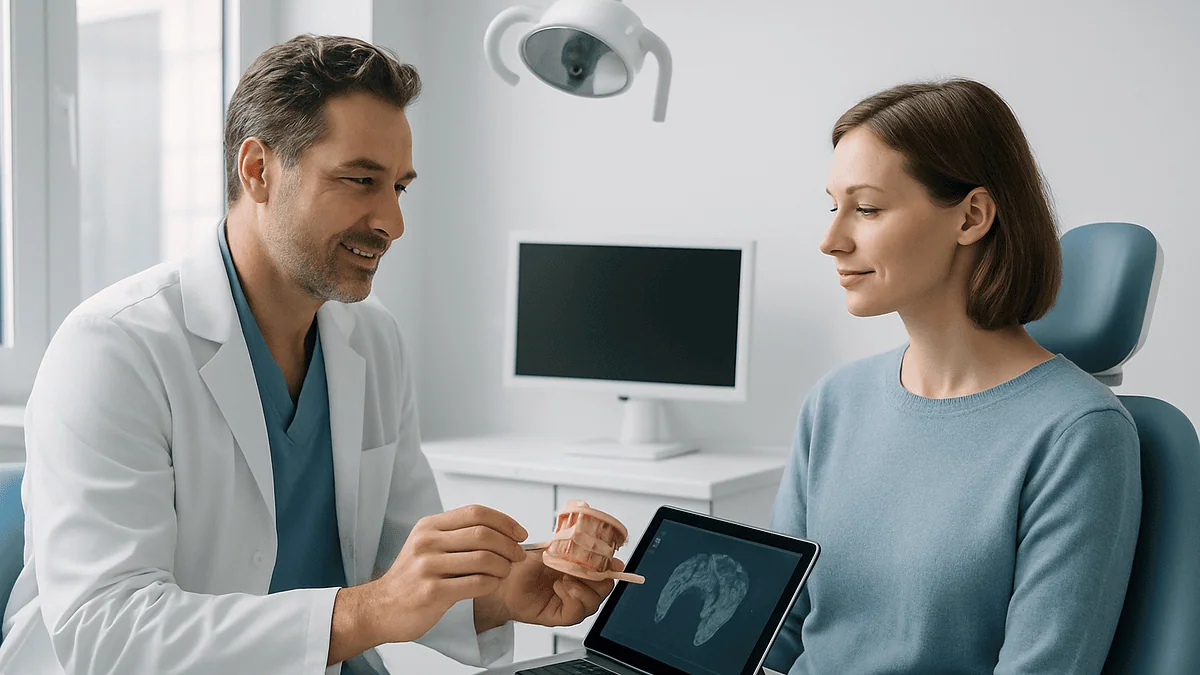
- An intraoral scanner captures the surface shape of teeth and gums; it cannot probe gum pockets, test whether a nerve is alive or detect decay between teeth.
- Scan accuracy is well supported for single crowns and short spans, but errors accumulate across full-arch scans, which is why final frameworks are still verified in the mouth.
- CBCT 3D imaging exposes patients to more radiation than a standard dental X-ray and should be justified by a specific clinical question, not routine.
- A surgical guide directs drill angle and depth, yet surgeons confirm each step and may abandon the guide if anatomy differs from the plan.
- Bone healing around an implant typically takes several months regardless of how the case was planned, according to Mayo Clinic.
- The "50-40-30 rule" has no single accepted definition in mainstream dental guidance and should be treated as an informal heuristic, not a standard.
No. An intraoral scanner records the shape of teeth and gums as a digital file; it does not examine, diagnose or decide. Scanners and 3D imaging can improve the precision of impressions and implant planning, but the evidence shows their accuracy depends on operator technique, tissue conditions and correct interpretation. The clinical examination, history and final judgment remain the dentist's responsibility.
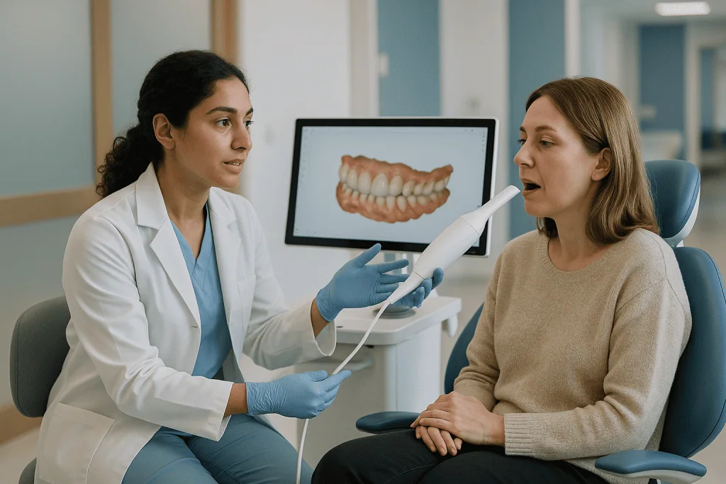
The wand hovered over her molars for about ninety seconds. On the screen beside the chair, a color model of her mouth assembled itself tooth by tooth, rotating slowly like a museum artifact. “So the computer plans the implant now?” she asked, half joking. The dentist smiled and said, “The computer draws. I decide.”
That exchange captures the real question behind digital dentistry limits. Patients weighing an implant, a crown or a set of aligners are often shown impressive software before anyone mentions what the software cannot see: a hairline crack under old filling material, a gum that bleeds too easily, a jaw joint that clicks on wide opening.
This explainer walks through what the scanner does, where the evidence supports it, where it runs out and why the person holding the wand still matters more than the wand itself.
What does an intraoral scanner actually do?
An intraoral scanner is a handheld camera that projects structured light or a laser pattern onto teeth and gums and captures thousands of small images per second. Software stitches those frames into a three-dimensional surface model, a digital version of the putty impression that many patients remember gagging on.
The output is a file, not a finding. It records the visible surface of enamel, existing restorations and soft tissue at the moment of capture. That file can be sent to a laboratory to design a crown, uploaded into implant-planning software or used to fabricate aligners. In each case, a human being still has to decide what the model means and what to do with it.
A few points about the capture itself matter for patients. The scanner reads only what it can see. Saliva, blood and reflective metal can distort frames. A margin hidden below the gumline, which is common when preparing a tooth for a crown, may need a retraction cord or a conventional impression to record cleanly. Movement, a tongue in the way or fogging of the lens forces the operator to rescan.
None of this makes the technology weak. It makes it a tool with operating conditions, much like an ultrasound probe. Dentists trained in scanning learn to manage tissue, control moisture and follow a scan path that limits the small distortions that accumulate across an arch. What the scanner cannot do, and was never designed to do, is take a history, probe a pocket, test a tooth for vitality or weigh a patient’s medical risks. Those tasks belong to the examination, which mainstream guidance from bodies such as the NHS still describes as the foundation of dental care.
How accurate are intraoral scanners? What the evidence shows
Accuracy in this field has two parts, and conflating them leads to inflated marketing. Trueness describes how close a scan comes to the real object. Precision describes how consistently repeated scans agree with one another. A device can be precise and still consistently wrong.
Published comparisons of digital and conventional impressions, indexed in PubMed, generally find that for single crowns and short spans the digital route performs comparably to well-made conventional impressions, with the advantage that distortions from tray removal and stone pouring disappear. The picture changes as the scanned span grows. Because the software stitches frames together, tiny errors compound across a full arch, and studies repeatedly flag full-arch and edentulous (toothless) scans as the situations where deviation is most likely to matter, particularly when several implants must be joined by a single rigid framework.
Several variables influence the result more than the brand of device:
- Operator experience and scan strategy.
- Moisture and bleeding control.
- Presence of metal restorations, which reflect light.
- Depth of the preparation margin below the gum.
- Number of remaining teeth to act as landmarks.
Notice that most items on that list are clinical, not technical. They are managed by the person, not the machine. This is why the honest answer to “how accurate are intraoral scanners” is “accurate enough for many indications when a skilled clinician controls the conditions, and not a substitute for verifying fit in the mouth.” A crown that looks perfect on screen is still tried in, checked with floss and articulating paper, and adjusted by hand. That final check is a judgment call, and it stays with the dentist.
Digital dentistry limits: where the scan ends and the exam begins
Think of the scan as a photograph of the shoreline. It shows the coast beautifully and tells you nothing about the tide, the current or the rocks below the surface. The examination is what maps the water.
A dentist examining a mouth is doing several things at once that no surface scan captures. Periodontal probing measures the depth of the pocket between gum and tooth, one of the main ways gum disease is assessed according to the NIH’s dental research institute. Percussion, air, cold and bite tests help distinguish a tooth that is sensitive from one whose nerve is dying. Palpation of the jaw, lymph nodes and floor of the mouth checks for swellings the camera cannot reach. A conversation about medications, diabetes, smoking, bisphosphonate use or radiation to the head and neck flags risks that change the plan entirely.
Radiographs fill in some of the gap, showing bone levels, decay between teeth and the position of roots. Yet an image also requires interpretation, and interpretation is where experience earns its keep. Two clinicians can look at the same cone-beam scan and reasonably disagree about whether bone volume is adequate, because “adequate” depends on the planned restoration, the patient’s bite and how much soft tissue will sit around the final crown.
The digital dentistry limits, then, are not glitches waiting for a software update. They are the boundary between recording and understanding. Scanners record extremely well. Understanding the whole patient, deciding whether treatment is wise at all and accepting responsibility for the outcome are not functions a file can perform. When a practice describes its workflow as “fully digital,” it usually means the impressions and planning are digital. The diagnosis is still made by a person.
Digital implant planning: what 3D imaging and guided surgery add (and don't)
Digital implant planning combines two files: the surface scan of the teeth and a cone-beam computed tomography (CBCT) scan, a low-dose 3D X-ray of the jaws. Software overlays them so the clinician can see bone, nerve canals, sinus floors and the intended crown in one view, then place a virtual implant where it will best support that crown.
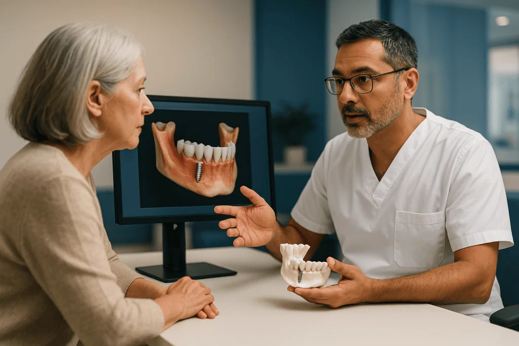
From that plan, a surgical guide can be printed: a stent that sits on the teeth or gums and directs the drill to the planned angle and depth. Mayo Clinic describes implant placement as a staged process in which the titanium post is placed in the jawbone and the bone is allowed to heal around it before the final tooth is attached, a healing period that typically spans several months.
What the digital step adds is foresight. Nerve proximity, thin ridges and sinus dips are visible before any incision. Restorative and surgical planning happen together, which supports the widely taught principle that the crown, not the bone, should dictate where an implant goes.
What it does not add is certainty. Guides can shift on soft tissue, especially in edentulous jaws. Bone density on a CBCT is an estimate, not a measurement of how the drill will feel. A patient’s mouth opening may not accommodate the guide and drill together at the back of the arch. Surgeons therefore verify each step directly, and they retain the option to abandon the guide and place the implant freehand if what they find differs from what the screen predicted.
Cleveland Clinic notes that implants carry risks including infection, injury to neighboring structures, nerve problems and sinus complications. Planning software lowers the chance of some of those errors by making anatomy visible. It does not remove the need for surgical skill, and it does not decide whether a given patient should have an implant at all.
Who a digital workflow usually suits, and who is usually asked to wait
Most adults having a single crown, a short bridge, aligners or one or two implants in a jaw with plenty of neighboring teeth are well served by a digital impression. Landmarks are abundant, the span is short and the reduced chair time and lack of impression material are genuine comforts, particularly for people with a strong gag reflex.
Guided implant surgery tends to be considered when anatomy is tight: an implant near the nerve in the lower jaw, close to the sinus in the upper jaw, or several implants that must line up for one prosthesis. It is also frequently used when a clinician wants to avoid raising a large flap of gum.
Some patients are usually asked to pause, not because the technology fails but because the underlying conditions are not ready:
- Active gum disease. The NIH’s dental research institute describes periodontitis as an infection that destroys the bone supporting teeth; placing implants into an infected environment is generally deferred until the disease is controlled.
- Uncontrolled diabetes or heavy smoking, both of which Mayo Clinic lists among factors that affect healing.
- Recent extraction sites still remodeling, where a scan today may not match the tissue in a few weeks.
- Certain medications that affect bone turnover, which require a medical conversation before any surgery.
- Growing adolescents, whose jaws have not finished developing.
In each case the scanner could capture a flawless model. The decision to wait comes from the history and examination. That is the point: eligibility is a clinical judgment, and the software offers no opinion on it. A patient who is told “not yet” has not been failed by digital dentistry; they have been protected by the part of dentistry that remains stubbornly human.
What are the downsides of digital dentistry?
Enthusiasts rarely lead with the downsides of digital dentistry, so here they are in plain language.
The first is false confidence. A crisp 3D render looks authoritative in a way a stone model never did, and both clinicians and patients can over-trust it. Error in a full-arch scan is invisible on screen; it shows up only when a framework does not seat.
The second is radiation. CBCT delivers a higher dose than a standard two-dimensional dental X-ray, and mainstream guidance holds that any imaging should be justified by a clinical question it will answer. Routine CBCT “because we have the machine” is not supported. Patients are entitled to ask why a 3D scan is needed for their case.
The third is the learning curve. Scanning technique, software planning and guide handling all take training. A clinician who is new to a workflow may produce worse results than an experienced one using conventional methods.
Fourth, workflows can hide hand-offs. A scan may travel to a design technician, a milling center and a printing service before anything returns to the chair. Each step is an opportunity for a communication error, and the treating dentist remains responsible for verifying the final fit and occlusion (the way the teeth meet).
Fifth, data. Scans and CBCT images are health records. Patients can reasonably ask how their files are stored, who can access them and how long they are retained.
Finally, there is scope creep. Software that can simulate a whitened, straightened, veneered smile in seconds makes it easy to propose treatment that was never clinically necessary. A neutral question to ask is simple: “What problem does this solve for my health, and what happens if I do nothing?” A good clinician will answer without the render.
Scanner versus clinician: who does what in a digital dental workflow
Seeing the division of labor side by side clears up most confusion about digital dentistry limits. The table below is a general summary drawn from how mainstream sources such as Mayo Clinic and Cleveland Clinic describe implant and restorative care, not a description of any particular product.
| Task | Digital tool | Clinician |
|---|---|---|
| Record tooth and gum surfaces | Intraoral scanner captures 3D surface | Controls moisture, tissue and scan path; judges whether the scan is usable |
| Assess bone, nerves, sinuses | CBCT displays anatomy in 3D | Decides if imaging is justified; interprets findings; estimates bone quality |
| Detect gum disease | No direct role | Probes pocket depths, checks bleeding, reviews bone loss on radiographs |
| Diagnose tooth pain | No direct role | History, vitality tests, percussion, examination |
| Plan implant position | Software merges scan and CBCT; proposes position | Sets the restorative goal; accepts, adjusts or rejects the proposal |
| Place the implant | Printed guide directs drill angle and depth | Verifies guide seating; decides whether to follow or abandon the guide |
| Fit the final crown | Design software and milling produce the restoration | Checks fit, contacts and bite in the mouth; adjusts by hand |
| Decide whether to treat at all | No role | Weighs medical history, risks, alternatives and patient goals |
Two patterns stand out. First, every row that involves diagnosis or a decision has no digital entry, because those steps are inherently clinical. Second, even the rows where technology leads still end with human verification. A guide directs; a surgeon confirms. Software proposes; a dentist disposes. When a practice shows you its scanner, the table is a useful reminder of what you are actually paying attention to: the judgment in the right-hand column.
What is the 50-40-30 rule in dentistry?
People search this phrase often, and the honest answer is that no mainstream clinical guideline defines a “50-40-30 rule” in dentistry. The numbers circulate online with different meanings depending on who is using them, which is a red flag in itself.
In some cosmetic contexts, ratio-based rules are used to describe how the widths of front teeth appear to shrink as they recede from the center of the smile, or how much of the upper teeth should show at rest and during a full smile. These are design heuristics for esthetics, not evidence-based standards for health, and they vary between authors. In other contexts the same numbers appear in practice-management material about scheduling or in informal rules of thumb about how much of a tooth can be lost before a crown is advised. Because there is no single accepted source, a patient who hears the phrase should simply ask what the clinician means by it and where it comes from.
Why does this matter in an article about scanners? Smile-design software often builds such ratios directly into its templates. A digital mock-up can make proposed veneers look mathematically “correct” while saying nothing about whether the underlying teeth are healthy, whether the bite will tolerate the new shapes or whether enamel must be sacrificed to achieve them. Proportion rules describe appearance. They do not assess disease, function or risk.
Mainstream sources such as the NHS emphasize that cosmetic treatments should follow a full examination and that patients should understand what is being removed from healthy teeth to achieve a look. Aesthetic guidelines have a place, and many skilled clinicians use them thoughtfully. Treat any numeric rule as a starting point for conversation, not a clinical recommendation, and ask the person, not the software, to justify the plan.
Teledentistry limitations: what a video visit can and cannot check
Teledentistry means dental advice delivered remotely, usually by video call or by reviewing photographs and records a patient sends in. It grew rapidly as a triage tool and it has real value: sorting urgent from routine problems, following up after surgery, reviewing aligner progress from photos and reaching people who live far from a dentist.
The major limitation is the one that runs through this whole article. A screen cannot examine. Over video, a clinician cannot probe a gum pocket, feel a swelling, tap a tooth, dry it and look for a crack, or take a radiograph. Lighting from a phone camera flattens color and hides shadows that would be obvious under a chair lamp. Decay between teeth is invisible without an X-ray. A tooth that hurts on video may be the neighbor of the real culprit, since dental pain often refers to nearby teeth.
Emergency management is the other hard boundary. Draining an abscess, stabilizing a knocked-out tooth, stopping bleeding after an extraction or relieving a lost temporary crown all require hands. The NHS advises that facial swelling, difficulty swallowing or breathing, or uncontrolled bleeding are situations for urgent in-person or emergency care, not a video call.
Where teledentistry limitations bite hardest is in the illusion of completeness. A patient who has “seen a dentist” online may reasonably feel reassured and delay a physical examination that would have caught early gum disease or a fractured root. Used well, a remote visit ends with a clear statement of what could not be assessed and when an in-person exam is needed. Used poorly, it becomes a substitute for care rather than a bridge to it. The same tool, the same ethics as any other digital instrument: it extends the clinician’s reach but not their eyes or hands.
Can a virtual dentist prescribe pain medicine?
Sometimes, within limits, and the limits are set by law and professional standards rather than by the technology. Whether a dentist can prescribe after a remote consultation depends on the jurisdiction, the type of medicine and whether a valid clinician-patient relationship has been established. Rules differ from state to state in the US and between countries, and they change; this article does not offer a legal summary.
Clinically, a few principles are consistent across mainstream guidance. Dental pain is usually a symptom of a problem that needs a physical fix: a nerve inflamed by deep decay, an abscess that needs drainage, a cracked tooth. Medicine can manage the pain for a short time, but the NHS and MedlinePlus both stress that persistent toothache requires a dentist to find and treat the cause. A remote clinician who cannot examine is unlikely to know the cause with confidence, which is why many will recommend over-the-counter options and an in-person visit rather than issuing a prescription.
Antibiotics deserve a specific word. Guidance from the CDC on antibiotic stewardship discourages prescribing antibiotics for dental pain without signs of spreading infection, because most toothaches are not treated by antibiotics and unnecessary use drives resistance. Requests for antibiotics over video are therefore often declined for good reason.
Controlled substances, including opioid pain relievers, face stricter rules almost everywhere and frequently require an in-person evaluation. Dental guidance has moved firmly toward non-opioid options for most dental pain.
Whatever is recommended, the decision to prescribe, what to prescribe and for how long rests with the prescribing clinician who knows your history. A virtual visit can be a sensible first step for pain that is manageable. Severe pain, swelling or fever calls for hands-on care, discussed in the red-flag section below.
What the following weeks look like after a digitally planned implant
The digital part of an implant is mostly finished before the first appointment ends. What follows is biology, and biology keeps its own schedule.
In the first few days after placement, Mayo Clinic describes swelling of the gums and face, bruising, minor bleeding and discomfort as expected. Soft foods, careful cleaning around the site and following the surgical team’s specific instructions are the standard advice. A surgical guide may have allowed a smaller incision, which some patients find eases the early days, but it does not change the fact that bone has been drilled.
Over the following weeks and months, the process called osseointegration takes place: bone grows onto the implant surface and locks it in place. Mayo Clinic notes this healing typically takes several months and that the timeline varies with the individual, the site and whether bone grafting was needed. No software shortens this stage. A CBCT can suggest how dense the bone is, but the body determines how quickly it heals.
Follow-up visits check that the tissue is settling and that no infection is developing. Once the surgeon judges integration adequate, an abutment (the connector between implant and crown) is placed, and the final crown is made, often from a new scan of the healed site. That second scan is a reminder that the mouth changed during healing, and the earlier digital model is no longer accurate.
Patients often ask whether a guided implant “heals faster.” There is no strong mainstream evidence that it does. Guided surgery aims for accurate position; it does not alter bone biology. Long-term maintenance, including daily cleaning and regular professional checks, matters more for keeping the implant healthy than any detail of how it was planned, a point Cleveland Clinic makes in its patient guidance.
What people often get wrong about digital dentistry limits
Myth: the scanner catches problems the dentist would miss. A surface scan shows shape. Decay under an old crown, a cracked root, a pocket of infection and early gum disease are not shape problems, and they are found by examination and radiographs.
Myth: a digital plan is a promise about the result. Software shows intent. Bone, gum and healing decide the outcome, and no reputable clinician will guarantee how an implant or crown will perform.
Myth: a 3D X-ray is always better than a 2D one. CBCT gives more information at a higher radiation dose. Mainstream imaging guidance calls for the lowest dose that answers the clinical question, which for many routine checks is a standard radiograph.
Myth: guided surgery removes human error. Guides can shift, deform or fail to seat, and the software plan can be wrong if the input scans were. Surgeons check every step because errors have not been abolished, only moved earlier where they are easier to catch.
Myth: a fully digital practice is a better practice. It may be more efficient and more comfortable for impressions. Quality of care still comes from diagnosis, communication, surgical skill and follow-up, none of which are digital.
Myth: teledentistry is the same as a check-up. It is a conversation with pictures. Without probing and radiographs, a remote visit cannot rule out the most common dental diseases.
Myth: numbers like the “50-40-30 rule” are clinical standards. They are informal esthetic or business heuristics with no single accepted definition.
The thread connecting all of these is the same. Digital tools extend what a dentist can record and rehearse. They do not extend what a dentist knows about you until the examination has been done, and they never take responsibility for the plan. Understanding digital dentistry limits is not skepticism about technology; it is clarity about who is accountable.
Questions to ask your care team before agreeing to a digital plan
A good consultation should survive scrutiny. These questions are neutral, and a thoughtful clinician will welcome them.
- What did the clinical examination find, separate from the scan? Ask specifically about gum health and any radiographic findings.
- Why is a 3D CBCT scan needed in my case, and what question will it answer that a standard X-ray cannot?
- For a full-arch or multi-implant case, how will the fit of the final framework be verified in my mouth before it is finalized?
- Will a surgical guide be used, and what will you do if it does not seat as planned?
- Who designs and manufactures the restoration, and who checks it before it is fitted?
- What are the alternatives to this plan, including doing nothing for now, and how do their risks compare?
- What in my medical history could affect healing, and does anything need to be addressed first?
- What is the expected sequence and rough timeline for healing, understanding that it varies?
- How are my scans and images stored, and who has access to them?
- If I have a problem outside office hours, how do I reach someone, and when should I go to an emergency department instead?
Listen for how the answers are framed. A clinician who explains what the scan showed and what the exam showed, in that order or the reverse, is demonstrating that both happened. A clinician who describes a plan entirely in terms of the software’s proposal, without reference to your gums, bone or history, is describing the render rather than you.
You are also entitled to a second opinion before irreversible treatment, and to time to consider. Mainstream guidance from the NHS on consent stresses that agreeing to treatment should follow a clear understanding of the benefits, risks and alternatives. A digital simulation can help that understanding. It cannot replace it.
When to call your doctor
Whether your care was planned on a screen or on a stone model, the warning signs after dental treatment are the same, and they call for a person, not a portal.
Contact your dental team promptly if you notice pain that worsens rather than eases after the first couple of days, swelling that continues to grow, a bad taste or discharge from a surgical site, a loose implant, abutment or crown, bleeding that does not settle with steady pressure, or numbness or tingling of the lip, chin or tongue that persists beyond the anesthetic wearing off. Mayo Clinic lists nerve injury, infection and sinus problems among recognized implant complications, and each is easier to manage when reported early.
Seek urgent or emergency care, rather than waiting for a routine appointment, if you develop facial swelling that spreads toward the eye or under the jaw, difficulty swallowing or breathing, a fever with dental pain, or heavy bleeding that soaks through gauze repeatedly. The NHS identifies these as situations where a dental infection may be spreading and where delay is dangerous.
If you have had a video consultation and were advised to be seen in person, treat that advice as time-sensitive. Remote clinicians cannot rule out the conditions above, and reassurance from a screen does not stand in for an examination.
Finally, if you are unsure whether a symptom is normal healing or a problem, call. Dental teams expect these calls, and describing what you feel to someone who knows your case is exactly the kind of judgment that no scanner, software or chatbot is positioned to make. The treating team decides what happens next.
Frequently asked questions
What are the downsides of digital dentistry?
The main downsides are over-confidence in polished 3D renders, accumulated error in full-arch scans, higher radiation from routine 3D imaging, a steep learning curve for clinicians, and more hand-offs between design and manufacturing steps. Digital simulations can also make elective cosmetic work look necessary. None of these are reasons to avoid the technology, but they are reasons to ask what the examination found alongside the scan.
How accurate are intraoral scanners compared with traditional impressions?
For single crowns and short spans, published comparisons indexed in PubMed generally find digital scans comparable to well-made conventional impressions. Accuracy declines as the scanned span lengthens, because small stitching errors compound across a full arch. Operator technique, moisture control and the number of remaining teeth influence the result more than the specific device, and the final fit is always checked in the mouth.
What is a major limitation of teledentistry?
The major limitation is that a clinician cannot examine you through a screen. Video and photographs do not allow probing of gum pockets, tapping or testing teeth, palpating swellings or taking radiographs, so common conditions such as decay between teeth and early gum disease cannot be ruled out. Teledentistry works well for triage and follow-up, but it should end with clear advice about when an in-person exam is needed.
Can a virtual dentist prescribe pain medication?
It depends on local law, the type of medicine and whether a valid clinician-patient relationship exists; rules vary by state and country. Controlled substances usually require in-person evaluation, and mainstream guidance discourages antibiotics for toothache without signs of spreading infection. Because dental pain usually needs a physical fix, remote clinicians often recommend over-the-counter relief and an in-person visit. The decision rests with the prescribing clinician.
What is the 50-40-30 rule in dentistry?
There is no accepted definition in mainstream clinical guidelines. The phrase appears online with different meanings, sometimes as a smile-design proportion heuristic for front teeth, sometimes in practice-management or informal restorative contexts. It is not an evidence-based standard for health. If a clinician uses the term, ask what they mean and how it applies to your teeth, and separate esthetic proportions from the clinical findings.
Does digital implant planning make surgery safer?
It can reduce certain errors by making nerves, sinuses and bone width visible before surgery and by directing the drill with a printed guide. It does not remove risk. Guides can shift, imaging can misjudge bone quality, and the plan depends on accurate input scans. Recognized complications such as infection, nerve injury and sinus problems remain possible, which is why surgeons verify each step and retain the option to work freehand.
Is a CBCT scan necessary before every dental implant?
Not necessarily. CBCT is a 3D X-ray that delivers more radiation than a standard two-dimensional dental image, and mainstream imaging guidance calls for the lowest dose that answers the clinical question. Many clinicians consider CBCT when anatomy is complex, such as near the lower jaw nerve or upper jaw sinus, or when several implants must align. Your dentist should be able to explain why it is or is not indicated for you.
Can an intraoral scanner detect cavities or gum disease?
No. A scanner records the visible surface of teeth and gums as a 3D model. Decay between teeth, under existing fillings or beneath the gumline, and the bone loss of gum disease, are found through clinical examination, probing and radiographs. Some scanners include add-on imaging modes marketed as caries aids, but these support rather than replace the dentist’s examination and interpretation.
How long does healing take after a digitally guided implant?
About the same as after conventionally placed implants. Mayo Clinic describes bone healing around the implant, called osseointegration, as typically taking several months, with variation depending on the site, the individual and whether bone grafting was needed. A guide may allow a smaller incision, but it does not change bone biology, and there is no strong mainstream evidence that guided placement speeds healing.
Should I choose a dentist because their practice is fully digital?
Equipment alone is a weak reason. Digital impressions are more comfortable for many patients and can improve planning, but the quality of care still comes from thorough examination, clear communication, clinical skill and follow-up. A more useful basis for choosing is how well the clinician explains what the exam found, why a treatment is proposed, what the alternatives are and what the risks involve.
References
This article is for general information only and is not a substitute for professional medical advice. Please consult a qualified doctor about your individual situation.

More from the Blog

Sinus Lift Complications Explained: Membrane Tears, Sinus Infection and Signs That Need a Call
Sinus lift complications are uncommon but real. The most frequently reported problem during surgery is a tear in the thin sinus membrane, which the…
Living With Dental Crowns: Foods to Approach Carefully, Night Guards and Flossing the Margins
Eating with dental crowns is mostly ordinary eating, with a few habits worth keeping. Once numbness fades and the cement has set, most foods…
What Happens on the Day of All On 4 Surgery: Extractions, Angled Implants and Temporary Teeth
On all on 4 surgery day, a surgeon typically removes any remaining teeth in one arch, smooths the bone, places four implants (two upright…
Dental Bridge Problems Explained: Decay Under the Support Teeth, Loosening and Gum Irritation
Most dental bridge problems trace back to three things: decay creeping under the crowns on the support teeth, cement or tooth failure that lets…
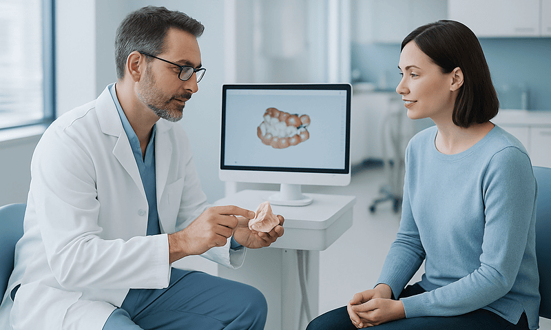
The Digital Dentistry Workflow Step by Step: Scan, Design, Milling and Fitting in Practice
A digital dentistry workflow replaces putty impressions and hand-poured models with data: an intraoral scanner captures the teeth, 3D X-ray imaging maps the jaw…

Zygomatic Implants Recovery: Swelling, Bruising and the Soft Diet Until the Final Bridge
Zygomatic implants recovery usually means cheek swelling and bruising that peak within about 2 to 3 days and fade over roughly 1 to 2…