

Follow-Up After Esophageal Cancer Treatment: Why Endoscopy and Imaging Are Repeated
Key Takeaways
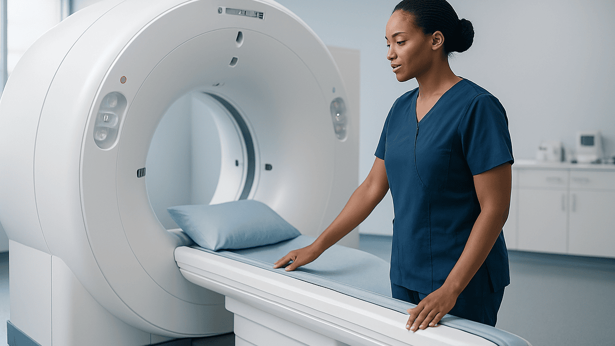
- Endoscopy and CT or PET-CT are combined after esophageal cancer treatment because the scope sees the lining and surgical join while imaging sees lymph nodes, liver and lungs, and neither covers the other's blind spots.
- Follow-up is concentrated in the first years after treatment because that is when recurrence is most often detected, with intervals usually lengthening later on.
- After esophagectomy, renewed difficulty swallowing is most often caused by a benign stricture at the anastomosis, which is treated by dilation through the endoscope rather than by cancer treatment.
- People treated with chemoradiation alone or with endoscopic resection usually need more frequent endoscopies with biopsies because the original site or lining remains in place.
- A PET-CT hot spot can be caused by healing or inflammation, so uncertain imaging findings are usually confirmed by endoscopy and biopsy before any conclusion is drawn.
- A steady unintended weight loss between visits is one of the most useful early signals that nutrition, a stricture or, less commonly, recurrence needs attention.
Endoscopy and imaging are repeated after esophageal cancer treatment because recurrence is most likely in the first few years and often begins without symptoms. Endoscopy checks the lining and surgical join directly and allows biopsies; CT or PET-CT looks for spread beyond the esophagus. Follow-up schedules vary with treatment type and stage and are set by the treating team.
The letter arrives three months after the last scan: another appointment, another camera, another day of not eating. A man who has already had part of his esophagus removed reads it at the kitchen table and asks the question nearly everyone asks at this point. If treatment went well, why keep looking?
It is a fair question, and the honest answer is more interesting than “just to be safe.” An esophageal cancer follow up endoscopy and a CT scan do different jobs, catch different problems, and are timed around how this particular cancer tends to behave. Some of what the team is watching for is cancer. A good deal of it is not: narrowing at the surgical join, reflux, weight loss, swallowing that has quietly become harder.
This explainer walks through what each test is for, why schedules differ from person to person, and what people most often misunderstand about the years after treatment.
Why is esophageal cancer follow up endoscopy repeated after treatment?
The esophagus is a muscular tube roughly ten inches long, and after treatment it is a changed landscape. Surgery may have removed most of it and pulled the stomach up into the chest; radiation may have scarred the lining; endoscopic therapy may have taken away a patch of early cancer and left healing tissue behind. Each of those situations creates a place where trouble can start again, and each is best inspected directly.
An endoscopy, formally an esophagogastroduodenoscopy or EGD, is a thin flexible tube with a camera passed through the mouth so a clinician can see the lining of the esophagus and stomach. It is the one test that shows color, texture and surface detail in real time, and the only one that allows a biopsy, a tiny tissue sample sent to a pathologist. MedlinePlus describes the examination itself as usually lasting about 5 to 20 minutes under sedation.
Repetition matters because early recurrence rarely announces itself. A small regrowth at the surgical join, or a fresh area of abnormal cells near the edge of a treated zone, can sit silently for months. By the time it causes swallowing difficulty, it is often larger. Scheduled looks are an attempt to find change while it is small and options are wider.
There is a second reason that is easy to overlook. The same scope that checks for cancer also checks for the ordinary consequences of treatment: a tight scar that needs stretching, inflammation from bile or acid reflux, or a slow-healing area. In many follow-up visits, these findings are what actually gets acted on. The team is not only asking “is the cancer back” but “is this esophagus working as well as it can.”
What actually happens during a follow-up endoscopy
Most people who have been through treatment have already had at least one endoscopy, usually the one that made the diagnosis. The follow-up version is similar in mechanics and often faster, because the team already knows the territory.
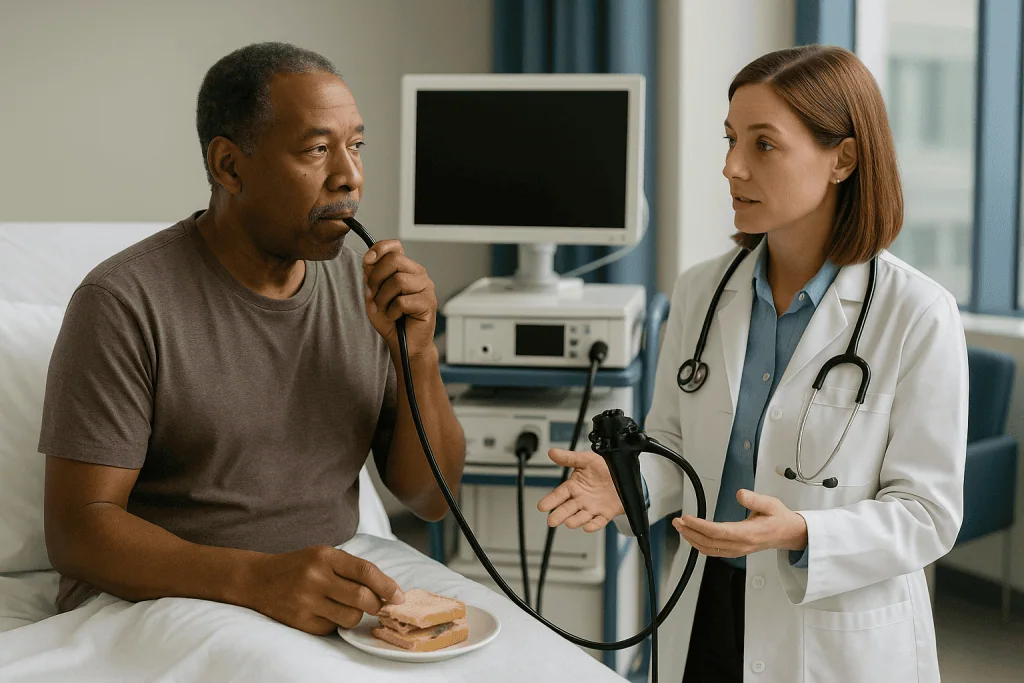
Preparation centers on an empty stomach. MedlinePlus advises no food for about 6 to 12 hours before the test, and the endoscopy unit will give exact instructions, including which regular medicines to take with a sip of water. A nurse places a small cannula in a vein for sedation, a numbing spray may be used on the throat, and a plastic guard protects the teeth. Sedation here is usually light to moderate; many people doze and remember little.
The endoscopist advances the scope slowly, often spending extra time at specific landmarks. After an esophagectomy, the key landmark is the anastomosis, the stitched or stapled join between the remaining esophagus and the stomach or bowel used to replace it. After chemoradiation, it is the site of the original tumor. After endoscopic resection, it is the scar and its margins. Modern scopes can switch to narrow-band light, which makes blood vessel patterns in the surface stand out and helps distinguish scar from suspicious tissue.
If anything looks uncertain, small biopsies are taken with forceps through the scope. This is painless and usually causes only a tiny spot of bleeding. If the join is narrowed, the endoscopist may pass a balloon or tapered dilator to stretch it during the same session, which is often why the appointment was made in the first place.
Afterward, there is a recovery bay, a drink once the throat numbness has worn off, and a short conversation about what was seen. Biopsy results typically take several days and are usually discussed at the next clinic visit or by phone.
Does esophageal cancer come back, and when is the risk highest?
People ask this directly, and it deserves a direct answer: yes, esophageal cancer can return after treatment, and follow-up schedules are built around that possibility. Both the NHS and Mayo Clinic describe regular check-ups after treatment precisely because recurrence is a recognized risk rather than a rare exception.
How likely it is for one person depends on things the treating team knows and a general article cannot: the stage at diagnosis, whether the cancer was adenocarcinoma (arising from gland cells, usually in the lower esophagus) or squamous cell carcinoma (arising from the flat surface cells), how many lymph nodes were involved, and how completely the tumor responded to treatment. Published percentages describe groups of patients, not individuals, so this article does not quote a single figure. Your oncologist can place your own situation within the evidence far more accurately.
What can be said in general terms is where and when recurrence tends to happen. It may be local, meaning at or near the original site or surgical join; regional, in nearby lymph nodes; or distant, in organs such as the liver, lungs or bones. Guidelines across major cancer bodies concentrate follow-up in the early years after treatment because this is when most recurrences are detected, with visits typically spaced further apart later on.
This explains the rhythm of the appointment letters. Frequent early, sparser later, and tailored to the type of treatment. It also explains why the follow-up plan after a large operation for a node-positive cancer looks different from the plan after endoscopic removal of a tiny early lesion. Neither plan is “more thorough”; each is matched to a different pattern of risk.
Surveillance after esophagectomy: what the team is looking for
An esophagectomy removes the diseased section of esophagus, usually with surrounding lymph nodes, and reconnects the digestive tract, most often by reshaping the stomach into a tube and bringing it up into the chest or neck. It is one of the larger operations in cancer surgery, and the follow-up afterward has three distinct threads.
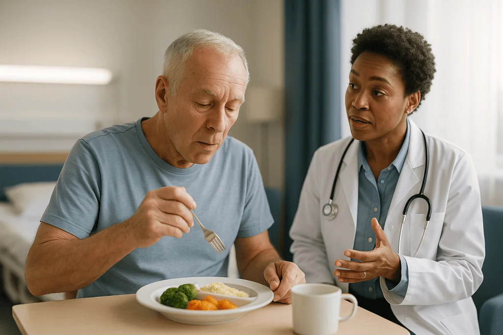
The first thread is the join. Scar tissue at the anastomosis can tighten over weeks to months, a problem called an anastomotic stricture. The classic sign is food sticking in the chest or a feeling of fullness after a few bites. Endoscopy both confirms the narrowing and treats it with dilation, sometimes in a series of sessions. Finding a stricture is not a sign that the cancer is back; it is a mechanical consequence of healing.
The second thread is the lining itself. The remaining esophagus above the join still has its original cells, and the pulled-up stomach is now exposed to a new environment. Reflux of acid and bile into the esophageal stump is common because the valve that normally guards the lower esophagus has been removed. Over time this can inflame the lining, and in some people can lead to Barrett’s esophagus, a change in the lining cells that itself warrants surveillance. The endoscopist looks for inflammation, ulcers and any new abnormal patches.
The third thread is recurrence, local or distant. Local regrowth at the join is checked by direct view and biopsy. Distant spread is beyond the reach of a scope, which is where imaging enters.
Between tests, the clinic visit matters as much as the technology. Weight trend, appetite, energy, coughing with meals and swallowing comfort are all recorded, and a change in any of them can bring an endoscopy or scan forward.
Follow-up after chemoradiation without surgery: why biopsies matter more
Some people are treated with chemotherapy and radiation together, called chemoradiation, and do not have surgery. This may be because the tumor is in the upper esophagus, because surgery carries too much risk for that person, or because the cancer responded so completely that the team and patient chose close monitoring instead of an operation. In this group, the esophagus is still in place, and so is the original tumor bed.
That changes the follow-up logic. After surgery, the tumor site is gone. After chemoradiation alone, the site remains and can harbor residual or regrowing cancer beneath a surface that looks healed. Radiation also leaves its own marks: pale scarred areas, fragile blood vessels, sometimes a narrowed segment. Telling scar from cancer by eye alone is genuinely difficult, which is why endoscopic surveillance in this setting leans heavily on biopsies, sometimes multiple samples from the treated zone even when nothing looks obviously wrong.
Endoscopic ultrasound, a scope with a small ultrasound probe on the tip, may be added. It lets the clinician look through the wall of the esophagus rather than just at its surface, and can sample lymph nodes next to it with a fine needle. Johns Hopkins describes this technique as part of the toolkit for assessing depth of involvement and nearby nodes.
Imaging is also woven in more tightly here. PET-CT, which combines a CT scan with a tracer that highlights metabolically active tissue, can suggest whether a treated area is still active, though radiation inflammation can light up too, so a hot spot on PET usually leads to a scope and biopsy rather than a conclusion on its own.
For people on an active surveillance pathway after a complete response, the stakes of each check are higher, since surgery remains an option if regrowth is found early. That is the trade being made, and it is a reasonable one for many, but it does ask for careful attendance.
Follow-up after endoscopic treatment of early cancer or Barrett's
Not every esophageal cancer needs an operation. Very early tumors confined to the innermost layers can sometimes be removed through the endoscope itself, using endoscopic mucosal resection (lifting and snaring the abnormal patch) or endoscopic submucosal dissection (cutting it out in one piece with fine instruments). Barrett’s esophagus with high-grade dysplasia, meaning cells that look markedly abnormal but have not yet invaded, is often treated the same way, sometimes followed by ablation, in which heat or cold destroys the remaining abnormal lining so normal cells can regrow.
These treatments preserve the esophagus, which is a large advantage for swallowing and quality of life. The price is a more intensive endoscopy schedule, at least at first. Three things are being watched.
The first is the resection site. The pathologist’s report on the removed tissue tells the team whether the margins were clear and how deep the cancer went. If the margins were close or involved, or the tumor reached deeper than expected, the team may recommend surgery or further endoscopic therapy, and surveillance will be closer.
The second is the rest of the Barrett’s segment. Abnormal cells can arise elsewhere in the same stretch of lining, so the whole segment is inspected and biopsied in a systematic pattern, often described as four-quadrant biopsies at regular intervals along its length. After ablation, the team checks that normal lining has returned and looks for buried glands, small islands of Barrett’s tissue hidden under the new surface.
The third is recurrence of Barrett’s itself after apparently successful ablation, which can happen and is the reason surveillance continues for years rather than stopping once the lining looks normal.
Imaging plays a smaller role for truly early disease, since the risk of distant spread is low. Here the scope does most of the work, and the schedule is set by the pathology, not the calendar alone.
CT scan after esophageal cancer: what imaging adds that a scope cannot
Endoscopy sees the inside of the tube. It cannot see through the wall into the chest or down into the liver, and most dangerous recurrences happen in exactly those places. That is the job of imaging.
A CT scan uses X-rays from many angles to build cross-sectional pictures of the chest, abdomen and sometimes pelvis. It is usually done with an injected contrast dye that makes blood vessels and organs stand out. On a follow-up CT, the radiologist compares every structure with the previous scan: the surgical bed, the lymph node stations along the esophagus and around the stomach, the liver, the lungs, the adrenal glands and the bones that appear in the field. A new nodule or an enlarging node is the kind of change that prompts further tests.
PET-CT adds a functional layer. The tracer, a sugar molecule tagged with a small amount of radioactivity, accumulates in tissues that are burning glucose quickly, which includes many cancers but also infection, inflammation and healing surgical sites. Mayo Clinic lists PET as one of the tools used to assess whether esophageal cancer has spread. In follow-up, it is often reserved for when a CT finding is unclear, when symptoms suggest recurrence but CT is unrevealing, or when a decision about further treatment hinges on the answer.
Which scan, how often, and whether imaging is scheduled routinely or triggered by symptoms varies between guidelines and between individuals. Some programs scan on a fixed timetable in the early years; others image mainly when something changes. Both approaches have evidence behind them, and the choice reflects stage, treatment and what would be done with the result.
Blood tests are part of the visit too, mostly to check nutrition, blood counts and organ function. There is no reliable blood tumor marker for esophageal cancer in routine follow-up, which is why nobody is relying on a single number from the lab.
Who is usually followed closely, and who is usually asked to wait
Follow-up intensity is not a reward or a worry index. It is matched to what the team expects to find and what they could do about it.
People who are usually followed more closely include those treated with chemoradiation alone, where the tumor site remains and salvage surgery might be possible if regrowth is caught early; those who had endoscopic resection or ablation, where the remaining lining needs systematic checking; those with a known stricture, reflux injury or Barrett’s segment that itself needs surveillance; and those whose pathology showed features linked to higher recurrence risk, such as involved lymph nodes or close margins. People who are candidates for further treatment if recurrence is found also tend to be scanned more regularly, because the information would change management.
People who may be asked to wait, or whose schedule is deliberately lighter, include those many years out from treatment with a stable series of normal tests; those with very early disease removed with clear margins, once the first intensive phase is complete; and, sometimes, those whose overall health means that aggressive further treatment would not be offered, where frequent scans would generate anxiety without changing decisions. In that last situation, follow-up often shifts toward symptom-led care and support for swallowing, nutrition and comfort, which is a legitimate and considered choice rather than neglect.
Timing of a specific test can also be deferred for good reasons. Endoscopy is usually postponed during an acute chest infection or soon after certain procedures. Contrast CT may be delayed if kidney function is temporarily reduced. A PET scan is less informative within weeks of surgery or radiation, when healing tissue is still metabolically active, so teams often wait for that inflammation to settle.
Whatever the schedule looks like, the decision sits with the treating team, and a schedule that has been thought through for you is more useful than one copied from someone else’s experience.
Esophageal cancer follow up endoscopy and imaging at a glance
The table below summarizes what each test is best at, what it cannot do, and when it tends to be used. Exact intervals are set by your team and depend on treatment type and stage.
| Test | What it shows well | What it misses | Typically used |
|---|---|---|---|
| Endoscopy (EGD) | Lining, surgical join, strictures, inflammation, Barrett’s; allows biopsy and dilation | Spread beyond the wall, lymph nodes, distant organs | After all treatment types; most frequent after endoscopic therapy and chemoradiation alone |
| Endoscopic ultrasound | Depth within the wall, adjacent lymph nodes; needle sampling | Distant disease; limited after heavy scarring | Selected cases, especially after chemoradiation without surgery |
| CT with contrast | Lymph nodes, liver, lungs, surgical bed, structural change over time | Small surface lesions in the lining; cannot biopsy | Routine or symptom-triggered in the early years, per team protocol |
| PET-CT | Metabolic activity suggesting active disease anywhere in the body | Can be falsely positive from inflammation or healing; small lesions | Clarifying uncertain CT findings or symptoms; before major treatment decisions |
| Clinic review and blood tests | Weight, swallowing, nutrition, organ function, new symptoms | Cannot detect recurrence on its own | Every follow-up visit |
Two things stand out. No single test covers everything, which is why they are combined rather than chosen between. And the clinic conversation, easily dismissed as the part before the real test, is where changes in weight or swallowing are picked up that bring a scope or scan forward. Treat it as a test in its own right and come prepared to describe what has changed since last time.
What the days around a follow-up test usually look like
Follow-up has a rhythm that becomes familiar, though rarely comfortable. A typical cycle runs over about a week.
The scan or scope comes first, often on a separate day from the clinic appointment so that results are available for discussion. For a CT with contrast, you may be asked to drink extra fluids beforehand and to have a recent kidney blood test on file. The scan itself takes minutes; the waiting takes longer. Contrast can cause a brief warm flush and a metallic taste, both normal. For endoscopy, the fasting window described earlier applies, and because of sedation you will need someone to take you home and should not drive, operate machinery or sign important documents for the rest of that day, as MedlinePlus advises.
The hours after an endoscopy usually bring a mildly sore throat and some bloating from air used to inflate the stomach. If a dilation was performed, there may be a sense of chest discomfort for a day. Eating usually restarts with soft foods once the throat is comfortable. After biopsies, a trace of blood in saliva is not unusual; more than that is a reason to call.
Then comes the gap. Radiology reports are usually ready within days; biopsy results take longer because tissue must be processed and examined. Many people describe this interval as the hardest part of the whole cycle, and that experience is common enough to have a name in the research literature. Planning something absorbing for those days is not avoidance; it is sensible.
The clinic visit closes the loop. Results are reviewed, weight is checked, swallowing and energy are discussed, and the next interval is agreed. If nothing has changed, the appointment may be brief, which can feel anticlimactic after a week of tension. Brief is good. Ask when the next tests are due and what, in the meantime, should prompt an earlier call.
Would a recurrence show up on endoscopy? Honest limits of each test
People often ask whether esophageal cancer would show up on endoscopy, and the answer is: usually, if it is on the lining and within reach of the scope. Endoscopy is the most sensitive way to find a surface recurrence or a new Barrett’s lesion, and biopsy makes the diagnosis definitive. It is the right tool for that question.
But recurrence does not always start on the surface. After esophagectomy, regrowth sometimes begins outside the join, in the tissue around it or in nearby lymph nodes, pressing inward only later. After chemoradiation, residual cancer can sit beneath a healed surface. A normal-looking lining on endoscopy therefore does not rule out recurrence, which is exactly why imaging is paired with it rather than replaced by it.
Imaging has its own blind spots. CT identifies abnormalities by size and shape, so a very small deposit may be too small to call, and a slightly enlarged lymph node may be reactive rather than cancerous. Scar tissue after surgery can look worrying on the first scan and then remain stable for years, which is why radiologists put so much weight on comparison with previous images. PET-CT improves on this by showing activity, but healing, infection and inflammation also take up the tracer, and uncertain PET findings frequently lead back to endoscopy and biopsy for confirmation.
Two practical consequences follow. First, a finding of “something that needs another look” is common and is not the same as a diagnosis of recurrence; many such findings turn out to be benign. Second, no test or combination of tests can promise that recurrence has been excluded. What a well-designed follow-up program does is shrink the window in which a problem can grow unnoticed, and give the team the best available chance of acting while options remain open.
Beyond scans: swallowing, weight, reflux and strictures
If you ask people living after esophageal cancer treatment what dominates daily life, many will not say scans. They will talk about eating. Follow-up that ignores this misses half the point.
After esophagectomy, the stomach has been reshaped into a narrow tube and no longer stores food the way it did. Small frequent meals replace three large ones. Some people experience dumping syndrome, in which food passes into the small bowel too quickly, causing cramping, sweating, light-headedness or diarrhea after eating, particularly after sugary foods. Dietitians are central here, and the Cleveland Clinic and NHS both describe nutritional support as a standard part of care after esophageal cancer treatment.
Reflux is nearly universal to some degree, because the valve at the lower end of the esophagus has been removed. Sleeping with the head of the bed raised and avoiding meals close to bedtime are the practical mainstays. Where medication is used, acid-suppressing medicines reduce the acid the stomach produces; whether one is appropriate, and for how long, is a question for the treating team, and it should never be started or stopped on the basis of an article.
Strictures deserve their own mention because they are so treatable and so often blamed on the cancer. A tightening join is a healing problem, and dilation through the endoscope, sometimes repeated over a few sessions, usually restores comfortable swallowing. Prompt reporting of food sticking means the problem is addressed before weight loss sets in.
Weight itself is tracked at every visit. A steady downward trend, even without obvious swallowing difficulty, is one of the more useful early signals that something needs attention, whether that is nutrition, a stricture or, less often, recurrence. Keeping a simple weekly weight note at home gives the team better data than a single clinic measurement.
What people often get wrong about follow-up after esophageal cancer
Several misunderstandings come up repeatedly in clinic, and clearing them away makes the whole process less frightening.
“If they keep scanning me, they must think it is coming back.” Scheduled follow-up is applied to everyone treated for this cancer, including those with the most favorable pathology. It is a standard of care, not a signal about your individual case.
“A clear scan means I am in the clear.” A clear scan means no abnormality was detectable at that moment with that test. It is reassuring and genuinely good news, but it does not change the schedule, because small changes can emerge between scans.
“The endoscopy is just a repeat of the scan.” They answer different questions. The scope sees the lining and the join and can biopsy and dilate; the scan sees nodes and organs. Skipping one because the other was normal leaves a gap.
“Difficulty swallowing again means the cancer is back.” After surgery, the most common reason is a benign stricture at the join, which is fixed by dilation. After radiation, scarring can do the same. Recurrence is one possibility among several, and endoscopy sorts them out.
“If I feel well I can delay the appointment.” Feeling well is the normal state in which early recurrence is found. Symptom-free surveillance exists precisely because waiting for symptoms often means waiting for a larger problem.
“Online survival statistics tell me my odds.” Published percentages describe large groups diagnosed years ago across every stage. They are a poor fit for any one person, and they lag behind current treatment. Your team can interpret the evidence against your own stage, pathology and response.
“There is a supplement or diet that prevents recurrence.” No dietary product has been shown in rigorous trials to prevent esophageal cancer from returning. Good nutrition after treatment matters for strength and recovery, which is a different and well-supported claim. Any supplement should be discussed with the team, since some interact with treatment.
Questions to ask your care team
Follow-up appointments can be short, and it is easy to leave with the questions you meant to ask still in your pocket. Writing them down beforehand helps. These are the ones that tend to produce the most useful answers.
- What is my follow-up schedule for the next year, and which tests are planned at each visit?
- Which of these tests are routine for everyone with my treatment, and which are specific to something in my pathology or scans?
- Was anything seen on the last endoscopy or scan that you are keeping an eye on, even if it is probably benign?
- If a recurrence were found, what would the options be, and does that affect how often you want to look?
- What symptoms should bring me back earlier than the next scheduled visit, and who do I contact?
- Do I have a stricture, reflux injury or Barrett’s that needs its own surveillance plan?
- Who is monitoring my weight and nutrition, and should I be seeing a dietitian between visits?
- How long will follow-up continue, and what would a decision to space out or stop tests be based on?
- Am I at risk of a second cancer elsewhere, for example in the head and neck or in the remaining esophagus, and is anything being checked for that?
- Is there support available for the anxiety around scans and results?
Bring a companion if you can; two memories are better than one, and the person who sits beside you often notices changes in eating or energy that you have stopped registering. Ask for a written summary of the plan. Most teams are glad to provide one, and it turns a series of appointment letters into something you can understand and anticipate.
When to call your doctor: esophageal cancer recurrence symptoms that need prompt review
Scheduled tests are the backbone of follow-up, but the plan assumes you will also report changes between visits. None of the signs below is a diagnosis; each is a reason for an earlier conversation and possibly an earlier endoscopy or scan. The cause is often benign and fixable, and finding out sooner is better either way.
Contact your care team promptly, rather than waiting for the next appointment, if you notice food sticking or swallowing becoming harder than it was, especially if it is progressing week by week; unintended weight loss or a steady downward drift on the scales; new or worsening pain in the chest, upper abdomen or back that does not settle; vomiting that is new or persistent, or vomiting blood; black, tarry stools, which can indicate bleeding higher in the digestive tract; a new hoarse voice lasting more than a couple of weeks; a persistent cough, particularly coughing when eating or drinking, which can suggest food entering the airway; or new shortness of breath.
Seek urgent same-day care, through emergency services if needed, for vomiting significant amounts of blood, passing large amounts of black or bloody stool, severe chest pain, sudden breathlessness, fever with chest pain after a recent endoscopy or dilation, or inability to swallow your own saliva. These can indicate bleeding, a tear in the esophagus or aspiration, all of which need assessment without delay.
Keep the clinic’s contact number and the name of your specialist nurse or coordinator somewhere easy to find. Teams expect these calls, and a call that turns out to be about a benign stricture is still a call worth making. Every decision about further tests or treatment rests with the clinicians who know your history, and the sooner they have the information, the more options they are likely to have.
Frequently asked questions
How often does esophageal cancer return after treatment?
Recurrence is a recognized risk after esophageal cancer treatment, which is why structured follow-up exists, but the likelihood varies widely with stage, cell type, lymph node involvement and response to treatment. Published figures describe groups rather than individuals, so they are not quoted here. Your oncology team can place your own pathology and scans against the evidence and give you a far more meaningful picture.
What is the 5-year survival rate for esophageal cancer?
Population survival statistics for esophageal cancer are strongly stage-dependent and are drawn from large registries of people diagnosed years ago, so a single headline number says little about any one person and lags behind current treatment. For that reason this article does not quote one. Ask your treating team how the evidence applies to your stage, treatment and response; that conversation is more accurate and more useful than a search result.
Would esophageal cancer show up on endoscopy?
Usually yes, if it involves the lining within reach of the scope, and biopsy during the same procedure can confirm it. Endoscopy is the most sensitive way to find a surface recurrence or a new Barrett’s lesion. It cannot see spread outside the esophageal wall, into lymph nodes or distant organs, which is why follow-up pairs it with CT or PET-CT rather than relying on either test alone.
How treatable is esophageal cancer if it comes back?
It depends on where and when it returns and on the person’s overall health. A small local regrowth found early at a surveillance visit may be treatable with further endoscopic therapy, surgery or radiation; widespread recurrence is usually managed with systemic treatment aimed at control and symptom relief. Finding change early widens the options, which is the central argument for attending scheduled follow-up even when you feel well.
Does esophageal cancer come back without any symptoms?
It can, and this is the main reason follow-up is scheduled rather than symptom-driven. Early local recurrence at the surgical join or a small lymph node deposit may cause no change in swallowing, weight or energy for months. Scheduled endoscopy and imaging are designed to find such changes during that silent window, when they are smaller and more options for treatment remain available.
How long does surveillance after esophagectomy continue?
Most programs follow people for several years, with the closest monitoring in the early period and visits spaced out as time passes without problems. Some aspects, such as checking the remaining esophagus for reflux damage or Barrett’s change, may continue longer. The length and intensity of follow-up are set by the treating team based on stage, pathology and what further treatment would be possible if something were found.
Is a CT scan after esophageal cancer treatment done routinely or only if something is wrong?
Both approaches are used, and the choice varies between guidelines and individuals. Some teams scan on a fixed timetable in the early years; others image mainly when symptoms, weight or endoscopy findings change. The deciding factors are stage, treatment type and whether a finding would change management. Ask your team which approach applies to you and why, so the schedule makes sense rather than feeling arbitrary.
Will I need biopsies at every follow-up endoscopy?
Not necessarily. After esophagectomy with a healthy-looking join, biopsies are often taken only if something appears abnormal. After chemoradiation alone or endoscopic treatment of Barrett’s esophagus, systematic biopsies of the treated area are more routine because scar and residual abnormal cells can look alike. Biopsies are quick and painless during the procedure, and results typically take several days to return from the pathology laboratory.
Why do I keep needing dilation after esophageal surgery?
Scar tissue at the anastomosis, the join between the remaining esophagus and the reconstructed stomach, can tighten as it heals and may re-tighten after being stretched. Dilation with a balloon or tapered dilator through the endoscope is often repeated over a few sessions until the opening stays comfortable. Needing dilation is a healing issue, not a sign of recurrence, and reporting food sticking early keeps it manageable.
Does the anxiety before every scan ever get easier?
For many people it eases over time as normal results accumulate, though a spike before results is common and well recognized in cancer care. Practical steps help: knowing exactly when results will be given, having a companion at the visit, and planning absorbing activities for the waiting days. If worry is affecting sleep, eating or daily life, tell your team; psychological support is a standard part of cancer follow-up.
References
- NHS: Oesophageal cancer
- MedlinePlus: EGD (esophagogastroduodenoscopy)
- Cleveland Clinic: Esophageal cancer
This article is for general information only and is not a substitute for professional medical advice. Please consult a qualified doctor about your individual situation.

More from the Blog
Recovering From Head and Neck Cancer Surgery: Breathing, Swallowing Therapy and Feeding
Recovery after head and neck cancer surgery usually means several days to two weeks in hospital, then weeks to months of rehabilitation. Breathing may…
IMRT Planning Day Explained: Simulation CT, Immobilization Mask and Skin Marks
An IMRT simulation planning CT is the mapping appointment before radiation treatment begins. You lie in the exact position you will use for every…

Why Chronic Lymphocytic Leukemia Is Often Watched First, and What Triggers Treatment
Chronic lymphocytic leukemia is often watched rather than treated at first because trials have not shown that starting treatment early helps people live longer,…
The Days Before Radiation Therapy Starts: Simulation, Skin Marks and a Checklist to Follow
Preparing for radiation therapy usually means attending a planning appointment called a simulation, where you lie in your treatment position for a CT scan,…
Bleeding, Fever or Pain After Endometrial Cancer Surgery: Which Signs Need a Call
After a hysterectomy for endometrial cancer, call your surgical team the same day for bleeding heavier than a light period or that suddenly restarts,…

When Does Leukemia Treatment Include a Stem Cell Transplant, and Who Is Assessed for It?
A stem cell transplant becomes part of leukemia treatment when the disease is judged likely to return after chemotherapy alone, or when it has…