

Graves Disease Work-Up: Thyroid Hormone Tests, Antibodies, Ultrasound and Scintigraphy

Key Takeaways
- A very low or undetectable TSH with raised free T4 or T3 confirms hyperthyroidism, but it does not by itself reveal whether Graves disease is the cause.
- TSH receptor antibodies (TRAb or TSI) are positive in most people with Graves disease, and a positive result often makes imaging unnecessary.
- A radioactive iodine uptake scan shows diffusely high uptake in Graves disease, low uptake in thyroiditis, and focal hot spots in toxic nodules.
- Scintigraphy is not performed in pregnancy, so Doppler ultrasound, which shows markedly increased blood flow in Graves disease, takes its place.
- High-dose biotin supplements and recent iodine contrast can distort thyroid blood tests and uptake scans, which is why surprising results are often repeated.
- TSH can stay suppressed for months after hormone levels normalize on treatment, so an early low TSH is not a sign the plan is failing.
Graves disease is usually diagnosed with blood tests first: a low or undetectable TSH with raised free T4 or T3 confirms an overactive thyroid, and a positive TSH receptor antibody (TRAb or TSI) test points to Graves as the cause. When antibodies are unclear, a radioactive iodine uptake scan or a thyroid ultrasound with Doppler helps separate Graves from thyroiditis or nodules. Your clinician interprets the pattern together.
The first clue is often a stranger’s comment. A colleague asks why your hands are shaking over a coffee cup. A friend notices that your eyes look wider than they used to. You have lost weight without trying, you are warm when everyone else is reaching for a sweater, and your heart seems to be running a race you did not enter.
By the time someone types graves disease diagnosis tests into a search bar, they usually already suspect what is going on. What they want to know is how a clinician turns a suspicion into a confirmed answer, which tests come first, which can wait, and why one person gets a scan while another does not.
The honest answer is that the work-up is a sequence, not a single tick box. Each test answers a different question: Is the thyroid overactive? Is the immune system driving it? Is the whole gland involved or just one lump? This explainer walks through that sequence in order.
What is Graves disease, and why is diagnosing it more than one blood test?
Graves disease is an autoimmune condition, meaning the immune system makes antibodies that mistakenly target the body’s own tissue. In this case the target is the thyroid, the butterfly-shaped gland at the front of the neck that sets the pace of metabolism. The antibodies lock onto the same receptor that the brain’s control hormone normally uses, and they press the accelerator continuously.
The result is hyperthyroidism, a state in which the gland releases more thyroid hormone than the body needs. According to the National Institute of Diabetes and Digestive and Kidney Diseases (NIDDK), Graves disease is the most common cause of hyperthyroidism in the United States, and women are affected far more often than men.
Here is the catch that shapes the whole work-up. Several unrelated problems can also make the thyroid overactive: an inflamed gland leaking stored hormone (thyroiditis), one or more nodules that produce hormone on their own, or taking too much thyroid hormone. A blood test showing hyperthyroidism tells your clinician that the pace is too fast. It does not, on its own, say why.
That distinction matters because the causes behave differently over time. Thyroiditis often settles on its own within months. Nodules do not. Graves disease can relapse and remit. Treatment conversations, follow-up schedules and even the safety of certain procedures depend on which cause is in play. So the diagnostic path is built as a series of questions: first confirm the overactivity, then look for the antibodies that define Graves, then, if the picture is still unclear, image the gland to see how it is behaving.
Thinking of the tests as a sequence rather than a shopping list also explains why two people with the same symptoms can leave their appointments with different orders in hand.
How Graves disease diagnosis tests work: the TSH feedback loop explained
To read thyroid results, it helps to know the plumbing. The pituitary, a pea-sized gland under the brain, releases thyroid-stimulating hormone (TSH). TSH travels to the thyroid and tells it to make two hormones: thyroxine, called T4, and triiodothyronine, called T3. When T4 and T3 rise, the pituitary senses it and turns TSH down. When they fall, TSH goes up. It is a thermostat.

In Graves disease the thermostat is bypassed. Antibodies stimulate the thyroid directly, so T4 and T3 climb regardless of what the pituitary wants. The pituitary responds the only way it can: it shuts TSH off almost completely. That is why the signature laboratory pattern, described by Mayo Clinic and the NHS, is a very low or undetectable TSH paired with a high free T4, a high T3, or both.
The word “free” is worth a sentence. Most thyroid hormone in the blood is bound to carrier proteins and inactive. Free T4 measures the small unbound fraction that actually acts on tissues, which is why clinicians prefer it to total T4, especially in pregnancy or in people taking estrogen, when carrier protein levels shift.
Because TSH is so sensitive, it is usually the first test run. The NHS describes a low TSH as the earliest and most reliable laboratory sign that the thyroid is overactive. If TSH is normal, true hyperthyroidism from the thyroid itself is very unlikely, and the search for the cause of symptoms moves elsewhere.
One more pattern deserves a mention: a low TSH with normal free T4 and T3. This is called subclinical hyperthyroidism, meaning the thermostat has reacted but hormone levels have not yet crossed the laboratory threshold. It is often rechecked after several weeks rather than acted on immediately, a decision that rests with the treating clinician.
Graves disease blood test: what the result pattern typically looks like
Most people receive their results as a list of numbers with reference ranges beside them. Reading the pattern is more useful than fixating on any single value, because laboratories use slightly different ranges and units.
| Result pattern | What it usually suggests | Typical next step |
|---|---|---|
| Low TSH, high free T4 and/or high T3 | Overt hyperthyroidism | Antibody testing to find the cause |
| Low TSH, normal free T4 and T3 | Subclinical hyperthyroidism | Repeat testing after an interval |
| Low TSH, high T3, normal free T4 | T3-predominant hyperthyroidism, which can occur early in Graves | Antibody testing; clinician review |
| Normal TSH, normal hormones | Thyroid overactivity unlikely | Look for other explanations |
| High TSH, high free T4 | Rare pituitary-driven pattern | Specialist assessment |
Mayo Clinic notes that in Graves disease, T3 is sometimes raised out of proportion to T4, so a clinician who orders only TSH and free T4 can occasionally miss the full picture. Many request all three when the suspicion is high.
A few things can distort these numbers without any thyroid disease at all. Acute illness, hospitalization and certain medicines temporarily lower TSH. High-dose biotin supplements, popular for hair and nails, can interfere with the laboratory method and produce results that mimic hyperthyroidism; the NIH Office of Dietary Supplements advises telling your clinician about biotin before blood draws. This is why a surprising result is often repeated before anyone acts on it.
Blood tests also give the clinician a baseline for later. Once treatment begins, the same panel is used to track response, so the first set of numbers is a reference point, not just a verdict.
TRAb antibody test and TSI test: what the antibodies actually prove
Once hyperthyroidism is confirmed, the next question is whether the immune system is responsible. Two closely related tests answer it.
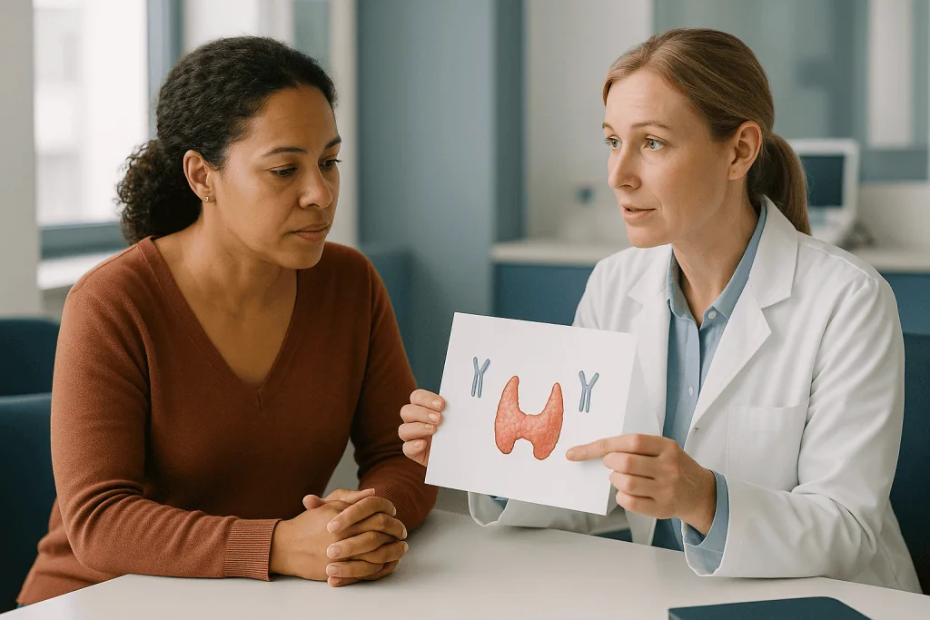
The TRAb test measures TSH receptor antibodies, meaning any antibody that binds to the receptor the pituitary’s hormone normally uses. The TSI test, short for thyroid-stimulating immunoglobulin, is a more specific version that measures only the antibodies that actively switch the receptor on. MedlinePlus describes both as blood tests used to help diagnose Graves disease and to distinguish it from other causes of an overactive thyroid.
Why does the distinction matter? TRAb assays detect stimulating antibodies but can also pick up blocking or neutral antibodies, which bind without accelerating the gland. In practice, when a person clearly has hyperthyroidism and a positive TRAb, the antibodies are almost always the stimulating kind, so many laboratories use TRAb as the routine test and reserve TSI for ambiguous cases. Your clinician chooses based on local availability and the clinical question.
A positive result in someone with confirmed hyperthyroidism is strong evidence for Graves disease, and it often allows the diagnosis to be made without any imaging. A negative result does not fully rule Graves out; a small proportion of people with the condition test negative, particularly early on or when the disease is mild, which is when a scan or ultrasound becomes useful.
Antibody testing has one more role. Mayo Clinic notes that TRAb levels are sometimes rechecked later in the course because high or rising levels can be relevant to decisions about continuing or stopping treatment. In pregnancy, antibody levels carry an added purpose: stimulating antibodies can cross the placenta, so clinicians monitor them to anticipate effects on the baby’s thyroid. That interpretation always belongs to the treating team.
A third antibody, thyroid peroxidase antibody (TPO), is often positive in Graves disease too, but it is also common in the underactive autoimmune condition Hashimoto thyroiditis, so it supports rather than confirms the diagnosis.
Thyroid scan uptake test: what scintigraphy shows that blood cannot
Scintigraphy is nuclear medicine imaging. A small amount of a radioactive tracer, usually a form of iodine or technetium, is swallowed or injected, and a gamma camera records where the thyroid takes it up. The thyroid is the only organ that actively concentrates iodine, which it uses to build hormone, so the tracer effectively maps how hard each part of the gland is working.
Two things are measured. The uptake is the percentage of tracer the gland absorbs, often recorded at a few hours and again at 24 hours, according to Mayo Clinic’s description of the radioactive iodine uptake test. The scan is the picture itself.
The patterns are distinctive. In Graves disease the whole gland is overactive, so uptake is high and the image is diffusely bright, both lobes glowing evenly. In toxic nodules, uptake concentrates in one or more hot spots while the rest of the gland appears dim, because excess hormone from the nodule has suppressed TSH and switched off the normal tissue. In thyroiditis the gland is leaking stored hormone rather than manufacturing it, so uptake is low even though blood hormone levels are high. The same is true when hyperthyroidism is caused by taking thyroid hormone tablets.
That contrast is why scintigraphy is so valuable when antibodies are negative or when a clinician feels a lump. It separates a gland that is overproducing from one that is overflowing.
The radiation dose is small, but it is not zero, and the tracer concentrates in the thyroid of a developing baby. Scintigraphy is therefore not performed in pregnancy, and breastfeeding usually needs to be paused or planned around the scan; the NIDDK and Mayo Clinic both flag this. Recent iodinated contrast from a CT scan, iodine-rich supplements and some heart medicines can flood the gland with ordinary iodine and make the uptake falsely low, so the test is often delayed for several weeks after such exposures.
Thyroid ultrasound in Graves disease: when sound replaces radiation
Ultrasound uses high-frequency sound waves, not radiation, to build a picture of the gland. A handheld probe glides over the neck with gel, the appointment takes minutes, and there is nothing to swallow or inject. For many people it is the gentlest step in the work-up.
What does it show? In Graves disease the thyroid is typically enlarged on both sides and looks darker and more uniform than a healthy gland, reflecting the dense, overworked tissue. The real diagnostic value comes from Doppler, a setting that displays blood flow in color. Graves disease produces markedly increased flow through the gland, a pattern sometimes nicknamed the “thyroid inferno” in radiology literature. Thyroiditis, by contrast, tends to show reduced or patchy flow because the inflamed tissue is not actively producing hormone.
Ultrasound is also the best tool for examining nodules. If a clinician feels a lump, or if scintigraphy shows a cold area that takes up no tracer, ultrasound characterizes its size, borders and texture and helps decide whether a biopsy is warranted. Graves disease and nodules can coexist, so finding one does not exclude the other.
Where ultrasound truly earns its place is in situations where scintigraphy is off the table. In pregnancy, during breastfeeding, after recent iodine contrast, or when a person simply prefers to avoid a radioactive tracer, Doppler ultrasound can provide much of the same discriminating information. The NHS lists it among the tests that may be used when the cause of an overactive thyroid is unclear.
Its limits are worth knowing. Ultrasound findings in Graves overlap with those of some other thyroid conditions, and the interpretation depends on the operator’s experience. It is best read alongside blood results and antibodies rather than in isolation. A normal-looking ultrasound does not rule out Graves disease, and an enlarged gland does not confirm it.
Graves disease diagnosis tests compared: which test answers which question
Patients often ask why they did not receive every test. The answer is that each test is designed for a specific question, and once that question is answered convincingly, the next test adds little. The table below summarizes what each one contributes.
| Test | Question it answers | Strength | Main limitation |
|---|---|---|---|
| TSH | Is the thyroid overactive? | Highly sensitive; first-line | Does not reveal the cause |
| Free T4 and T3 | How overactive, and in what pattern? | Grades severity; tracks treatment | Affected by illness, biotin, some medicines |
| TRAb / TSI | Is the immune system driving it? | Positive result strongly indicates Graves | Negative in a minority with Graves |
| Radioactive iodine uptake and scan | Is the gland overproducing or leaking? Whole gland or a nodule? | Clearly separates Graves, nodules, thyroiditis | Not used in pregnancy; iodine exposure distorts it |
| Ultrasound with Doppler | What does the gland look like, and how is blood flowing? | No radiation; excellent for nodules | Operator-dependent; findings overlap |
Reading across the rows, a typical path emerges. A low TSH with raised hormones and a positive TRAb settles the diagnosis in most people, and clinicians frequently stop there. A negative antibody result, a lump on examination, or a confusing hormone pattern sends the work-up to imaging. Pregnancy or recent contrast steers that imaging toward ultrasound rather than scintigraphy.
The Mayo Clinic and NHS both describe this stepwise approach. What looks like inconsistency between patients is usually the sequence doing its job: stopping when the answer is clear, continuing when it is not. If you are unsure why a particular test was or was not ordered, that is a fair and useful question for your clinician.
Who is tested right away, and who is usually asked to wait or test differently?
Not everyone follows the same timetable. Several groups are prioritized, and several are routed around particular tests.
People with a very fast or irregular heartbeat, marked weight loss, or eye changes are usually tested promptly, because untreated hyperthyroidism strains the heart and bones. Older adults deserve particular attention: the NIDDK notes that hyperthyroidism in later life can present with fatigue, a new heart rhythm problem or weight loss rather than the classic restlessness, so clinicians often have a lower threshold to check TSH.
Pregnant and breastfeeding women are tested quickly but differently. Blood tests and antibodies proceed as usual, and free T4 is interpreted against pregnancy-specific ranges because normal pregnancy shifts the numbers. Scintigraphy is avoided entirely, as both Mayo Clinic and the NIDDK state, and ultrasound takes its place when imaging is needed.
Those who recently had a CT scan with iodinated contrast, took iodine-containing supplements or started an iodine-rich heart medicine are typically asked to wait before a radioactive iodine uptake test, because the gland is already saturated with ordinary iodine and the tracer reading would be misleadingly low. The waiting period is judged by the clinician based on the exposure.
People taking high-dose biotin are generally asked to pause the supplement for a period before repeat blood work, following the interference caution from the NIH Office of Dietary Supplements, so the laboratory reading reflects true hormone levels.
Someone recovering from a serious illness or hospital stay may be asked to retest after a few weeks rather than be labeled with a thyroid condition straight away, since acute illness can suppress TSH temporarily.
Finally, a person with subclinical results, a low TSH but normal hormones and no symptoms, is often observed with repeat testing rather than imaged immediately. The interval and the decision belong to the treating team, who weigh age, heart health and bone health in choosing how actively to pursue a diagnosis.
What happens in the days and weeks after a Graves disease diagnosis?
A confirmed diagnosis usually leads to a conversation about three broad approaches, which the NIDDK, Mayo Clinic and NHS all describe: antithyroid medicines, radioactive iodine treatment and surgery to remove the gland. The choice depends on age, eye involvement, pregnancy plans, gland size and personal preference, and it is made with the treating team, never from an article.
Many people also receive a beta-blocker early on. This class of medicine does not change thyroid hormone levels; it blunts the hormone’s effect on the heart and nervous system, so the racing pulse, tremor and anxiety ease while the underlying treatment takes hold. Clinicians usually review its use as hormone levels come down.
Antithyroid medicines belong to a class called thionamides. They work by blocking the enzyme the thyroid uses to attach iodine to hormone, slowing production at the source. Because the gland holds a reserve of pre-made hormone, people do not feel the effect overnight. The NHS describes symptoms typically improving over about one to two months, and a standard course often lasts in the range of 12 to 18 months according to Mayo Clinic, after which the team reassesses.
Follow-up blood tests are the rhythm of those months. Free T4 and T3 are rechecked at intervals the clinician sets, often every few weeks at first, to guide adjustments. TSH is a laggard: Mayo Clinic notes it can remain suppressed for months even after hormone levels normalize, because the pituitary takes time to recover. A still-low TSH early in treatment is therefore not necessarily a sign of failure.
Clinicians also watch for the known side effects of thionamides, including rash, joint pain and, rarely, a sharp drop in infection-fighting white cells, which is why people are told to report fever or a sore throat promptly. Radioactive iodine and surgery follow their own timelines and typically lead to an underactive thyroid that is then managed with replacement hormone. Each path has trade-offs the team will walk through.
Does Graves disease make you more emotional? Mood, eyes and other clues clinicians look for
Yes, and there is a mechanism behind it. Thyroid hormone sets the sensitivity of the nervous system, and excess amounts leave people irritable, anxious, tearful or unable to sleep. Mayo Clinic and the NIDDK both list anxiety, irritability and mood changes among the common features of hyperthyroidism. People sometimes describe feeling as if they have had far too much coffee for months, with emotions arriving faster and larger than usual. Partners and friends often notice before the person does.
This matters diagnostically for a sobering reason: emotional symptoms are easy to attribute to stress, burnout or a mental health condition, and the thyroid can go unchecked. A TSH test is simple and is often part of a sensible assessment when anxiety or mood change appears without an obvious trigger, particularly alongside weight loss, heat intolerance or palpitations. Treating the thyroid usually eases the mood symptoms over the same weeks that hormone levels settle, although anyone with ongoing distress deserves support in its own right.
The eyes are the other distinctive clue. Thyroid eye disease, also called Graves ophthalmopathy, is an immune reaction in the tissues behind the eye that causes swelling, a staring or bulging appearance, grittiness and sometimes double vision. Mayo Clinic estimates that roughly 30 percent of people with Graves disease develop some degree of it. Because it is specific to Graves rather than to hyperthyroidism in general, its presence helps clinicians feel confident in the diagnosis even before antibodies return, and it influences treatment choice, since radioactive iodine can worsen eye disease in some people.
Clinicians also look at the neck for a smooth, symmetrical enlargement called a goiter, listen for the whoosh of increased blood flow over the gland, and check the shins for a thickened, reddish skin change called pretibial dermopathy, which is uncommon but, when present, is nearly unique to Graves disease. None of these findings replaces the tests. They are the context that tells the clinician which tests to order first.
Diet and exercise with Graves disease: Mediterranean eating, iodine and the right kind of movement
Two of the most common questions after diagnosis are about food and exercise, and both deserve honest answers rather than enthusiasm.
Is a Mediterranean diet good for Graves disease? There is no trial evidence that any diet treats Graves disease or lowers thyroid antibodies, and claims that a specific eating pattern will reverse the condition are not supported by mainstream evidence. That said, a Mediterranean-style pattern built around vegetables, legumes, whole grains, fish and olive oil is a well-studied approach to heart health, as the American Heart Association describes, and heart health is relevant because hyperthyroidism places extra strain on the cardiovascular system. People who have lost weight and muscle during untreated disease also benefit from adequate protein and calcium, since excess thyroid hormone accelerates bone turnover. The sensible framing is that good general nutrition supports recovery; it does not substitute for treatment.
Iodine deserves a specific caution. The thyroid uses iodine to make hormone, and high-dose iodine supplements or kelp products can worsen hyperthyroidism and distort uptake tests, as noted earlier. The NIH Office of Dietary Supplements advises against exceeding recommended intakes, and anyone with Graves disease should discuss supplements with their clinician before taking them.
What is the best type of exercise for Graves disease? While hormone levels are still high, the heart is already working harder than normal at rest, and intense exercise adds further load. Clinicians commonly suggest gentle activity such as walking, stretching or light strength work until levels are controlled, then a gradual return to usual intensity. Weight-bearing and resistance exercise are valuable later because they help rebuild bone and muscle lost during the overactive phase. Anyone with palpitations, chest discomfort or breathlessness during activity should stop and report it. The threshold for resuming vigorous exercise is a decision for the treating team, informed by heart rate and repeat blood tests rather than by how a person feels on a given day.
What people often get wrong about Graves disease testing
Misunderstandings cluster around a handful of ideas, and correcting them saves anxiety.
The first is that a normal TSH on a routine panel years ago rules out thyroid disease today. Graves disease can begin at any age and often appears after a trigger such as a stressful period, pregnancy or a viral illness; a past normal result says nothing about the present.
The second is that “my T4 is normal, so my thyroid is fine.” As described earlier, some people with Graves have a T3-predominant pattern with a normal free T4, and TSH is the more sensitive marker in any case. Reading one number in isolation is a common route to a missed diagnosis.
The third is that antibodies must be positive for the diagnosis to stand. A minority of people with genuine Graves disease test negative, especially early. Clinicians use imaging and clinical features, like eye disease, to reach the answer in those cases. Conversely, a positive antibody test with a normal TSH and no symptoms does not mean active disease; antibodies can be present at low levels in people who never develop hyperthyroidism.
The fourth is that the radioactive scan is dangerous. The tracer dose used for diagnosis is small and is cleared within days. It is withheld in pregnancy and planned around breastfeeding out of appropriate caution for the baby’s thyroid, not because it harms adults. The scan is a different procedure from radioactive iodine treatment, which uses a far larger activity to deliberately shrink the gland.
The fifth is that diet or supplements can replace testing or treatment. No food lowers thyroid antibodies, and some supplements, particularly iodine and biotin, actively interfere with the work-up.
The last is that Graves disease is a problem only of the thyroid. It is an immune condition that can affect the eyes, skin, heart and bones, which is why the work-up is designed to characterize the cause and not merely confirm the overactivity.
Questions to ask your care team about your Graves disease work-up
Appointments are short and results come with jargon. Arriving with specific questions turns a monologue into a conversation. Consider asking some of the following, adapted to your situation.
- Which of my results confirmed that my thyroid is overactive, and which one points to Graves disease as the cause?
- Were TSH receptor antibodies (TRAb or TSI) measured, and what did the result show? If they were negative, how was the diagnosis reached?
- Do I need a radioactive iodine uptake scan or an ultrasound, and if not, why is the diagnosis already clear enough without imaging?
- Is there anything I am taking, including biotin, iodine supplements or herbal products, that could have affected my results or that I should pause before repeat tests?
- Have I had any recent contrast scans or iodine exposure that might change the timing of further testing?
- Are there signs of thyroid eye disease, and does that affect which treatment options you would consider for me?
- Which blood tests will be repeated during treatment, how often, and which number will you be watching most closely?
- How long should I expect before symptoms begin to ease, and what would tell you that the treatment is not working as hoped?
- What side effects of the medicine should I report immediately, and whom do I contact outside office hours?
- I am planning a pregnancy, or I am pregnant or breastfeeding: how does that change the tests and the treatment plan?
- What activity level is reasonable for me right now, and when would you expect me to return to more vigorous exercise?
- Is there a nurse, pharmacist or patient education resource I can turn to with questions between visits?
You are entitled to a copy of your results, and it is reasonable to ask for the actual values alongside the laboratory’s reference ranges so you can follow your own trend over time. Writing down the answers, or bringing someone with you, helps on days when the diagnosis itself is a lot to absorb.
When to call your doctor: red-flag signs during a Graves disease work-up
Most of the Graves disease journey unfolds at a measured pace, with tests and reviews spaced weeks apart. A small number of situations should not wait for the next scheduled appointment.
Seek emergency care immediately if you develop a very rapid or irregular heartbeat with chest pain, breathlessness, fainting or confusion, or a high fever with agitation, vomiting or diarrhea. These can signal thyroid storm, a rare but life-threatening surge of thyroid hormone that the NIDDK and Mayo Clinic describe as a medical emergency, sometimes triggered by infection, surgery or stopping treatment abruptly.
Call your clinician the same day if you have started an antithyroid medicine and develop a fever, sore throat or mouth ulcers. These can be the first signs of agranulocytosis, a rare but serious drop in white blood cells that the NHS specifically advises reporting without delay so a blood count can be checked. The same urgency applies to yellowing of the skin or eyes, dark urine or severe abdominal pain, which may indicate a liver reaction.
Contact the team promptly, within a day or two, for sudden worsening of eye symptoms: new double vision, eye pain, a rapid increase in bulging, or any change in color vision or sharpness. Thyroid eye disease can occasionally threaten sight and responds best when assessed early. Marked swelling in the neck that affects swallowing or breathing also warrants a quick call.
Let your clinician know, even if it feels minor, if you become pregnant during the work-up or treatment, if you were given iodinated contrast for another scan, if you start a new supplement, or if a scheduled scan or blood test was missed. Each of these can change the interpretation of results or the safety of a planned test.
If you are unsure whether a symptom counts, the safe default is to call and describe it. Clinicians would far rather hear about a false alarm than learn later that something was missed.
Frequently asked questions
What is the first graves disease blood test a doctor usually orders?
TSH is almost always the first test. It is the most sensitive marker of an overactive thyroid because the pituitary shuts it down as soon as thyroid hormone levels rise. If TSH is low, the laboratory usually adds free T4 and T3 to confirm and grade the hyperthyroidism, then TSH receptor antibodies to find out whether Graves disease is the cause.
What does the TRAb antibody test show, and is it the same as the TSI test?
The TRAb test detects antibodies against the TSH receptor, which is the hallmark of Graves disease. The TSI test is a more specific version that measures only the antibodies that actively stimulate the receptor. In someone with confirmed hyperthyroidism, a positive result on either strongly supports Graves disease. Clinicians choose between them based on availability and the clinical question.
Can Graves disease be diagnosed without a thyroid scan uptake test?
Yes, frequently. When blood tests show hyperthyroidism and TSH receptor antibodies are positive, many clinicians consider the diagnosis settled without imaging. A radioactive iodine uptake scan is most useful when antibodies are negative, when a nodule is felt, or when the hormone pattern is confusing. In pregnancy the scan is avoided and ultrasound is used instead.
Does Graves disease make you more emotional or anxious?
It commonly does. Excess thyroid hormone heightens the nervous system, producing anxiety, irritability, tearfulness and poor sleep, which Mayo Clinic and the NIDDK list among typical features. These symptoms are easily mistaken for stress alone, so a TSH test is often reasonable when mood changes appear alongside weight loss or palpitations. Mood usually settles as hormone levels are brought under control.
Is the Mediterranean diet good for people with Graves disease?
No diet has been shown to treat Graves disease or reduce thyroid antibodies, so it should not replace medical care. A Mediterranean-style pattern is well supported for heart health, which matters because hyperthyroidism strains the cardiovascular system, and adequate protein and calcium help rebuild muscle and bone. Avoid high-dose iodine supplements and kelp, which can worsen hyperthyroidism and distort tests.
What is the best type of exercise for Graves disease?
While hormone levels remain high, gentle activity such as walking, stretching or light strength work is usually suggested, because the heart is already working harder than normal at rest. Once levels are controlled, a gradual return to usual intensity is typical, and weight-bearing or resistance exercise helps restore bone and muscle. Stop and report palpitations, chest discomfort or breathlessness during activity.
Why can biotin or a recent CT scan affect my Graves disease test results?
High-dose biotin interferes with the laboratory method used for many thyroid tests and can produce results that mimic hyperthyroidism, which is why the NIH Office of Dietary Supplements advises telling your clinician about it before blood draws. Iodinated contrast from a CT scan saturates the thyroid with ordinary iodine, making a radioactive iodine uptake test falsely low for weeks afterward.
How long does it take for Graves disease tests to normalize after treatment starts?
Free T4 and T3 usually begin falling within weeks, and the NHS describes symptoms typically improving over about one to two months on antithyroid medicine. TSH recovers much more slowly and, according to Mayo Clinic, can remain suppressed for months after hormone levels normalize. Clinicians therefore rely on free T4 and T3 in the early phase rather than TSH.
Can I have a tsi test for graves during pregnancy, and which tests are avoided?
Yes. Antibody tests, TSH and free T4 are all safe in pregnancy, and free T4 is interpreted using pregnancy-specific ranges. Stimulating antibodies are also monitored because they can cross the placenta. The radioactive iodine uptake scan is avoided throughout pregnancy and planned carefully around breastfeeding; if imaging is needed, ultrasound with Doppler is used instead.
Is there financial assistance available for people with Graves disease?
Support options vary by country and insurance arrangement, so the most reliable route is to ask your clinic’s social worker, financial counselor or pharmacist what programs apply to you. Many health systems offer assistance for testing and medicines based on income, and patient advocacy groups often maintain lists of resources. Your care team can point you to what is relevant locally.
References
- NIDDK (NIH): Graves' Disease
- NHS: Overactive thyroid (hyperthyroidism), Diagnosis
- MedlinePlus: Thyroid Antibodies
- Cleveland Clinic: Graves' Disease
This article is for general information only and is not a substitute for professional medical advice. Please consult a qualified doctor about your individual situation.

More from the Blog
How to Read a Longevity Report: Cardiovascular, Metabolic and Cancer Risk Markers Explained
A longevity report explained simply is a bundle of standard laboratory tests, blood pressure readings and sometimes imaging, grouped into cardiovascular, metabolic and cancer…

Why Is Thyroid Disease Monitored More Closely in Pregnancy, and How Is Care Coordinated?
Thyroid disease in pregnancy is monitored more closely because pregnancy changes how the thyroid works and because the baby depends on the mother's thyroid…

Questions to Ask Your Endocrine Surgeon Before a Parathyroidectomy
Before a parathyroidectomy, ask your endocrine surgeon which gland or glands are thought to be overactive, whether a focused or four-gland operation is planned,…
Endoscopic Pituitary Tumor Surgery Explained: The Transsphenoidal Route and What It Avoids
Transsphenoidal surgery is an operation that reaches the pituitary gland through the nose and the sphenoid sinus, an air pocket behind the nasal cavity…

Surgery, Medication or Radiotherapy for Acromegaly: How Do Specialists Combine the Three?
Specialists usually start with transsphenoidal surgery to remove the pituitary tumor when it can be reached safely, then add medicines such as somatostatin analogs,…

Total or Partial Thyroidectomy: How Do Surgeons Decide Whether the Whole Gland Comes Out?
Surgeons choose between total and partial thyroidectomy mainly on what the gland is doing and why it needs surgery. Removing one lobe (hemithyroidectomy) is…