

How a Dental Bone Graft Is Done: Ridge Exposure, Graft Placement, Membranes and Closure
Key Takeaways
- A bone graft is a scaffold, not the finished bone: the material is gradually absorbed while the patient's own cells lay down living jawbone in its place.
- The membrane placed over the graft is what stops fast-growing gum tissue from filling the space before slow-growing bone can, which is why an exposed membrane matters.
- Cleveland Clinic gives a typical soft-tissue recovery of one to two weeks but a bone-maturation window of three to nine months before an implant can be placed.
- Most implant-related grafts use donated, animal-derived or synthetic material; hip bone harvest is reserved for very large reconstructions.
- Suction from straws or smoking and pressure from an unadjusted denture are the everyday habits most likely to dislodge a fresh graft.
- Pain that worsens after the third day, fever, pus or a visible gap in the gum are not normal healing and warrant a same-day call to the surgical team.
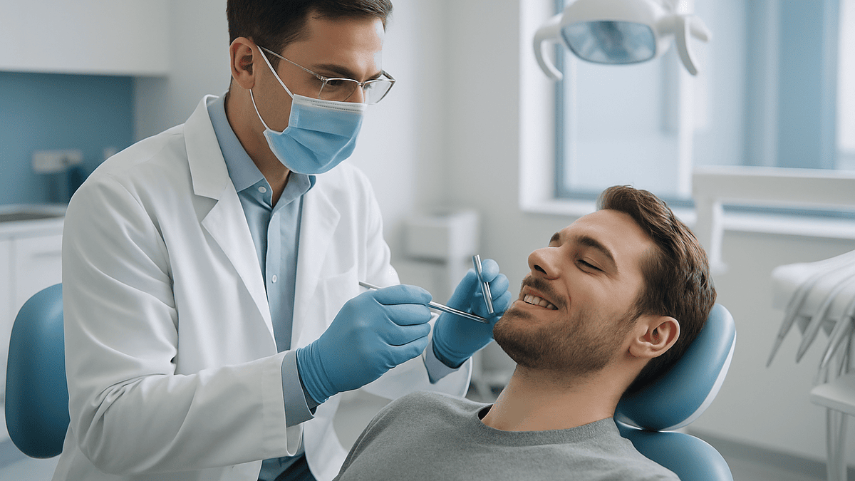
A dental bone graft procedure adds bone or bone-like material to a thinned area of the jaw so it can later hold an implant or support the gums. Under local anesthesia, the surgeon opens the gum to expose the bony ridge, cleans the site, packs in graft granules or a block, covers it with a protective membrane, and stitches the gum closed. Over several months the graft is gradually replaced by the patient's own bone.
The X-ray is up on the screen, and the spot where the molar used to be looks like a small valley. The surgeon taps it with a pen. “There isn’t enough bone here for an implant yet,” she says. “We’d need to build it first.” For a lot of people, that is the moment they hear the words dental bone graft procedure for the first time, and the questions arrive all at once: build it with what, exactly? Does it hurt? How long does it take before anything can go in?
The honest answer is that bone grafting in the mouth is both less dramatic and more interesting than it sounds. It is usually a short outpatient appointment done with the same numbing used for a filling. What makes it work is not the surgeon’s hands alone but a piece of biology: bone, given the right scaffold and left undisturbed, rebuilds itself.
This explainer walks through each stage, from the first incision to the last stitch, and then through the weeks that follow, correcting a few myths along the way.
What is a dental bone graft procedure, in plain language?
A dental bone graft procedure is minor oral surgery in which a surgeon places bone or a bone-like material into a part of the jaw that has become too thin, too short or too hollow to do its job. The job is usually one of two things: anchoring a dental implant, or holding the gum and neighboring teeth in a stable position after a tooth has been lost.
The graft itself is not a permanent part. Think of it as scaffolding rather than brick. The material, whether it comes from the patient, a donor or a synthetic source, acts as a framework that the body’s own bone-forming cells can crawl into. Over months those cells lay down new bone along the scaffold while the graft material is slowly broken down and absorbed. The end result, when healing goes well, is living jawbone that belongs to the patient. Cleveland Clinic describes this process as the graft acting as a “placeholder” while the body regenerates its own tissue.
Two technical words come up often, so here they are in one sentence each. Osseointegration is the process by which bone grows tightly against a titanium implant so the implant becomes fixed. Osteoconduction is the ability of a graft material to serve as a passive scaffold that guides that new bone growth. A bone graft is essentially an osteoconduction step that makes osseointegration possible later.
Most grafts in the jaw are done under local anesthesia in a dental or oral surgery chair, and Mayo Clinic notes that they are commonly performed on an outpatient basis. The appointment often takes under an hour for a small site. The grafted area is then left alone while biology does the slow part, which is why the calendar, not the procedure itself, is what most patients end up asking about.
Why does the jaw lose bone in the first place?
Jawbone behaves like muscle in one respect: it stays strong where it is used and shrinks where it is not. The bone that surrounds tooth roots, called the alveolar ridge, exists to hold teeth. Every time you bite, the roots transmit tiny forces into that bone, and the bone responds by staying dense. Remove the tooth and the signal stops. The ridge begins to narrow and lower, most noticeably in the first months after an extraction, and the process continues more slowly for years. Cleveland Clinic points out that this loss starts as soon as a tooth is gone, which is why some grafts are placed on the same day as the extraction.

Gum disease is the other major thief. Periodontitis is a chronic bacterial infection of the tissues around the teeth that, according to the National Institute of Dental and Craniofacial Research, destroys the bone supporting the teeth if it is not controlled. Patients who lose teeth to advanced gum disease often arrive with less bone than those who lose a tooth to a fracture, because the infection has already been eating away at the ridge.
Several other situations leave thin bone behind:
- Long-term wear of a removable denture, which presses on the ridge without stimulating it.
- Trauma to the face or jaw that fractures or removes a section of bone.
- Teeth that never developed, leaving a ridge that was never fully formed.
- Cysts or benign growths that had to be removed along with surrounding bone.
- The upper back jaw, where the maxillary sinus sits just above the molar roots and naturally leaves little bone height.
Understanding the cause matters because it shapes the plan. A ridge thinned by disuse behaves differently from one hollowed by infection, and the surgeon will want the infection fully controlled before any graft goes in.
Who is usually a candidate for a bone graft for dental implant treatment, and who is asked to wait?
The most common reason for a bone graft for dental implant treatment is simple geometry. An implant is a small titanium post, and it needs bone around it on all sides, much like a fence post needs soil packed around it rather than balanced on a crumbling edge. When imaging shows the ridge is too narrow, too short or too close to the sinus or a nerve, the surgeon has a choice: pick a shorter or narrower implant, angle it differently, or add bone. Mayo Clinic notes that a graft is recommended when the jawbone is too soft or not thick enough to support an implant.
People who are commonly offered grafting include those planning an implant after an extraction, those with old extraction sites that have shrunk, those with gum disease that has been treated and stabilized, and those whose upper molars sit beneath a large sinus.
Some people are asked to wait, and the reasons are about healing rather than eligibility. The treating team may pause grafting when:
- Active gum disease or an untreated dental infection is present nearby.
- Blood sugar is poorly controlled, since high glucose slows wound healing.
- The person smokes heavily, because smoking restricts blood flow to the gums and is linked with graft failure in Cleveland Clinic’s patient guidance.
- Certain medicines that affect bone turnover, or a history of radiation to the jaw, raise the risk of poor bone healing. This is a conversation for the prescribing clinician, never a reason to stop a medicine on your own.
- A recent illness or surgery has left the body with less reserve for healing.
Being asked to wait is not a refusal. It is usually a plan: treat the infection, stabilize the medical condition, arrange smoking support, then return. The decision about timing always sits with the surgeon and, where relevant, the physician managing the underlying condition.
Where does the graft material come from?
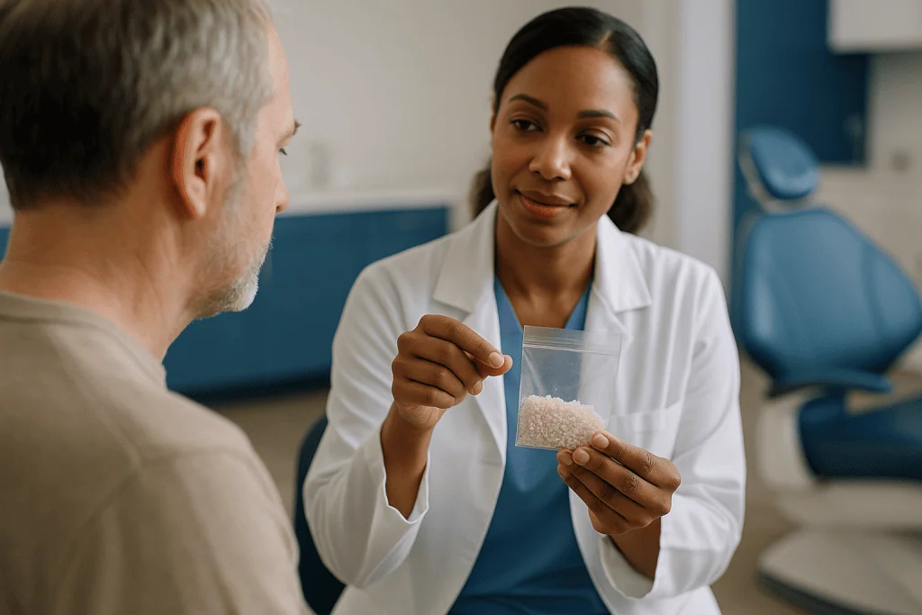
Patients are often surprised that there are four broad families of graft material, and that the surgeon may mix them. Each has a trade-off between how quickly it becomes living bone and what it costs the patient in terms of an extra surgical site. Cleveland Clinic and Mayo Clinic describe all four as accepted options; none is universally “best,” and the choice depends on the size of the defect and the surgeon’s judgment.
| Type | Source | Main advantage | Main trade-off |
|---|---|---|---|
| Autograft | The patient’s own bone, often from the chin, back of the lower jaw or, for large defects, the hip | Contains the patient’s living cells and growth signals | Requires a second surgical site, with its own soreness |
| Allograft | Donated human bone, processed and sterilized by a tissue bank | No second site; widely available in granule or block form | Cells are removed, so it acts mainly as a scaffold |
| Xenograft | Animal-derived bone, usually bovine, processed to remove organic material | Resorbs slowly, holding volume well | Some people decline for personal or religious reasons |
| Alloplast | Synthetic mineral such as calcium phosphate or bioactive glass | No human or animal tissue; consistent supply | Scaffold only; performance varies by product and site |
A tissue bank is a regulated facility that screens, processes and stores donated human tissue. Donor bone used in dentistry goes through screening and sterilization steps designed to remove cells and reduce infection risk, and Cleveland Clinic notes that disease transmission from processed allograft is considered very rare.
Which material a surgeon reaches for depends on the defect. A small socket may be filled with granules of allograft or xenograft; a large vertical loss may call for a block of the patient’s own bone screwed into place. Patients who feel strongly about avoiding human or animal tissue should say so at the consultation, because a synthetic alternative usually exists.
Step one: numbing, incision and ridge exposure
The appointment begins much like any other in the dental chair. The surgeon reviews the imaging, usually a cone-beam CT scan, which is a three-dimensional X-ray that shows bone width and height in millimeters. Some people are offered sedation in addition to local anesthesia; that decision is made with the team based on anxiety, the size of the graft and medical history.
Local anesthetic is placed around the site. Once the area is fully numb, the surgeon makes an incision along the top of the ridge and sometimes small release cuts at the sides. The gum, together with the thin membrane under it that supplies blood to the bone, is gently lifted away from the bone. This lifted section is called a flap, and the membrane beneath it is the periosteum, a living layer that feeds the outer surface of bone and helps it heal.
With the flap raised, the surgeon can see the actual shape of the ridge for the first time. This step, called ridge exposure, often reveals details the scan hinted at: a knife-edge crest, a dent on the cheek side where a root used to be, or a thin shell of bone over a void. The surgeon measures again here, because plans sometimes change once the bone is visible.
Patients typically feel pressure and vibration during this stage but not sharp pain, because the anesthetic blocks the nerve signals. Cleveland Clinic advises patients to expect a sensation of pushing and pulling rather than pain, and to raise a hand if anything feels sharp so more anesthetic can be added. The whole exposure step often takes only a few minutes for a single-tooth site.
Step two: preparing the site and placing the graft
Bone does not grow well onto a dirty or inert surface, so the exposed ridge is prepared before anything is added. The surgeon removes any remaining soft tissue, infected material or fragments from the old socket. In many cases small holes are then drilled through the dense outer layer of bone, a step called decortication, which means opening tiny channels into the marrow so blood and bone-forming cells can reach the graft. Bleeding from bone is welcome here; it carries the cells that will do the rebuilding.
The graft material is then placed. Granules are moistened with saline or the patient’s own blood so they hold together, then packed into the defect and shaped until the ridge has the contour the implant will need. A block graft is fitted against the ridge and fixed with one or two small titanium screws so it cannot shift while it heals. The surgeon is aiming for a slightly over-built shape, because some shrinkage during healing is expected.
Where the ridge is being widened rather than filled, the technique is often called guided bone regeneration, or GBR, which simply means using a barrier to guide where new bone forms. Where a socket is filled at the time of extraction, it is called socket preservation. Where bone is added to the floor of the upper sinus, it is a sinus lift. The packing step looks similar in each, but the anatomy and risks differ, which the next sections address.
Some surgeons draw a small blood sample beforehand, spin it in a centrifuge and place the resulting concentrate of platelets and proteins over the graft. Cleveland Clinic describes this as an option some surgeons use; the evidence on whether it speeds healing is mixed, and it is best understood as an adjunct rather than a requirement. Patients can reasonably ask whether it is planned and why.
Step three: the bone graft membrane, sutures and closure
Left uncovered, a bed of graft granules would be invaded by fast-growing gum cells before slow-growing bone cells could claim it. The result would be soft tissue, not bone. The answer is a bone graft membrane: a thin sheet placed over the graft that acts as a fence, holding the granules in place and keeping gum tissue out while bone fills in underneath.
Membranes come in two broad kinds. Resorbable membranes, usually made of collagen, dissolve on their own over weeks to months and need no second procedure. Non-resorbable membranes, often made of a dense synthetic material, sometimes reinforced with titanium, hold their shape better for large defects but must be removed at a later visit. Cleveland Clinic lists both as standard options; which one is used depends on how much the graft needs to be protected from collapse.
The membrane is trimmed to size, tucked under the edges of the flap and sometimes pinned with tiny tacks. The flap is then brought back over everything. Because the ridge is now wider or taller than before, the gum often no longer meets in the middle. The surgeon releases the periosteum with a shallow scoring cut so the flap can stretch, then closes the incision with sutures placed without tension. Tension is the enemy of grafts: a tight flap pulls apart during healing and exposes the membrane.
Sutures may be dissolvable or may need removal at a follow-up visit. A gauze pad is placed, and the patient bites gently for a period to control bleeding. Cleveland Clinic notes that most people leave the same day and that the first day or two involve swelling and some oozing, both expected. This closure stage is where surgical skill shows most, because the fate of the graft over the next months depends heavily on whether the gum stays sealed.
Socket preservation, ridge augmentation and sinus lift: how the dental bone graft procedure differs by site
The phrase dental bone graft procedure covers several operations that share the same steps but vary in where the bone goes and what sits nearby.
Socket preservation is the simplest. The tooth is removed as gently as possible to keep the thin socket walls intact, the empty socket is cleaned and filled with granules, a small membrane or collagen plug is placed over the top, and a stitch or two holds it. Because the socket walls already exist, there is no need for a large flap. Cleveland Clinic describes this as a way to prevent the shrinkage that would otherwise follow extraction, giving the future implant site a head start.
Ridge augmentation rebuilds a ridge that has already shrunk. This is the full sequence described above: flap, decortication, granules or block, membrane, tension-free closure. Widening a ridge is generally more predictable than adding height, because gravity and the pull of the lip work against vertical gains. Surgeons often quote longer healing for vertical augmentation.
Sinus lift applies to the upper back jaw. The maxillary sinus is an air-filled cavity behind the cheekbone that sits directly above the roots of the upper molars. When those teeth are lost, the sinus floor often drops and the bone beneath it becomes very thin. The surgeon opens a small window in the side wall of the jaw, lifts the delicate sinus lining upward like raising a tent, and packs graft under it to create new floor height. Mayo Clinic describes this as one of the specialized grafting approaches used before upper-jaw implants.
The sinus lift carries a specific instruction set afterward: no forceful nose-blowing, sneezing with the mouth open, and no drinking through straws for a period the surgeon will specify, because pressure changes can tear the lining or push graft into the sinus. Patients with chronic sinus problems should mention them at the consultation.
Is a dental bone graft painful? What the evidence and experience actually show
“How painful is it?” is the most-searched question about this surgery, and it deserves a straight answer. During the procedure, with adequate local anesthesia, patients report pressure and vibration rather than pain. Afterward, the picture is closer to a tooth extraction than to a major operation. Cleveland Clinic states that most people describe discomfort in the days following a bone graft as manageable and comparable to having a tooth removed, with swelling and tenderness peaking around the second or third day and then easing.
Several factors shape how sore a given person will be:
- Size of the site: a single socket preservation is generally milder than a block graft or sinus lift.
- Second surgical site: an autograft taken from the chin or back of the jaw adds a second sore area, and hip harvests, which are uncommon in dentistry, involve more pain and a limp for some days.
- Swelling: the cheek over a large graft may swell noticeably; this is inflammation, not infection, and it is expected.
- Individual variation: pain thresholds and anxiety levels differ, and both influence the experience.
Pain control after grafting usually relies on over-the-counter anti-inflammatory or acetaminophen-type medicines, with the surgeon advising what is appropriate given the patient’s medical history and other prescriptions. Anti-inflammatory medicines work by reducing the chemical signals that cause swelling and soreness, which is why they are often favored for dental surgery. Any decision about which medicine, and whether a stronger prescription is warranted, belongs to the treating clinician. Ice applied to the cheek in the first day, a soft diet and rest do a surprising amount of the work.
One honest caveat: pain that worsens after the third day rather than improving, or pain accompanied by a bad taste, fever or pus, is not normal soreness. That pattern belongs in the “when to call” list further down.
Dental bone graft healing time: what the following days and weeks usually look like
Dental bone graft healing time has two clocks running at once. The first is the soft-tissue clock, which is what the patient feels. The second is the bone clock, which only imaging can read.
On the soft-tissue clock, Cleveland Clinic gives a typical range of one to two weeks for the gum to close and for swelling, tenderness and diet restrictions to ease. The first 24 to 48 hours bring the most swelling and some oozing of blood-tinged saliva. Bruising on the cheek or under the jaw can appear on day two or three and fade over a week. Sutures dissolve or are removed at the follow-up visit, usually within the first two weeks. Small white or gray granules may occasionally work their way out through the gum in this period; a few escaping particles are common and not a sign of failure, but a steady stream of them or a visible gap in the gum warrants a call.
On the bone clock, the timeline is much longer. Cleveland Clinic states that a graft typically needs three to nine months before it is mature enough to accept an implant, and Mayo Clinic describes the wait as “several months” for the transplanted bone to grow enough new bone. Where in that range a given person falls depends on graft size, location, material and general health. A small socket preservation sits at the shorter end; a large sinus lift or vertical augmentation sits at the longer end.
The team will schedule a repeat scan toward the end of the expected window. Only then, with bone volume confirmed on imaging, is the implant appointment booked. Occasionally a smaller implant can be placed at the same time as the graft when enough native bone exists to hold it, which shortens the overall path. The surgeon will say which applies, and no one can promise the calendar in advance.
What can you not do after a dental bone graft?
The restrictions after a graft exist for one reason: to keep the graft still and sealed while it knits. Most of them last one to two weeks, matching the soft-tissue healing window Cleveland Clinic describes, though the surgeon will give exact timings for the specific procedure.
Things patients are usually asked to avoid in the early period:
- Disturbing the site. No touching with fingers or tongue, no pulling the lip to look, no vigorous rinsing or spitting in the first day. Gentle saltwater rinses are usually introduced after that.
- Straws and suction. Sucking creates negative pressure that can lift the clot and membrane. This is also why smoking is discouraged: the suction and the nicotine both work against healing.
- Hard, crunchy or seedy food on that side. Nuts, chips, popcorn and small seeds can lodge in the incision. Soft, cool foods are the norm for the first days.
- Strenuous exercise. Heavy lifting or running raises blood pressure and can restart bleeding or increase swelling in the first few days.
- Nose-blowing and sneezing with a closed mouth, specifically after a sinus lift, because pressure can tear the sinus lining.
- Wearing a removable denture or partial over the site unless the surgeon has adjusted it, because pressure on the graft can cause it to fail.
- Very hot drinks and alcohol in the first day or two, which can increase bleeding and interfere with some medicines.
Brushing the other teeth continues as normal, and gently around the site once the team says so; a clean mouth is protective, not a threat. If a specific medicine is prescribed, such as an antibiotic or a prescription mouth rinse, it is taken as directed by the prescriber. Patients on blood thinners should never change them on their own; the surgeon and the prescribing physician coordinate that before the appointment.
How serious is a dental bone graft? Risks and alternatives in neutral terms
Grafting is classed as minor oral surgery, but “minor” describes the scale of the operation, not the seriousness with which it is planned. Cleveland Clinic and Mayo Clinic list the same core risks, all of which are uncommon but worth knowing:
- Infection at the graft site, which can cause the graft to be lost.
- Graft exposure, where the gum opens and the membrane or granules become visible. Small exposures often heal with care; larger ones can compromise the graft.
- Bleeding and swelling beyond the expected range, more likely in people on anticoagulants.
- Nerve injury in the lower jaw, where the nerve supplying the lip and chin runs close to the graft area, causing numbness that is usually temporary but can occasionally persist.
- Sinus membrane tear during a sinus lift, which may require pausing the procedure.
- Graft failure, meaning the material does not turn into usable bone and the procedure must be repeated or the plan changed. Cleveland Clinic identifies smoking, uncontrolled diabetes and poor oral hygiene as the main modifiable risk factors.
Alternatives exist, and a good consultation lays them out. Shorter or narrower implants can sometimes avoid grafting in borderline cases. Tilted implants may reach denser bone in the front of the jaw. A fixed bridge on neighboring teeth or a well-made removable denture replaces the tooth without any implant at all. Each option has its own trade-offs in longevity, cleaning and effect on neighboring teeth, and none is right for everyone.
The seriousness question, then, has a measured answer. For a healthy non-smoker, a single-site graft carries a low risk of complication and a high likelihood of producing usable bone, though no surgeon can guarantee the outcome. For someone with several risk factors, the calculation is different, which is exactly why the treating team may suggest waiting or choosing another route.
What people often get wrong about bone grafting
Some of the most common beliefs about grafting are half true, and the half that is wrong causes needless worry.
“They take bone from my hip.” Hip harvests are reserved for very large reconstructions, often after trauma or tumor surgery. For the great majority of implant-related grafts, the material is donated, animal-derived or synthetic, or a small amount of the patient’s own bone from inside the mouth. Cleveland Clinic lists all of these as routine sources.
“The graft is the new bone.” The graft is a scaffold. What ends up under the implant is the patient’s own bone, laid down by their own cells over months while the graft is slowly absorbed. This is why healing cannot be rushed.
“Donor bone can give me a disease.” Human bone used in dentistry is screened and processed by regulated tissue banks to remove cells, and Cleveland Clinic notes that transmission of disease through processed allograft is very rare. Patients with concerns can choose a synthetic material.
“A few granules coming out means it failed.” A small number of particles working through the gum in the first week or two is common. Continuous loss, a gap in the gum or exposed membrane is different and should be reported.
“If I can’t feel it, it’s healed.” The gum heals in one to two weeks, but the bone beneath needs three to nine months by Cleveland Clinic’s estimate. Feeling fine at week three says nothing about whether the site is ready for an implant.
“The graft guarantees my implant will work.” It improves the conditions. Implant outcomes depend on many things, including bite forces, hygiene and general health, and no procedure carries a guarantee.
“Grafts only matter if I want an implant.” Socket preservation also supports the gum line and adjacent teeth, and can matter for the appearance of a future bridge or denture.
Questions to ask your care team before the appointment
A grafting consultation is short, and patients often leave with questions they meant to ask. Bringing a written list changes that. The following are the questions that tend to matter most, grouped by theme.
About the plan
- What type of graft is planned for me: socket preservation, ridge widening, height gain or sinus lift?
- Which material will you use, and why that one for my site? Is there a synthetic option if I prefer to avoid human or animal tissue?
- Will a membrane be used, and will it need to be removed later?
- Is there any chance the implant can be placed at the same visit, or is a separate stage definitely needed?
About my health
- Do any of my medicines, including blood thinners or bone medicines, need to be discussed with my physician before surgery? (Never adjust these on your own.)
- How does my diabetes, smoking or gum health affect the expected outcome, and what could I do beforehand to improve it?
About the day itself
- Will this be under local anesthesia alone, or with sedation? Do I need someone to drive me?
- How long should I plan to be in the chair?
About afterward
- What is the typical healing window you expect for my case, and when will you re-scan to check the bone?
- What exactly should I avoid, and for how long? Can I wear my denture?
- What would make you consider the graft unsuccessful, and what would the plan be then?
- Who do I call after hours if something worries me?
None of these questions is a challenge to the surgeon. Clinicians generally welcome them, because a patient who understands the plan follows the aftercare more closely, and aftercare is half the outcome.
When to call your doctor
Most people recover from a dental bone graft without any drama beyond a swollen cheek and a few days of soft food. Still, certain signs mean the site needs to be looked at rather than waited out. Both Cleveland Clinic and Mayo Clinic advise contacting the surgical team promptly if any of the following occur.
Call the same day if you notice:
- Bleeding that does not slow with firm gauze pressure after about half an hour, or bleeding that restarts heavily after the first day.
- Pain that gets worse after the third day instead of better, or pain that is not relieved by the medicines the team advised.
- Swelling that keeps increasing after day three, spreads toward the eye or down the neck, or makes it hard to open the mouth.
- Fever, chills, or a persistent bad taste or foul smell from the site.
- Pus or thick discharge from the incision.
- Visible membrane, a gap in the gum, or a steady stream of graft granules coming out.
- Numbness of the lip, chin or tongue that persists beyond the expected wearing-off time of the anesthetic.
- After a sinus lift: nosebleeds, a feeling of air or fluid moving between mouth and nose, or granules appearing in nasal discharge.
Seek urgent or emergency care if:
- Swelling causes difficulty breathing or swallowing.
- You develop a high fever with rapidly spreading facial swelling.
- Bleeding is heavy and cannot be controlled.
These situations are uncommon, but they are time-sensitive. Surgical teams generally prefer a phone call about something that turns out to be normal over a delayed call about something that is not. Keep the after-hours number provided at your appointment somewhere easy to find, and do not attempt to self-treat an exposed graft or suspected infection with leftover medicines. Every decision about what to do next belongs to the treating team, who know the specific graft, material and anatomy involved.
Frequently asked questions
How painful is a bone grafting procedure?
During the procedure itself, with adequate local anesthesia, patients feel pressure and vibration rather than pain. Afterward, Cleveland Clinic describes the soreness as comparable to a tooth extraction, peaking around the second or third day and easing over the first week. Larger grafts, sinus lifts and grafts taken from a second site in the mouth tend to be sorer. Pain that worsens after day three rather than improving should be reported to the surgeon.
How serious is a dental bone graft?
It is classed as minor oral surgery, usually done under local anesthesia as an outpatient. Risks listed by Cleveland Clinic and Mayo Clinic include infection, graft exposure, bleeding, nerve irritation in the lower jaw, sinus lining tears during a sinus lift and graft failure. These are uncommon, and smoking, uncontrolled diabetes and poor oral hygiene are the main modifiable factors. Your surgeon will weigh your individual risk before recommending the procedure.
Why doesn't dental insurance cover bone grafts?
Coverage varies widely by plan rather than following a single rule. Some plans classify grafting done to prepare for an implant as part of implant treatment, which many dental plans limit or exclude, while grafting performed to treat gum disease or following trauma may be handled differently. Medical plans occasionally cover grafts linked to injury or tumor surgery. The only reliable answer comes from a pre-treatment estimate submitted by the surgeon’s office to your specific plan.
What can you not do after a dental bone graft?
For roughly the first one to two weeks, patients are usually asked to avoid straws, smoking, vigorous rinsing or spitting, hard or seedy foods on that side, strenuous exercise, and touching the site. After a sinus lift, forceful nose-blowing and sneezing with a closed mouth are also restricted. Removable dentures should not be worn over the site unless the surgeon has adjusted them. The surgeon provides exact timings for the specific procedure.
Is a dental bone graft painful when the anesthetic wears off?
Most people notice a dull ache and swelling as the numbness fades, similar to the hours after an extraction. Cleveland Clinic advises that this discomfort is usually managed with medicines the surgeon recommends based on your health history, along with ice on the cheek and a soft diet. The peak is typically the second or third day, then it subsides. Sharp, escalating or throbbing pain after that point is a reason to call.
How long does dental bone graft healing time take before an implant?
Cleveland Clinic gives a typical range of three to nine months for the graft to mature into bone dense enough to hold an implant, and Mayo Clinic describes the wait as several months. Small socket preservation grafts sit at the shorter end; large sinus lifts and vertical ridge gains sit at the longer end. A repeat scan confirms bone volume before the implant appointment is scheduled. Individual health, smoking and graft size all shift the timeline.
What is a bone graft membrane and does it have to be removed?
A bone graft membrane is a thin barrier sheet placed over the graft to hold the material in place and keep fast-growing gum tissue out while bone forms underneath. Resorbable collagen membranes dissolve on their own over weeks to months and need no removal. Non-resorbable membranes, used for larger defects, hold their shape better but require a short second visit to remove. Your surgeon will tell you which type is planned for your site.
Can a bone graft for dental implant treatment be done at the same time as the implant?
Sometimes. When enough native bone exists to hold the implant firmly, the surgeon may place the implant and pack graft around any exposed threads in one visit. When the ridge is too thin or short to stabilize an implant, Mayo Clinic notes the graft must heal first, typically over several months, before the implant is placed in a second stage. The scan and what the surgeon sees once the ridge is exposed determine which path applies.
Where does the bone for a dental graft come from?
There are four sources: the patient’s own bone (autograft), usually from inside the mouth; donated human bone processed by a tissue bank (allograft); animal-derived bone, usually bovine (xenograft); and synthetic minerals such as calcium phosphate (alloplast). Cleveland Clinic lists all four as accepted options. Hip harvests are reserved for very large reconstructions. Patients who wish to avoid human or animal tissue can ask about a synthetic alternative.
What are the signs that a dental bone graft is failing?
Warning signs include pain that worsens after the third day, persistent swelling, fever, pus or a foul taste, a gap in the gum with visible membrane, a steady stream of granules coming out, or mobility of a fixed block graft. A few particles escaping in the first week are common and not by themselves a failure. Any of the other signs should prompt a same-day call so the surgeon can examine the site and decide on treatment.
References
- Cleveland Clinic – Dental Bone Graft
- Cleveland Clinic – Dental Implants
- NIH National Institute of Dental and Craniofacial Research – Periodontal (Gum) Disease
This article is for general information only and is not a substitute for professional medical advice. Please consult a qualified doctor about your individual situation.

More from the Blog
How Inlays and Onlays Are Placed: Shaping, Impressions, Lab Fabrication and Bonding Explained
An inlay onlay procedure usually takes two visits. First, the dentist numbs the tooth, removes decay or an old filling, and shapes the cavity…
Imaging in Implantology: How Panoramic X-Rays and 3D Scans Guide Implant Position and Depth
Implantology imaging combines two-dimensional dental X-rays, most often a panoramic film, with a three-dimensional cone beam CT (CBCT) scan when the clinician needs to…

Why Jaw Growth Decides When Implantology Can Place an Implant in a Younger Patient
Dental implants for teenagers are usually postponed until the jaw has stopped growing, because an implant fuses to bone and cannot move with the…
How Mini Implants Are Placed: Flapless Insertion, Same-Day Denture Fitting and What to Expect
A mini implant procedure places one or more narrow titanium posts into the jawbone, usually through a small pierce in the gum rather than…

All On 4 Healing Timeline: Soft Diet Weeks, Bone Integration and the Final Bridge Switch
All-on-4 healing usually unfolds in stages. Swelling and soreness peak in the first two to three days and settle over one to two weeks,…

Mini Implant Problems Explained: Loosening, Wear of Denture Attachments and When to Call
Mini implant problems most often fall into three groups: the implant itself loosening because bone has not bonded to it or has later receded,…