

How a Kidney Biopsy Is Done: Ultrasound Guidance, the Needle and What You Feel

Key Takeaways
- Most kidney biopsies are done under local anesthetic with the patient awake, lying face-down, because the doctor needs you to hold your breath while the needle is fired.
- Ultrasound guides the needle to the lower pole of the kidney, the region farthest from the organ's largest blood vessels, which is the main reason serious bleeding is uncommon.
- The spring-loaded core needle captures a cylinder of tissue narrower than a matchstick, and two or three cores are usually taken because the laboratory examines them by three different microscopy methods.
- The NIDDK describes the appointment as taking about an hour, followed by several hours lying on your back so the puncture through the kidney capsule can clot.
- A pink tinge in the urine for a day or two is expected; bright red urine, clots, inability to urinate, or faintness are the signs that need urgent attention.
- Heavy lifting and strenuous exercise are typically avoided for about two weeks afterward, according to NIDDK guidance, because a sudden rise in blood pressure can restart bleeding at the biopsy site.
A kidney biopsy is usually done with you lying on your stomach while a doctor uses ultrasound to locate the lower part of one kidney, numbs the skin and deeper tissue with local anesthetic, and passes a thin spring-loaded needle through a tiny incision to collect one or more small cores of tissue. Most people feel pressure and a brief click rather than sharp pain, and lie flat for several hours afterward.
The instructions arrive on a single sheet: nothing to eat after midnight, wear something loose, bring someone who can drive you home. What the sheet does not say is what it is like to lie face-down on a table while a person you met twenty minutes ago slides a needle toward an organ you have never seen.
That gap between the paperwork and the experience is what this article fills. If you have been told you need to find out how a kidney biopsy is done, you probably already know the why: your blood or urine tests are showing something the doctors cannot fully explain from the outside. What follows is the inside view, from the ultrasound gel on your back to the sound the needle makes to the strange, mild ache that may sit in your flank that evening.
None of it replaces the conversation with your own nephrologist, the kidney specialist guiding your care. It should, however, make that conversation shorter and far less frightening.
How is a kidney biopsy done? The short version first
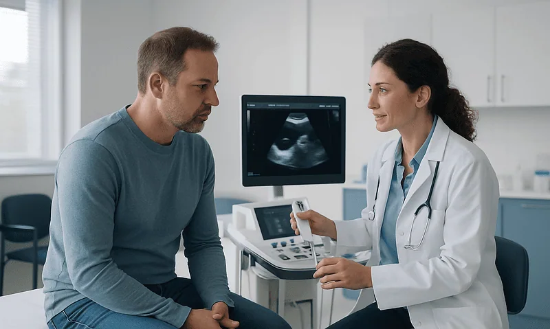
Strip away the medical vocabulary and the procedure is surprisingly compact. You lie on your stomach, sometimes with a rolled towel or firm pillow under your abdomen so the kidneys press gently backward toward the skin. A doctor, usually a nephrologist or a radiologist trained in image-guided procedures, places an ultrasound probe on your lower back and finds the kidney, typically aiming for the lower outer edge where there are fewer large blood vessels.
The skin is cleaned and numbed. A local anesthetic, a medicine that blocks pain signals in one small area while you stay fully awake, is injected first into the skin and then along the path the needle will follow. A nick a few millimeters wide is made so the biopsy needle passes cleanly.
Then comes the part people remember most: the spring-loaded needle. Guided in real time on the ultrasound screen, it is advanced to the surface of the kidney. You are asked to take a breath and hold still. The doctor fires the device, which produces a sharp click, and a slender core of kidney tissue roughly the diameter of pencil lead is captured inside the needle. The needle comes out, the core is dropped into preservative, and the pass may be repeated once or twice to be sure the pathologist has enough to work with.
The National Institute of Diabetes and Digestive and Kidney Diseases (NIDDK) describes the whole appointment, from arrival to the start of recovery, as taking about an hour, with the needle portion itself being a matter of minutes. Afterward you roll onto your back and stay lying down, usually for several hours, while nurses check your blood pressure, pulse and urine for signs of bleeding. Most people go home the same day.
Why doctors order a kidney biopsy when blood and urine tests are not enough
Kidneys are quiet organs. They can lose a great deal of function before you feel anything, which is why so much kidney disease is picked up on routine tests rather than symptoms. Those tests are good at showing that something is wrong: protein leaking into the urine, blood cells where there should be none, a creatinine level that climbs from one visit to the next. What they cannot do is show the pattern of injury inside the filtering units.

Each kidney contains roughly a million of those units, called glomeruli, tiny tufts of blood vessels that act as the first sieve for your blood. Dozens of different diseases can damage them, and many look identical from the outside. An immune condition that deposits antibodies on the filter, a scarring process, an inflammatory disease affecting several organs, or damage from long-standing diabetes can all produce protein in the urine. Treatments for these conditions differ sharply, sometimes involving medicines that suppress the immune system and sometimes involving no medicine at all beyond blood pressure control.
A biopsy lets a pathologist, a doctor who examines tissue under a microscope, look directly at the glomeruli, the small tubules that carry filtered fluid, the blood vessels and the supporting tissue. The Mayo Clinic lists the usual reasons: unexplained blood or protein in the urine, a kidney function decline without an obvious cause, checking how a known kidney disease is progressing, and evaluating a transplanted kidney that is not working as expected.
The honest framing is this: a biopsy does not make you better. It makes the next decision better informed. For many people it is the difference between a treatment plan built on a best guess and one built on what the tissue actually shows.
Who a kidney biopsy is usually for, and who is usually asked to wait
Not everyone with abnormal kidney tests needs a biopsy, and the decision is rarely automatic. Your team weighs how much the result would change management against the risk of putting a needle into an organ rich in blood vessels.
A biopsy is more likely to be recommended when the cause of kidney damage is genuinely unclear, when the amount of protein in the urine is substantial, when kidney function is falling quickly, or when a specific diagnosis would open the door to a specific treatment. It is also standard in transplant care, where a biopsy is often the only way to distinguish rejection from infection, drug toxicity or a recurrence of the original disease.
Some people are asked to wait, or a biopsy is not offered at all. Common reasons include a clotting problem that has not yet been corrected, blood pressure that is too high on the day, an active urinary or skin infection near the site, or kidneys that are already small and heavily scarred on imaging. In that last situation, the sample would mostly show old scar tissue and would not change the plan. Having only one working kidney raises the stakes of any complication, so the discussion becomes more careful, though it is not an absolute barrier.
Medicines matter too. Anticoagulants, drugs that slow clotting, and antiplatelet drugs such as aspirin, which make blood cells less sticky, both increase bleeding risk. Your team will tell you whether and when to pause them; that call depends on why you take them, and it is never a decision to make on your own. Similarly, if a person has a condition that makes lying still on their stomach impossible, alternative approaches or positions are considered rather than pressing ahead.
Preparing for a kidney biopsy: the week before and the night before
Preparation is mostly about bleeding risk, because bleeding is the complication that matters most. In the days before, you will have blood drawn to check your clotting ability, your hemoglobin (the oxygen-carrying protein in red blood cells, used as a baseline in case of later blood loss) and your kidney function. A urine sample rules out infection. Many centers also want a recent ultrasound or scan to confirm the kidneys’ size, position and whether there are two of them.

You will be asked to list every medicine and supplement you take. This is not bureaucracy. Some over-the-counter pain relievers, certain herbal products and fish oil supplements can all affect how well blood clots. The NIDDK advises telling your team about all of them so a plan can be made; the instruction about what to stop and when comes from the prescribing clinician, not from a leaflet.
Fasting is usual for several hours beforehand, partly in case sedation is needed and partly because an empty stomach is more comfortable when you lie face-down. Blood pressure is checked on arrival; if it is markedly elevated, the procedure may be postponed because high pressure in the kidney’s vessels makes bleeding more likely.
Practical details deserve attention. You will need someone to take you home, since sedation, even a mild dose, impairs judgment for the rest of the day. Bring reading material or headphones for the recovery hours. Wear something you can pull on without twisting at the waist. If you are anxious about the needle, say so during the consent conversation; some teams offer a light sedative, given by mouth or into a vein, that leaves you awake enough to hold your breath on command but noticeably calmer.
How ultrasound guidance finds the kidney and steers the needle
Ultrasound uses high-frequency sound waves, well above what the ear can hear, bounced off tissues and turned into a moving picture. It involves no radiation, which is one reason it has become the default way to guide kidney biopsies. The kidney shows up clearly against surrounding muscle and fat: an oval organ with a bright central area of collecting tubes and blood vessels, wrapped in a darker outer band called the cortex, where most of the glomeruli live.
The cortex is the target. The doctor tilts the probe until the lower pole of the kidney, its bottom tip, comes into view. This region is favored because the largest vessels enter and leave the kidney nearer its middle, so a needle at the lower pole is less likely to meet one.
Once the path is chosen, the doctor watches the needle on the screen as it advances. Modern probes can hold a guide that keeps the needle in the imaging plane, so its tip appears as a bright moving dot. The doctor can see it reach the kidney capsule, the thin fibrous skin around the organ, and stop there before firing.
Breathing is the variable to manage. Kidneys move up and down by a couple of centimeters with each breath, riding along beneath the diaphragm. That is why you are asked to hold your breath, sometimes at the end of a breath in and sometimes after breathing out, for the few seconds of each pass. Holding still is not a formality; it keeps the target where the ultrasound said it was.
Where ultrasound cannot see well, for example in people with a great deal of abdominal tissue or unusual anatomy, a CT scanner may be used instead. It gives sharper cross-sectional images at the cost of some radiation exposure.
The needle: what a spring-loaded biopsy device actually does
The instrument used for most kidney biopsies is a core needle. Unlike the fine needle used to draw blood or aspirate fluid, a core needle removes an intact cylinder of tissue so the pathologist can see the architecture, the way cells are arranged, rather than a loose scatter of cells.
The device works in two parts. An inner needle has a small notch cut into its side, a recess where tissue will settle. Around it slides an outer cutting sheath. When the doctor squeezes the trigger, a spring drives the inner needle forward a short, fixed distance into the kidney; a fraction of a second later the sheath snaps forward over it, slicing the tissue that has dropped into the notch and sealing it inside. That is the click you hear. The whole cycle takes far less time than it takes to describe.
The core that emerges is thin, narrower than a matchstick, and typically a centimeter or two long. The doctor may inspect it with a magnifier to confirm it contains cortex rather than fat or scar. Because pathology laboratories examine kidney tissue in three different ways, two or three cores are usually taken, each from a slightly different angle through the same numbed tract. The Mayo Clinic notes that the needle may need to be inserted more than once for this reason.
A worry people voice is that the needle might go through the kidney or into the bowel or lung. The fixed firing distance and real-time imaging are the safeguards: the doctor sets the tip at the capsule and knows exactly how far the spring will carry it. Injury to neighboring organs is described in the literature as rare, and the lower-pole approach keeps the needle well away from the lungs and the largest vessels.
Is a kidney biopsy painful? What you feel, minute by minute
The most common question, and the one with the most reassuring answer if you know what to expect. Almost everyone reports the same sequence.
The ultrasound gel is cold. The cleaning solution is colder. The local anesthetic is the sharpest moment: a brief sting as the first small needle enters the skin, then a spreading, slightly burning pressure as the deeper tissue is numbed. Within a minute or two the area feels thick and distant, the way your lip does after dental work.
As the biopsy needle advances you may feel pushing or pressure, a sensation of something moving inside your back, but not the sharp pain the imagination supplies. The kidney itself has few pain fibers; most sensation comes from the muscle and the capsule, and the anesthetic covers those. The click of the device is louder than you expect and can make you flinch. Some people describe a dull, deep thump at that instant, like a firm knock on a door from the inside. It passes quickly.
Afterward, when the numbness fades over the following hours, a bruised ache at the site is normal. The Mayo Clinic and NIDDK both describe soreness at the biopsy site lasting a few days. Nurses typically offer a pain reliever that does not affect clotting; which one is up to them, and you should not take anything from your own cabinet without asking.
The hardest part, most people say in retrospect, is not the needle at all. It is the hours of lying on your back afterward, unable to sit up or roll onto your side while the puncture site clots. Bring something to occupy your mind. The stiffness is the price of avoiding the complication everyone is watching for.
What happens to the tissue after the needle comes out
The moment the core leaves your body, a second procedure begins in the laboratory, and it explains why results take as long as they do.
Kidney tissue is examined in three complementary ways. The first is light microscopy: the core is preserved, sliced into sections thinner than a human hair and stained with dyes that make the glomeruli, tubules and blood vessels visible in color. This shows scarring, inflammation and the overall pattern of damage. The second is immunofluorescence, a technique that uses antibodies tagged with glowing markers to reveal whether immune proteins have been deposited on the filters, and in what arrangement. That pattern often points to a specific diagnosis. The third is electron microscopy, which magnifies the filter’s membranes tens of thousands of times to show structural changes invisible by other means.
Each method needs tissue handled differently, which is why more than one core is taken and why the samples are divided at the bedside into separate containers. A renal pathologist, a specialist in kidney tissue specifically, then integrates all three views with your clinical history and lab results before writing a report.
Timing varies. The Mayo Clinic advises that a preliminary or full report is often available within about a week, though specialized stains and electron microscopy can extend that. Your nephrologist, not the laboratory, will explain what the findings mean, because the same microscopic picture can carry different implications depending on your blood pressure, kidney function and other conditions.
Occasionally the sample is inadequate, containing too few glomeruli or mostly scar tissue. This is uncommon with ultrasound guidance, but if it happens your team may discuss whether repeating the biopsy would change the plan enough to justify it.
Kidney biopsy recovery time: the first hours and the following two weeks
Recovery has two distinct phases, and understanding the reason for each makes them easier to tolerate.
The first phase is the observation period. After the needle comes out, pressure is held on the site, a dressing is applied, and you are turned onto your back. Lying on the puncture compresses it against the table, helping a clot form where the needle passed through the kidney capsule. The NIDDK and Mayo Clinic describe this rest lasting several hours, during which nurses check your pulse and blood pressure regularly, looking for the fast heart rate or falling pressure that would signal bleeding. You will be asked to urinate before leaving so the color can be checked. A small amount of visible blood in the first urine is common; heavy, persistent bleeding is not, and would extend your stay. Some centers keep people overnight, particularly after transplant biopsies or if bleeding risk is higher.
The second phase is at home. For the first day or two, expect a bruised feeling in the flank and perhaps a small bruise on the skin. Mild pink tinge in the urine can persist briefly; the Mayo Clinic notes it usually clears within a few days. Drinking normally helps flush the urinary tract.
The important restriction is on physical strain. The NIDDK advises avoiding heavy lifting and strenuous exercise for about two weeks after the procedure. The reason is mechanical: the kidney is a soft organ that now has a small wound, and a sudden rise in blood pressure from lifting or impact could restart bleeding before the tract has fully healed. Ordinary walking, desk work and light housework are generally fine within a day or two, but your team’s written instructions take precedence over anything general.
Kidney biopsy risks and complications, honestly graded
Any honest account has to begin with bleeding, because the kidney receives a remarkable share of the heart’s output for its size and the needle unavoidably crosses small vessels.
Minor bleeding is the norm rather than the exception. Blood may collect in a small pool around the kidney, called a hematoma, or appear in the urine. Most of these events are self-limiting, cause little more than a sore back, and are discovered only if a follow-up scan is done. The Mayo Clinic and NIDDK both describe this kind of minor bleeding as common and usually resolving on its own within days.
More significant bleeding, enough to require a blood transfusion or a procedure to seal the vessel, is uncommon. When it does occur, radiologists can often pass a fine tube through a groin artery and block the bleeding vessel from the inside, avoiding surgery. Loss of the kidney because of uncontrollable bleeding is described in patient guidance as rare.
A less familiar complication is an arteriovenous fistula: an abnormal connection between a small artery and vein created by the needle. Many close on their own; a few need the same sealing procedure. Infection at the site is possible but uncommon because the puncture is tiny and the skin is sterilized. Injury to nearby organs is rare with imaging guidance.
Pain, in this hierarchy, sits at the bottom. It is expected, usually mild to moderate and temporary.
The alternative, remember, is not zero risk. It is treating a kidney disease without knowing exactly what it is, which can mean months on medicines with real side effects that turn out not to be needed, or delay in treating something that responds well when caught early. That trade-off is what the consent conversation is for.
Native kidney, transplant kidney and other biopsy approaches compared
The face-down, ultrasound-guided approach described so far is the workhorse for biopsies of a person’s own kidneys, often called native kidneys. Other situations call for variations, and the table below lays them out.
| Approach | How it is done | Anesthesia | Typically used when |
|---|---|---|---|
| Percutaneous, native kidney | Face-down; ultrasound or CT guides a core needle through the back into the lower pole | Local, sometimes with mild sedation | Most diagnostic biopsies of a person’s own kidneys |
| Percutaneous, transplant kidney | Lying on the back; the transplanted kidney sits just under the skin of the lower abdomen and is reached from the front | Local | Suspected rejection, unexplained rise in creatinine, scheduled surveillance |
| Transvenous (transjugular) | A catheter is threaded from a neck vein down to the kidney vein; the sample is taken from inside the organ | Local with sedation | Severe bleeding disorders, very high bleeding risk, or when a liver biopsy is needed at the same time |
| Open or laparoscopic surgical | A surgeon removes a small wedge of tissue under direct vision, either through an incision or with a camera | General | Percutaneous approaches are unsafe or have failed; sometimes combined with another operation |
Transplant biopsies deserve a word because they feel different. The new kidney is placed in the pelvis, close to the surface, so you lie on your back, the ultrasound probe rests on your lower abdomen, and the needle travels a shorter distance. Many transplant programs perform these biopsies at planned intervals even when everything looks fine, because rejection can begin silently.
The transvenous route is reserved for people whose blood does not clot well enough for a needle through the back. Because the sample is taken from within the kidney’s own vein, any bleeding tends to flow back into the bloodstream rather than around the organ. It is technically more complex and offered in centers with the right expertise; whether it suits you is a decision for your team.
What people often get wrong about kidney biopsies
Misunderstandings cluster around a few themes, and clearing them up tends to lower anxiety more than any reassurance about the needle.
The first is the assumption that a biopsy means cancer. In kidney medicine it usually does not. The overwhelming majority of native kidney biopsies are done to diagnose inflammatory, immune or scarring diseases of the filters. Suspicious kidney masses are more often evaluated by imaging, and when a mass is sampled it is a different conversation with a different specialist.
The second is that you will be asleep. Most kidney biopsies are done under local anesthetic with you awake, because the doctor needs you to hold your breath on command. Sedation, when offered, is light. General anesthesia is reserved for surgical approaches and for young children.
The third is that the needle removes a large chunk of kidney. The cores are slender, a tiny fraction of an organ containing roughly a million filtering units. The kidney does not lose measurable function from the sampling itself.
The fourth is that a biopsy always gives a definite answer. Usually it narrows the diagnosis considerably and often names it, but some findings are nonspecific, and the report has to be read alongside your blood tests, urine results and history. A result that says “consistent with” rather than “diagnostic of” is not a failure; it is honest pathology.
The fifth is that results arrive the same day. Preparing and staining tissue for three types of microscopy takes days, and the Mayo Clinic’s guidance of around a week reflects that. Ask your team how and when you will hear, so you are not left refreshing a patient portal.
Finally, some people believe blood in the urine afterward means something has gone badly wrong. A pink tinge in the first day or so is expected. Heavy, clotted or persistent bleeding is the concern, and that distinction is covered below.
Questions to ask your care team before a kidney biopsy
A good consent conversation is a dialogue, and the questions that help most are the ones tied to your specific situation rather than to biopsies in general. Take a list; anxiety erases memory.
- What do you expect the biopsy to show, and how would each likely result change my treatment?
- What would we do instead if I decided not to have it, and what would we lose by waiting?
- Which of my medicines and supplements need to be paused, exactly when, and when do I restart them?
- Will I be awake, and can I have something to help me relax if I need it?
- How many samples do you plan to take, and what happens if the first ones do not contain enough tissue?
- How long will I need to lie flat afterward, and is there any chance I will stay overnight?
- Who do I call, at any hour, if I have bleeding or severe pain at home, and what number do I use?
- When and how will I get the results, and who will explain them to me?
- Does my having only one kidney, or a transplanted kidney, change the approach or the risks?
- Is there anything about my anatomy on the scan that makes ultrasound guidance harder?
Two further points are worth raising if they apply. If you have had a reaction to local anesthetics or sedatives before, say so early. If lying on your stomach is difficult because of breathing problems, back pain or pregnancy, mention it at the planning visit rather than on the table, so positioning or an alternative approach can be arranged.
Write down the answers. The instructions about medicines in particular are the ones people most often misremember, and they are the ones with the most direct bearing on bleeding risk.
When to call your doctor after a kidney biopsy
You will go home with a written sheet of warning signs. Read it before you leave, while a nurse is still there to answer questions, and keep it where you can find it. The signs below are drawn from the NIDDK and Mayo Clinic patient guidance and apply to the days after a percutaneous biopsy.
Seek urgent care, calling emergency services if you cannot reach your team quickly, for any of the following:
- Urine that is bright red, contains clots, or remains visibly bloody beyond the first day or two, or bleeding that becomes heavier rather than lighter.
- Being unable to pass urine at all, which can happen if a clot blocks the flow.
- Pain in the back, flank or abdomen that is severe, worsening, or not eased by the pain relief your team advised.
- Feeling faint, lightheaded or dizzy on standing, a racing heartbeat, or looking unusually pale; these can be early signs of internal bleeding before it shows in the urine.
- Fever, chills, or increasing redness, warmth, swelling or discharge at the biopsy site.
- Shortness of breath or chest pain.
Contact your team during working hours, without waiting for the follow-up visit, if a pink tinge to the urine lasts longer than they told you to expect, if the bruised feeling in your flank is not improving after a few days, or if you are uncertain whether to restart a medicine that was paused.
Do not restart anticoagulants or antiplatelet drugs on your own timetable; the day to resume them is a decision for the prescribing clinician, who will weigh bleeding risk against the reason you take them. Equally, do not stop any regular medicine because of the procedure unless you were specifically told to.
Trust your instincts. Nurses on biopsy units would far rather take a call about a symptom that turns out to be nothing than hear afterward that someone waited.
Frequently asked questions
How long does a kidney biopsy take from start to finish?
The appointment itself, from positioning and numbing through the final needle pass, is described by the NIDDK as taking about an hour, with the needle portion lasting only minutes. The longer part is the observation period afterward, when you lie on your back for several hours while nurses monitor for bleeding. Most people leave the same day, though some centers keep patients overnight, particularly after transplant biopsies or when bleeding risk is higher.
Is a kidney biopsy painful, or just uncomfortable?
Most people describe pressure and a startling click rather than sharp pain. The sharpest moment is usually the sting of the local anesthetic being injected; once the area is numb, the biopsy needle produces a sensation of pushing and a deep thump when fired. Afterward, a bruised ache in the flank lasting a few days is normal. Your team can offer a pain reliever that does not affect clotting; avoid taking your own without asking.
What is kidney biopsy recovery time for returning to work and exercise?
Light activity such as walking and desk work is usually possible within a day or two, once the flank soreness eases. The NIDDK advises avoiding heavy lifting and strenuous exercise for about two weeks, because physical strain raises blood pressure and can reopen the small wound in the kidney before it has healed. Your own team’s written instructions may be more specific and should override any general timeline.
What are the main kidney biopsy risks and complications?
Bleeding is the principal risk. Minor bleeding, showing as a pink tinge in the urine or a small collection of blood around the kidney, is common and usually settles on its own within days. Bleeding severe enough to need a transfusion or a procedure to seal the vessel is uncommon, and loss of the kidney is rare. Other uncommon complications include an abnormal artery-to-vein connection at the needle site, infection, and injury to nearby organs.
Will I be asleep during a kidney biopsy?
Usually not. Most percutaneous kidney biopsies use local anesthetic with the patient awake, because the doctor needs you to hold your breath on command so the kidney stays still under the ultrasound. Light sedation may be offered if you are anxious, leaving you relaxed but responsive. General anesthesia is generally reserved for surgical biopsy approaches and for young children who cannot cooperate with breath-holding.
Why do I have to lie on my stomach?
Your own kidneys sit toward the back of the abdomen, just below the ribs, so the shortest and safest path for the needle is through the muscles of the lower back. Lying face-down, sometimes with a firm pillow under the abdomen, pushes the kidneys backward toward the skin and stabilizes them. Transplanted kidneys are different: they are placed near the surface of the lower abdomen, so those biopsies are done lying on your back.
How much kidney tissue is removed, and does it damage the kidney?
Very little. Each core is a slender cylinder narrower than a matchstick and typically a centimeter or two long, and two or three are usually taken. Against an organ containing roughly a million filtering units, this is a tiny fraction, and the sampling itself does not cause a measurable loss of kidney function. The tissue is divided at the bedside so the laboratory can examine it by light microscopy, immunofluorescence and electron microscopy.
When will I get my kidney biopsy results?
The Mayo Clinic notes that a report is often available within about a week, although specialized stains and electron microscopy can extend that. Preparing kidney tissue for three types of examination takes days rather than hours. Your nephrologist, rather than the laboratory, will explain the findings, because the same microscopic picture can mean different things depending on your blood pressure, kidney function and medical history. Ask in advance how you will be contacted.
Can I have a kidney biopsy if I take blood thinners?
Often yes, but with planning. Anticoagulants and antiplatelet drugs increase bleeding risk, so your team will usually ask you to pause them for a period before the procedure and tell you when to restart. The exact timing depends on which medicine you take and why, and is decided by the prescribing clinician, never independently. For people whose clotting cannot be safely corrected, a transvenous approach through a neck vein may be considered.
Is blood in my urine after a kidney biopsy normal?
A pink or slightly red tinge during the first day or two is common and expected; the Mayo Clinic notes it usually clears within a few days. What is not normal is urine that is bright red, contains clots, becomes heavier rather than lighter, or an inability to pass urine at all. Those signs, along with dizziness, a racing pulse or severe pain, need urgent contact with your team or emergency services.
References
- Kidney Biopsy: National Institute of Diabetes and Digestive and Kidney Diseases (NIH)
- Renal biopsy: MedlinePlus Medical Encyclopedia
- Biopsy: NHS
This article is for general information only and is not a substitute for professional medical advice. Please consult a qualified doctor about your individual situation.

More from the Blog

How Do a Bladder Diary and Urodynamic Testing Pinpoint Female Bladder Problems?
A bladder diary records, over roughly three days, what a woman drinks, when she passes urine, how much, and when leaks or urgency occur,…

Is Your Nephrostomy Tube Not Draining or Leaking? What Is Normal and When to Call the Team
A nephrostomy tube that stops draining is most often kinked, clamped, blocked by a clot or debris, or pulled out of position, and it…

Fluid Management With Heart or Kidney Disease: Why the Drinking Plan Is Individually Set
Fluid management in heart and kidney patients is individually set because the safe amount of fluid depends on how well the heart pumps, how…
How a Ureteral Stone Is Located Before Treatment: CT, Ultrasound and Kidney Function Tests
A ureteral stone is usually located with a non-contrast CT scan of the abdomen and pelvis, which shows the stone's size and exact position…

Bowel and Urinary Preparation Before Minimally Invasive Urology: What the Instructions Mean
Bowel prep before urology surgery ranges from nothing beyond standard fasting to a full colonoscopy-style clean-out, depending on whether the surgeon will work near…

Inherited Kidney Disease Care: How Alport Syndrome and Similar Conditions Are Monitored
Hereditary kidney disease treatment for conditions such as Alport syndrome and polycystic kidney disease centers on lifelong monitoring rather than a single procedure: regular…